|
Marginal Artery Of The Colon
In human anatomy, the marginal artery of the colon, also known as the marginal artery of Drummond, the artery of Drummond, and simply as the marginal artery, is an artery that connects the inferior mesenteric artery with the superior mesenteric artery. It is sometimes absent, as an anatomical variant. Structure The marginal artery runs in the mesentery close to the large intestine as part of the vascular arcade that connects the superior mesenteric artery and the inferior mesenteric artery. It provides an effective anastomosis between these two arteries for the large intestine. Variation The marginal artery is almost always present, and its absence should be considered a variant. Clinical significance Removal of the inferior mesenteric artery Along with branches of the internal iliac arteries, it is usually sufficiently large to supply the oxygenated blood to the large intestine. This means that the inferior mesenteric artery does not have to be re-implanted (re-attached) ... [...More Info...] [...Related Items...] OR: [Wikipedia] [Google] [Baidu] |
Abdominal Aorta
In human anatomy, the abdominal aorta is the largest artery in the abdominal cavity. As part of the aorta, it is a direct continuation of the descending aorta (of the thorax). Structure The abdominal aorta begins at the level of the diaphragm, crossing it via the aortic hiatus, technically behind the diaphragm, at the vertebral level of T12. It travels down the posterior wall of the abdomen, anterior to the vertebral column. It thus follows the curvature of the lumbar vertebrae, that is, convex anteriorly. The peak of this convexity is at the level of the third lumbar vertebra (L3). It runs parallel to the inferior vena cava, which is located just to the right of the abdominal aorta, and becomes smaller in diameter as it gives off branches. This is thought to be due to the large size of its principal branches. At the 11th rib, the diameter is 122mm long and 55mm wide and this is because of the constant pressure. The abdominal aorta is clinically divided into 2 segments: ... [...More Info...] [...Related Items...] OR: [Wikipedia] [Google] [Baidu] |
Open Aortic Surgery
Open aortic surgery (OAS), also known as open aortic repair (OAR), describes a technique whereby an abdominal, thoracic or retroperitoneal surgical incision is used to visualize and control the aorta for purposes of treatment, usually by the replacement of the affected segment with a prosthetic graft. OAS is used to treat aneurysms of the abdominal and thoracic aorta, aortic dissection, acute aortic syndrome, and aortic ruptures. Aortobifemoral bypass is also used to treat atherosclerotic disease of the abdominal aorta below the level of the renal arteries. In 2003, OAS was surpassed by endovascular aneurysm repair (EVAR) as the most common technique for repairing abdominal aortic aneurysms in the United States. Depending on the extent of the aorta repaired, an open aortic operation may be called an Infrarenal aortic repair, a Thoracic aortic repair, or a Thoracoabdominal aortic repair. A thoracoabdominal aortic repair is a more extensive operation than either an isolated infra ... [...More Info...] [...Related Items...] OR: [Wikipedia] [Google] [Baidu] |
University Of Manitoba
The University of Manitoba (U of M, UManitoba, or UM) is a Canadian public research university in the province of Manitoba.''University of Manitoba Act'', C.C.S.M. c. U60. Retrieved on July 15, 2008 Founded in 1877, it is the first of . Both by total student enrolment and campus area, the U of M is the largest university in the province of Manitoba and the 17th-largest in all of Canada. Its main campus is located in the [...More Info...] [...Related Items...] OR: [Wikipedia] [Google] [Baidu] |
Marginal Artery (other) , a branch of the circumflex artery, traveling along the left margin of heart
{{disambig ...
Marginal artery can refer to: * Marginal artery of the colon, also known as the artery of Drummond * Right marginal branch of right coronary artery, a branch of the right coronary artery that follows the acute margin of the heart * Left marginal artery The left marginal artery (or obtuse marginal artery) is a branch of the circumflex artery, originating at the left atrioventricular sulcus, traveling along the left margin of heart towards the apex of the heart. See also * Right marginal branch ... [...More Info...] [...Related Items...] OR: [Wikipedia] [Google] [Baidu] |
Marginal Branch Of The Right Coronary Artery
In the blood supply of the heart, the right coronary artery (RCA) is an artery originating above the right cusp of the aortic valve, at the right aortic sinus in the heart. It travels down the right coronary sulcus, towards the crux of the heart. It supplies the right side of the heart, and the interventricular septum. Structure The right coronary artery originates above the right aortic sinus above the aortic valve. It passes through the right coronary sulcus (right atrioventricular groove), towards the crux of the heart. It gives off many branches, including the posterior interventricular artery, the right marginal artery, the conus artery, and the sinoatrial nodal artery. Segments * Proximal: starting at RCA origin, spanning half the distance to the acute margin * Middle: from proximal segment to the acute margin * Distal: from middle segment to origination point of the posterior interventricular artery, where the posterior interventricular sulcus meets the atrioven ... [...More Info...] [...Related Items...] OR: [Wikipedia] [Google] [Baidu] |
Ischemic Colitis
Ischemic colitis (also spelled ischaemic colitis) is a medical condition in which inflammation and injury of the large intestine result from inadequate blood supply. Although uncommon in the general population, ischemic colitis occurs with greater frequency in the elderly, and is the most common form of bowel ischemia. http://www.guideline.gov/summary/summary.aspx?ss=15&doc_id=3069&nbr=2295 Causes of the reduced blood flow can include changes in the systemic circulation (e.g. low blood pressure) or local factors such as constriction of blood vessels or a blood clot. In most cases, no specific cause can be identified. Ischemic colitis is usually suspected on the basis of the clinical setting, physical examination, and laboratory test results; the diagnosis can be confirmed by endoscopy or by using sigmoid or endoscopic placement of a visible light spectroscopic catheter (see Diagnosis). Ischemic colitis can span a wide spectrum of severity; most patients are treated supportively ... [...More Info...] [...Related Items...] OR: [Wikipedia] [Google] [Baidu] |
Left Colic Artery
The left colic artery is a branch of the inferior mesenteric artery distributed to the descending colon, and left part of the transverse colon. It ends by dividing into an ascending branch and a descending branch; the terminal branches of the two branches go on to form anastomoses with the middle colic artery, and a sigmoid artery (respectively). Structure The left colic artery usually represents the dominant arterial supply to the left colic flexure. Course The left colic artery passes to the left posterior to the peritoneum. After a short but variable course, it divides into an ascending branch and a descending branch. Branches and anastomoses Ascending branch The ascending branch passes superior-ward. It passes anterior to the (ipsilateral) psoas major muscle, gonadal vessels, ureter, and kidney; it passes posterior to the inferior mesenteric vein. Its terminal branches form anastomoses with those of the middle colic artery; it also forms anastomoses with the desc ... [...More Info...] [...Related Items...] OR: [Wikipedia] [Google] [Baidu] |
Middle Colic Artery
The middle colic artery is an artery of the abdomen; a branch of the superior mesenteric artery distributed to parts of the ascending and transverse colon. It usually divides into two terminal branches - a left one and a right one - which go on to form anastomoses with the left colic artery, and right colic artery (respectively), thus participating in the formation of the marginal artery of the colon. Parts of the artery may be removed in different types of hemicolectomy. Structure The middle colic artery supplies the superior/distal part of the ascending colon and right/proximal two-thirds of the transverse colon. Origin The middle colic artery is a branch of the superior mesenteric artery, branching off from its right aspect. Its origin is situated just inferior the neck of the pancreas. It may share a common origin with the right colic artery. Course The middle colic artery passes anterosuperiorly between the layers of the transverse mesocolon just right of t ... [...More Info...] [...Related Items...] OR: [Wikipedia] [Google] [Baidu] |
Blood
Blood is a body fluid in the circulatory system of humans and other vertebrates that delivers necessary substances such as nutrients and oxygen to the cells, and transports metabolic waste products away from those same cells. Blood in the circulatory system is also known as ''peripheral blood'', and the blood cells it carries, ''peripheral blood cells''. Blood is composed of blood cells suspended in blood plasma. Plasma, which constitutes 55% of blood fluid, is mostly water (92% by volume), and contains proteins, glucose, mineral ions, hormones, carbon dioxide (plasma being the main medium for excretory product transportation), and blood cells themselves. Albumin is the main protein in plasma, and it functions to regulate the colloidal osmotic pressure of blood. The blood cells are mainly red blood cells (also called RBCs or erythrocytes), white blood cells (also called WBCs or leukocytes) and platelets (also called thrombocytes). The most abundant cells in vertebrate blood a ... [...More Info...] [...Related Items...] OR: [Wikipedia] [Google] [Baidu] |
Inferior Mesenteric Artery
In human anatomy, the inferior mesenteric artery, often abbreviated as IMA, is the third main branch of the abdominal aorta and arises at the level of L3, supplying the large intestine from the distal transverse colon to the upper part of the anal canal. The regions supplied by the IMA are the descending colon, the sigmoid colon, and part of the rectum. Structure Proximally, its territory of distribution overlaps (forms a watershed) with the middle colic artery, and therefore the superior mesenteric artery. The SMA and IMA anastomose via the marginal artery of the colon (artery of Drummond) and via Riolan's arcade (also called the "meandering artery", an arterial connection between the left colic artery and the middle colic artery). The territory of distribution of the IMA is more or less equivalent to the embryonic hindgut. Branches The IMA branches off the anterior surface of the abdominal aorta below the renal artery branch points, 3-4 cm above the aortic bifurcation ( ... [...More Info...] [...Related Items...] OR: [Wikipedia] [Google] [Baidu] |
Internal Iliac Artery
The internal iliac artery (formerly known as the hypogastric artery) is the main artery of the pelvis. Structure The internal iliac artery supplies the walls and viscera of the pelvis, the buttock, the reproductive organs, and the medial compartment of the thigh. The vesicular branches of the internal iliac arteries supply the bladder. It is a short, thick vessel, smaller than the external iliac artery, and about 3 to 4 cm in length. Course The internal iliac artery arises at the bifurcation of the common iliac artery, opposite the lumbosacral articulation, and, passing downward to the upper margin of the greater sciatic foramen, divides into two large trunks, an anterior and a posterior. It is posterior to the ureter, anterior to the internal iliac vein, anterior to the lumbosacral trunk, and anterior to the piriformis muscle. Near its origin, it is medial to the external iliac vein, which lies between it and the psoas major muscle. It is above the obturator nerve. ... [...More Info...] [...Related Items...] OR: [Wikipedia] [Google] [Baidu] |
Mesentery
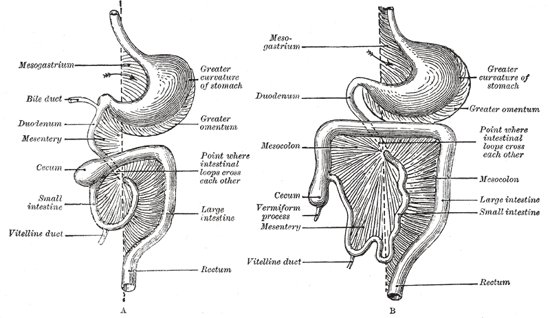
The mesentery is an organ that attaches the intestines to the posterior abdominal wall in humans and is formed by the double fold of peritoneum. It helps in storing fat and allowing blood vessels, lymphatics, and nerves to supply the intestines, among other functions. The mesocolon was thought to be a fragmented structure, with all named parts—the ascending, transverse, descending, and sigmoid mesocolons, the mesoappendix, and the mesorectum—separately terminating their insertion into the posterior abdominal wall. However, in 2012, new microscopic and electron microscopic examinations showed the mesocolon to be a single structure derived from the duodenojejunal flexure and extending to the distal mesorectal layer. Thus, the mesentery is an internal organ. Structure The mesentery of the small intestine arises from the root of the mesentery (or mesenteric root) and is the part connected with the structures in front of the vertebral column. The root is narrow, about ... [...More Info...] [...Related Items...] OR: [Wikipedia] [Google] [Baidu] |
.gif)


