|
Left Colic Artery
The left colic artery is a branch of the inferior mesenteric artery distributed to the descending colon, and left part of the transverse colon. It ends by dividing into an ascending branch and a descending branch; the terminal branches of the two branches go on to form anastomoses with the middle colic artery, and a sigmoid artery (respectively). Structure The left colic artery usually represents the dominant arterial supply to the left colic flexure. Course The left colic artery passes to the left posterior to the peritoneum. After a short but variable course, it divides into an ascending branch and a descending branch. Branches and anastomoses Ascending branch The ascending branch passes superior-ward. It passes anterior to the (ipsilateral) psoas major muscle, gonadal vessels, ureter, and kidney; it passes posterior to the inferior mesenteric vein. Its terminal branches form anastomoses with those of the middle colic artery; it also forms anastomoses with the descen ... [...More Info...] [...Related Items...] OR: [Wikipedia] [Google] [Baidu] |
Inferior Mesenteric Artery
In human anatomy, the inferior mesenteric artery, often abbreviated as IMA, is the third main branch of the abdominal aorta and arises at the level of L3, supplying the large intestine from the distal transverse colon to the upper part of the anal canal. The regions supplied by the IMA are the descending colon, the sigmoid colon, and part of the rectum. Structure Proximally, its territory of distribution overlaps (forms a watershed) with the middle colic artery, and therefore the superior mesenteric artery. The SMA and IMA anastomose via the marginal artery of the colon (artery of Drummond) and via Riolan's arcade (also called the "meandering artery", an arterial connection between the left colic artery and the middle colic artery). The territory of distribution of the IMA is more or less equivalent to the embryonic hindgut. Branches The IMA branches off the anterior surface of the abdominal aorta below the renal artery branch points, 3-4 cm above the aortic bifurcation ... [...More Info...] [...Related Items...] OR: [Wikipedia] [Google] [Baidu] |
Kidney
The kidneys are two reddish-brown bean-shaped organs found in vertebrates. They are located on the left and right in the retroperitoneal space, and in adult humans are about in length. They receive blood from the paired renal arteries; blood exits into the paired renal veins. Each kidney is attached to a ureter, a tube that carries excreted urine to the bladder. The kidney participates in the control of the volume of various body fluids, fluid osmolality, acid–base balance, various electrolyte concentrations, and removal of toxins. Filtration occurs in the glomerulus: one-fifth of the blood volume that enters the kidneys is filtered. Examples of substances reabsorbed are solute-free water, sodium, bicarbonate, glucose, and amino acids. Examples of substances secreted are hydrogen, ammonium, potassium and uric acid. The nephron is the structural and functional unit of the kidney. Each adult human kidney contains around 1 million nephrons, while a mouse kidney ... [...More Info...] [...Related Items...] OR: [Wikipedia] [Google] [Baidu] |
Colorectal Cancer
Colorectal cancer (CRC), also known as bowel cancer, colon cancer, or rectal cancer, is the development of cancer from the colon or rectum (parts of the large intestine). Signs and symptoms may include blood in the stool, a change in bowel movements, weight loss, and fatigue. Most colorectal cancers are due to old age and lifestyle factors, with only a small number of cases due to underlying genetic disorders. Risk factors include diet, obesity, smoking, and lack of physical activity. Dietary factors that increase the risk include red meat, processed meat, and alcohol. Another risk factor is inflammatory bowel disease, which includes Crohn's disease and ulcerative colitis. Some of the inherited genetic disorders that can cause colorectal cancer include familial adenomatous polyposis and hereditary non-polyposis colon cancer; however, these represent less than 5% of cases. It typically starts as a benign tumor, often in the form of a polyp, which over time becomes ... [...More Info...] [...Related Items...] OR: [Wikipedia] [Google] [Baidu] |
Abdominal Surgery
The term abdominal surgery broadly covers surgical procedures that involve opening the abdomen (laparotomy). Surgery of each abdominal organ is dealt with separately in connection with the description of that organ (see stomach, kidney, liver, etc.) Diseases affecting the abdominal cavity are dealt with generally under their own names (e.g. appendicitis). Types The most common abdominal surgeries are described below. *Appendectomy: surgical opening of the abdominal cavity and removal of the appendix. Typically performed as definitive treatment for appendicitis, although sometimes the appendix is prophylactically removed incidental to another abdominal procedure. *Caesarean section (also known as C-section): a surgical procedure in which one or more incisions are made through a mother's abdomen (laparotomy) and uterus ( hysterotomy) to deliver one or more babies, or, rarely, to remove a dead fetus. * Inguinal hernia surgery: the repair of an inguinal hernia. *Exploratory laparot ... [...More Info...] [...Related Items...] OR: [Wikipedia] [Google] [Baidu] |
Ligature (medicine)
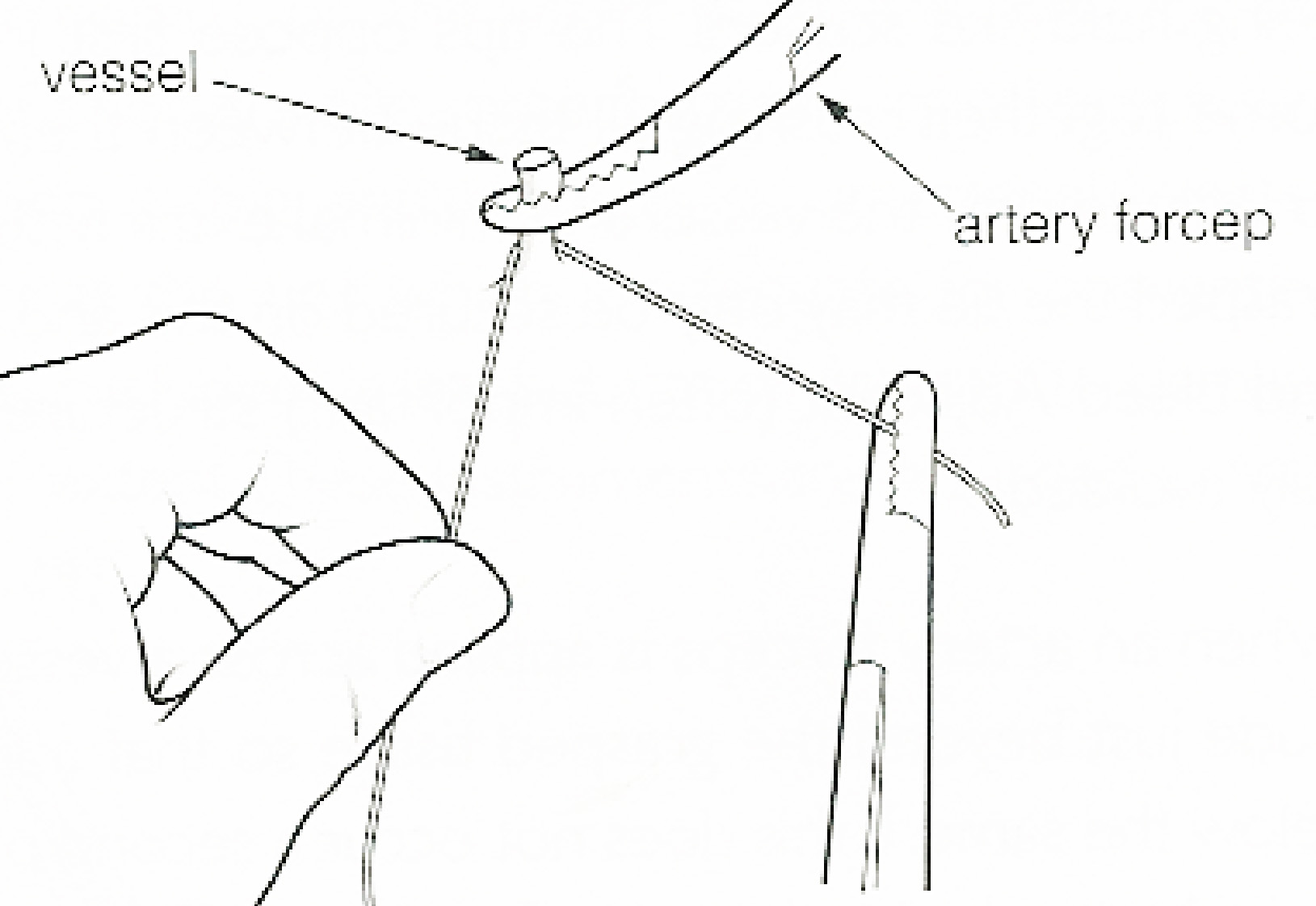
In surgery or medical procedure, a ligature consists of a piece of thread ( suture) tied around an anatomical structure, usually a blood vessel or another hollow structure (e.g. urethra) to shut it off. History The principle of ligation is attributed to Hippocrates and Galen. In ancient Rome, ligatures were used to treat hemorrhoids. The concept of a ligature was reintroduced some 1,500 years later by Ambroise Paré, and finally it found its modern use in 1870–80, made popular by Jules-Émile Péan. Procedure With a blood vessel the surgeon will clamp the vessel perpendicular to the axis of the artery or vein with a hemostat, then secure it by ligating it; i.e. using a piece of suture around it before dividing the structure and releasing the hemostat. It is different from a tourniquet in that the tourniquet will not be secured by knots and it can therefore be released/tightened at will. Ligature is one of the remedies to treat skin tag, or acrochorda. It is done by tying s ... [...More Info...] [...Related Items...] OR: [Wikipedia] [Google] [Baidu] |
Jejunal Arteries
The jejunal arteries are branches of the superior mesenteric artery which supply blood to the jejunum The jejunum is the second part of the small intestine in humans and most higher vertebrates, including mammals, reptiles, and birds. Its lining is specialised for the absorption by enterocytes of small nutrient molecules which have been previou .... External links Arteries of the abdomen {{circulatory-stub ... [...More Info...] [...Related Items...] OR: [Wikipedia] [Google] [Baidu] |
Sigmoid Artery
The sigmoid arteries are 2-5 branches of the inferior mesenteric artery that are distributed to the distal descending colon and the sigmoid colon. Anatomy Course and relations The sigmoid arteries course obliquely inferior-ward and to the left, passing posterior to the peritoneum and in anterior to the psoas major, ureter, and gonadal vessels. Anastomoses The sigmoid arteries anastomose with the left colic superiorly, and with the superior rectal artery The superior rectal artery (superior hemorrhoidal artery) is an artery that descends into the pelvis to supply blood to the rectum. Structure The superior rectal artery is the continuation of the inferior mesenteric artery. It descends into the ... inferiorly. References External links * - "Intestines and Pancreas: Branches of the Inferior Mesenteric Artery" * Arteries of the abdomen {{circulatory-stub ... [...More Info...] [...Related Items...] OR: [Wikipedia] [Google] [Baidu] |
Marginal Artery Of The Colon
In human anatomy, the marginal artery of the colon, also known as the marginal artery of Drummond, the artery of Drummond, and simply as the marginal artery, is an artery that connects the inferior mesenteric artery with the superior mesenteric artery. It is sometimes absent, as an anatomical variant. Structure The marginal artery runs in the mesentery close to the large intestine as part of the vascular arcade that connects the superior mesenteric artery and the inferior mesenteric artery. It provides an effective anastomosis between these two arteries for the large intestine. Variation The marginal artery is almost always present, and its absence should be considered a variant. Clinical significance Removal of the inferior mesenteric artery Along with branches of the internal iliac arteries, it is usually sufficiently large to supply the oxygenated blood to the large intestine. This means that the inferior mesenteric artery does not have to be re-implanted (re-attached) ... [...More Info...] [...Related Items...] OR: [Wikipedia] [Google] [Baidu] |
Sigmoid Arteries
The sigmoid arteries are 2-5 branches of the inferior mesenteric artery that are distributed to the distal descending colon and the sigmoid colon. Anatomy Course and relations The sigmoid arteries course obliquely inferior-ward and to the left, passing posterior to the peritoneum and in anterior to the psoas major, ureter, and gonadal vessels. Anastomoses The sigmoid arteries anastomose with the left colic superiorly, and with the superior rectal artery The superior rectal artery (superior hemorrhoidal artery) is an artery that descends into the pelvis to supply blood to the rectum. Structure The superior rectal artery is the continuation of the inferior mesenteric artery. It descends into the ... inferiorly. References External links * - "Intestines and Pancreas: Branches of the Inferior Mesenteric Artery" * Arteries of the abdomen {{circulatory-stub ... [...More Info...] [...Related Items...] OR: [Wikipedia] [Google] [Baidu] |
Inferior Mesenteric Vein
In human anatomy, the inferior mesenteric vein (IMV) is a blood vessel that drains blood from the large intestine. It usually terminates when reaching the splenic vein, which goes on to form the portal vein with the superior mesenteric vein (SMV). Structure The inferior mesenteric vein merges with the splenic vein, posterior to the middle of the body of the pancreas. The splenic vein then merges with the superior mesenteric vein to form the portal vein. Tributaries Tributaries of the inferior mesenteric vein drain the large intestine, sigmoid colon and rectum. These include: * left colic vein * sigmoid veins * superior rectal vein * rectosigmoid veins Variation Anatomical variations include the inferior mesenteric vein draining into the confluence of the superior mesenteric vein ''and'' splenic vein and the inferior mesenteric vein draining in the superior mesenteric vein. Clinical significance The inferior mesenteric vein may be damaged during surgery on the body a ... [...More Info...] [...Related Items...] OR: [Wikipedia] [Google] [Baidu] |
Ureter
The ureters are tubes made of smooth muscle that propel urine from the kidneys to the urinary bladder. In a human adult, the ureters are usually long and around in diameter. The ureter is lined by urothelial cells, a type of transitional epithelium, and has an additional smooth muscle layer that assists with peristalsis in its lowest third. The ureters can be affected by a number of diseases, including urinary tract infections and kidney stone. is when a ureter is narrowed, due to for example chronic inflammation. Congenital abnormalities that affect the ureters can include the development of two ureters on the same side or abnormally placed ureters. Additionally, reflux of urine from the bladder back up the ureters is a condition commonly seen in children. The ureters have been identified for at least two thousand years, with the word "ureter" stemming from the stem relating to urinating and seen in written records since at least the time of Hippocrates. It is, ho ... [...More Info...] [...Related Items...] OR: [Wikipedia] [Google] [Baidu] |
Sigmoid Colon
The sigmoid colon (or pelvic colon) is the part of the large intestine that is closest to the rectum and anus. It forms a loop that averages about in length. The loop is typically shaped like a Greek letter sigma (ς) or Latin letter S (thus ''sigma'' + '' -oid''). This part of the colon normally lies within the pelvis, but due to its freedom of movement it is liable to be displaced into the abdominal cavity. Structure The sigmoid colon begins at the superior aperture of the lesser pelvis, where it is continuous with the iliac colon, and passes transversely across the front of the sacrum to the right side of the pelvis. It then curves on itself and turns toward the left to reach the middle line at the level of the third piece of the sacrum, where it bends downward and ends in the rectum. Its function is to expel solid and gaseous waste from the gastrointestinal tract. The curving path it takes toward the anus allows it to store gas in the superior arched portion, enabling ... [...More Info...] [...Related Items...] OR: [Wikipedia] [Google] [Baidu] |


