|
Lunate Bone
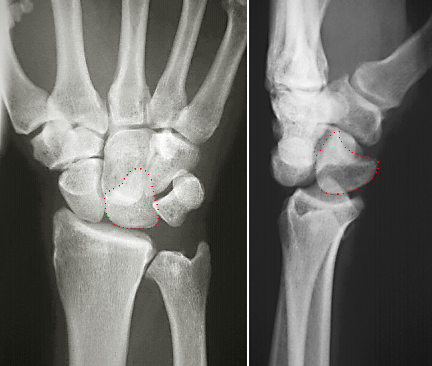
The lunate bone (semilunar bone) is a carpal bone in the human hand. It is distinguished by its deep concavity and crescentic outline. It is situated in the center of the proximal row carpal bones, which lie between the ulna and radius and the hand. The lunate carpal bone is situated between the lateral scaphoid bone and medial triquetral bone. Structure The lunate is a crescent-shaped carpal bone found within the hand. The lunate is found within the proximal row of carpal bones. Proximally, it abuts the radius. Laterally, it articulates with the scaphoid bone, medially with the triquetral bone, and distally with the capitate bone. The lunate also articulates on its distal and medial surface with the hamate bone. The lunate is stabilised by a medial ligament to the scaphoid bone and a lateral ligament to the triquetral bone. Ligaments between the radius and carpal bone also stabilise the position of the lunate, as does its position in the lunate fossa of the radius. Bone Th ... [...More Info...] [...Related Items...] OR: [Wikipedia] [Google] [Baidu] |
Radius (bone)
The radius or radial bone is one of the two large bones of the forearm, the other being the ulna. It extends from the lateral side of the elbow to the thumb side of the wrist and runs parallel to the ulna. The ulna is usually slightly longer than the radius, but the radius is thicker. Therefore the radius is considered to be the larger of the two. It is a long bone, prism-shaped and slightly curved longitudinally. The radius is part of two joints: the elbow and the wrist. At the elbow, it joins with the capitulum of the humerus, and in a separate region, with the ulna at the radial notch. At the wrist, the radius forms a joint with the ulna bone. The corresponding bone in the lower leg is the fibula. Structure The long narrow medullary cavity is enclosed in a strong wall of compact bone. It is thickest along the interosseous border and thinnest at the extremities, same over the cup-shaped articular surface (fovea) of the head. The trabeculae of the spongy t ... [...More Info...] [...Related Items...] OR: [Wikipedia] [Google] [Baidu] |
Hamate Bone
The hamate bone (from Latin hamatus, "hooked"), or unciform bone (from Latin '' uncus'', "hook"), Latin os hamatum and occasionally abbreviated as just hamatum, is a bone in the human wrist readily distinguishable by its wedge shape and a hook-like process ("hamulus") projecting from its palmar surface. Structure The hamate is an irregularly shaped carpal bone found within the hand. The hamate is found within the distal row of carpal bones, and abuts the metacarpals of the little finger and ring finger. Adjacent to the hamate on the ulnar side, and slightly above it, is the pisiform bone. Adjacent on the radial side is the capitate, and proximal is the lunate bone. Surfaces The hamate bone has six surfaces: * The ''superior'', the apex of the wedge, is narrow, convex, smooth, and articulates with the lunate. * The ''inferior'' articulates with the fourth and fifth metacarpal bones, by concave facets which are separated by a ridge. * The ''dorsal'' is triangular and rough for ... [...More Info...] [...Related Items...] OR: [Wikipedia] [Google] [Baidu] |
Upper Limb Anatomy
Upper may refer to: * Shoe upper or ''vamp'', the part of a shoe on the top of the foot * Stimulant, drugs which induce temporary improvements in either mental or physical function or both * ''Upper'', the original film title for the 2013 found footage film ''The Upper Footage ''The Upper Footage'' (also known as ''Upper'') is a 2013 found footage film written and directed by Justin Cole. First released on January 31, 2013 to a limited run of midnight theatrical screenings at Landmark’s Sunshine Cinema in New York Cit ...'' See also {{Disambiguation ... [...More Info...] [...Related Items...] OR: [Wikipedia] [Google] [Baidu] |
Wrist
In human anatomy, the wrist is variously defined as (1) the carpus or carpal bones, the complex of eight bones forming the proximal skeletal segment of the hand; "The wrist contains eight bones, roughly aligned in two rows, known as the carpal bones." (2) the wrist joint or radiocarpal joint, the joint between the radius and the carpus and; (3) the anatomical region surrounding the carpus including the distal parts of the bones of the forearm and the proximal parts of the metacarpus or five metacarpal bones and the series of joints between these bones, thus referred to as ''wrist joints''. "With the large number of bones composing the wrist (ulna, radius, eight carpas, and five metacarpals), it makes sense that there are many, many joints that make up the structure known as the wrist." This region also includes the carpal tunnel, the anatomical snuff box, bracelet lines, the flexor retinaculum, and the extensor retinaculum. As a consequence of these various definitions, fract ... [...More Info...] [...Related Items...] OR: [Wikipedia] [Google] [Baidu] |
Carpal Bone
The carpal bones are the eight small bones that make up the wrist (or carpus) that connects the hand to the forearm. The term "carpus" is derived from the Latin carpus and the Greek καρπός (karpós), meaning "wrist". In human anatomy, the main role of the wrist is to facilitate effective positioning of the hand and powerful use of the extensors and flexors of the forearm, and the mobility of individual carpal bones increase the freedom of movements at the wrist.Kingston 2000, pp 126-127 In tetrapods, the carpus is the sole cluster of bones in the wrist between the radius and ulna and the metacarpus. The bones of the carpus do not belong to individual fingers (or toes in quadrupeds), whereas those of the metacarpus do. The corresponding part of the foot is the tarsus. The carpal bones allow the wrist to move and rotate vertically. Structure Bones The eight carpal bones may be conceptually organized as either two transverse rows, or three longitudinal columns. When consid ... [...More Info...] [...Related Items...] OR: [Wikipedia] [Google] [Baidu] |
Latin
Latin (, or , ) is a classical language belonging to the Italic branch of the Indo-European languages. Latin was originally a dialect spoken in the lower Tiber area (then known as Latium) around present-day Rome, but through the power of the Roman Republic it became the dominant language in the Italian region and subsequently throughout the Roman Empire. Even after the fall of Western Rome, Latin remained the common language of international communication, science, scholarship and academia in Europe until well into the 18th century, when other regional vernaculars (including its own descendants, the Romance languages) supplanted it in common academic and political usage, and it eventually became a dead language in the modern linguistic definition. Latin is a highly inflected language, with three distinct genders (masculine, feminine, and neuter), six or seven noun cases (nominative, accusative, genitive, dative, ablative, and vocative), five declensions, four ... [...More Info...] [...Related Items...] OR: [Wikipedia] [Google] [Baidu] |
Teisen Classification
Teisen classification is a system of categorizing fractures of the lunate bone The lunate bone (semilunar bone) is a carpal bone in the human hand. It is distinguished by its deep concavity and crescentic outline. It is situated in the center of the proximal row carpal bones, which lie between the ulna and radius and the han ..., which is located in the hand. Classification References Orthopedic classifications Injuries of wrist and hand {{Orthopedics-stub ... [...More Info...] [...Related Items...] OR: [Wikipedia] [Google] [Baidu] |
Carpal Coalition
Carpal coalition is the abnormal fusion of two or more carpal bones when they fail to segment during intrauterine development. First described by Eduard Sandifort in 1779, carpal coalitions are often an isolated issue which connect two carpal bones in the same row of the wrist. These issues are congenital and occur at various rates throughout the population. Signs and symptoms Patients with carpal coalition often offer no clinical significance and patients rarely have any associated issues. Though infrequent, some patients may complain of pain. Causes Carpal coalition result from an incomplete separation of a common embryological carpal precursor in utero, during the fifth to eighth weeks. Subtypes The lunate and triquetral bones are the most common carpal bones to fuse together, resulting in a lunotriquetral coalition in 1% of people. 60% of patients with a lunotriquetral coalition will have it bilaterally. Among isolated incidents the capitate and hamate bones are the next mo ... [...More Info...] [...Related Items...] OR: [Wikipedia] [Google] [Baidu] |
Ossification
Ossification (also called osteogenesis or bone mineralization) in bone remodeling is the process of laying down new bone material by cells named osteoblasts. It is synonymous with bone tissue formation. There are two processes resulting in the formation of normal, healthy bone tissue: Intramembranous ossification is the direct laying down of bone into the primitive connective tissue (mesenchyme), while endochondral ossification involves cartilage as a precursor. In fracture healing, endochondral osteogenesis is the most commonly occurring process, for example in fractures of long bones treated by plaster of Paris, whereas fractures treated by open reduction and internal fixation with metal plates, screws, pins, rods and nails may heal by intramembranous osteogenesis. Heterotopic ossification is a process resulting in the formation of bone tissue that is often atypical, at an extraskeletal location. Calcification is often confused with ossification. Calcificati ... [...More Info...] [...Related Items...] OR: [Wikipedia] [Google] [Baidu] |
Scapholunate Ligament
The scapholunate ligament is a ligament of the wrist. Rupture of the scapholunate ligament causes scapholunate instability, which, if untreated, will eventually cause a predictable pattern of wrist osteoarthritis called scapholunate advanced collapse (SLAC). Anatomy The scapholunate ligament is an intraarticular ligament binding the scaphoid and lunate bones of the wrist together. It is divided into three areas, dorsal, proximal and palmar, with the dorsal segment being the strongest part. It is the main stabilizer of the scaphoid. In contrast to the scapholunate ligament, the lunotriquetral ligament is more prominent on the palmar side. Instability Complete rupture of this ligament leads to wrist instability. The main type of such instability is dorsal intercalated segment instability (DISI) deformity, where the lunate angulates to the posterior side of the hand. A ''dynamic scapholunate instability'' is where the scapholunate ligament is completely ruptured, but secondary ... [...More Info...] [...Related Items...] OR: [Wikipedia] [Google] [Baidu] |

_-_animation02.gif)


.jpg)