|
Lumbricals Of The Hand
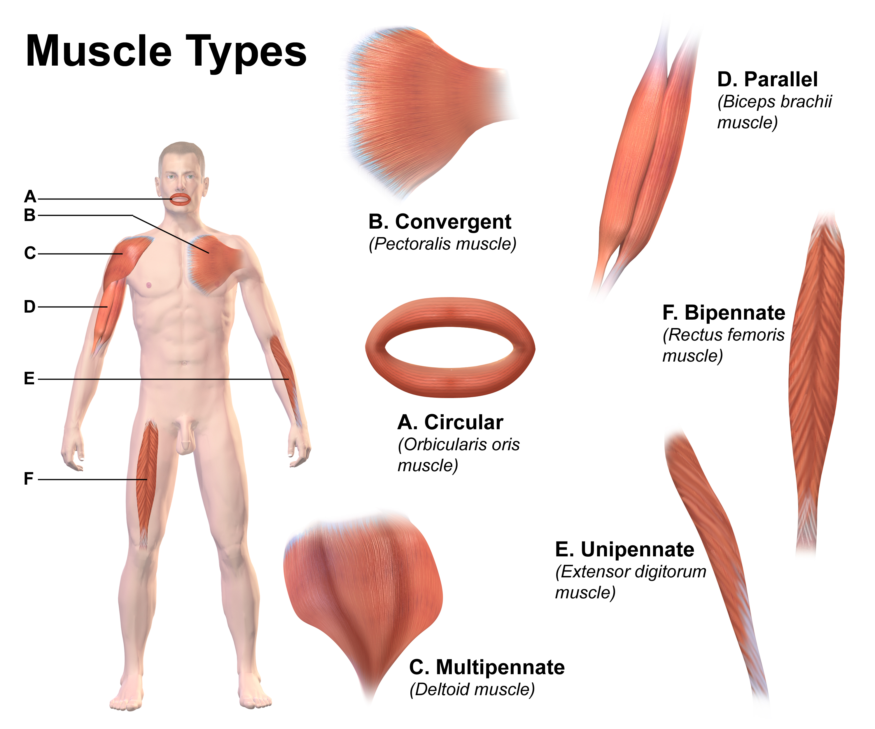
The lumbricals are intrinsic muscles of the hand that flex the metacarpophalangeal joints, and extend the interphalangeal joints. p. 97 The lumbrical muscles of the foot also have a similar action, though they are of less clinical concern. Structure The lumbricals are four, small, worm-like muscles on each hand. These muscles are unusual in that they do not attach to bone. Instead, they attach proximally to the tendons of flexor digitorum profundus, and distally to the extensor expansions. The first and second lumbricals are unipennate, while the third and fourth lumbricals are bipennate. Nerve supply The first and second lumbricals (the most radial two) are innervated by the median nerve. The third and fourth lumbricals (most ulnar two) are innervated by the deep branch of ulnar nerve. This is the usual innervation of the lumbricals (occurring in 60% of individuals). However 1:3 (median:ulnar - 20% of individuals) and 3:1 (median:ulnar - 20% of individuals) also exist ... [...More Info...] [...Related Items...] OR: [Wikipedia] [Google] [Baidu] |
Flexor Digitorum Profundus
The flexor digitorum profundus is a muscle in the forearm of humans that flexes the fingers (also known as digits). It is considered an extrinsic hand muscle because it acts on the hand while its muscle belly is located in the forearm. Together the flexor pollicis longus, pronator quadratus, and flexor digitorum profundus form the deep layer of ventral forearm muscles.Platzer 2004, p 162 The muscle is named . Structure Flexor digitorum profundus originates in the upper 3/4 of the anterior and medial surfaces of the ulna, interosseous membrane and deep fascia of the forearm. The muscle fans out into four tendons (one to each of the second to fifth fingers) to the palmar base of the distal phalanx. Along with the flexor digitorum superficialis, it has long tendons that run down the arm and through the carpal tunnel and attach to the palmar side of the phalanges of the fingers. Flexor digitorum profundus lies deep to the superficialis, but it attaches more distally. Therefore, ... [...More Info...] [...Related Items...] OR: [Wikipedia] [Google] [Baidu] |
Extension (kinesiology)
Motion, the process of movement, is described using specific anatomical terms. Motion includes movement of organs, joints, limbs, and specific sections of the body. The terminology used describes this motion according to its direction relative to the anatomical position of the body parts involved. Anatomists and others use a unified set of terms to describe most of the movements, although other, more specialized terms are necessary for describing unique movements such as those of the hands, feet, and eyes. In general, motion is classified according to the anatomical plane it occurs in. ''Flexion'' and ''extension'' are examples of ''angular'' motions, in which two axes of a joint are brought closer together or moved further apart. ''Rotational'' motion may occur at other joints, for example the shoulder, and are described as ''internal'' or ''external''. Other terms, such as ''elevation'' and ''depression'', describe movement above or below the horizontal plane. Many anatomica ... [...More Info...] [...Related Items...] OR: [Wikipedia] [Google] [Baidu] |
Worm
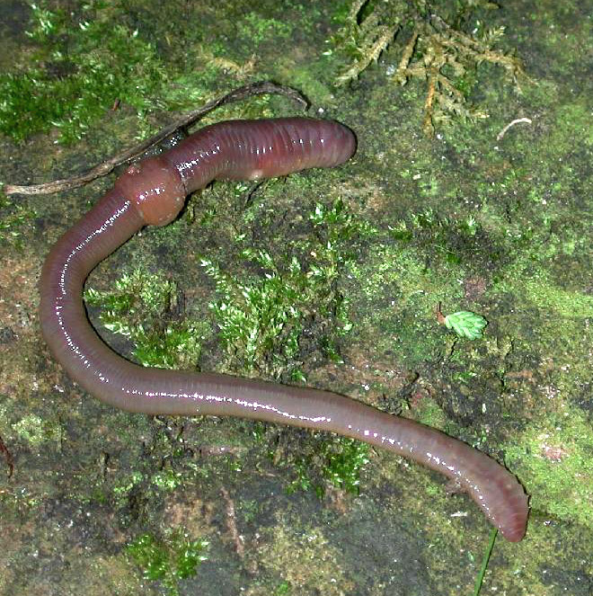
Worms are many different distantly related bilateral animals that typically have a long cylindrical tube-like body, no limbs, and no eyes (though not always). Worms vary in size from microscopic to over in length for marine polychaete worms (bristle worms); for the African giant earthworm, ''Microchaetus rappi''; and for the marine nemertean worm (bootlace worm), ''Lineus longissimus''. Various types of worm occupy a small variety of parasitic niches, living inside the bodies of other animals. Free-living worm species do not live on land but instead live in marine or freshwater environments or underground by burrowing. In biology, "worm" refers to an obsolete taxon, ''vermes'', used by Carolus Linnaeus and Jean-Baptiste Lamarck for all non-arthropod invertebrate animals, now seen to be paraphyletic. The name stems from the Old English word ''wyrm''. Most animals called "worms" are invertebrates, but the term is also used for the amphibian caecilians and the slowworm '' A ... [...More Info...] [...Related Items...] OR: [Wikipedia] [Google] [Baidu] |
Latin
Latin (, or , ) is a classical language belonging to the Italic branch of the Indo-European languages. Latin was originally a dialect spoken in the lower Tiber area (then known as Latium) around present-day Rome, but through the power of the Roman Republic it became the dominant language in the Italian region and subsequently throughout the Roman Empire. Even after the fall of Western Rome, Latin remained the common language of international communication, science, scholarship and academia in Europe until well into the 18th century, when other regional vernaculars (including its own descendants, the Romance languages) supplanted it in common academic and political usage, and it eventually became a dead language in the modern linguistic definition. Latin is a highly inflected language, with three distinct genders (masculine, feminine, and neuter), six or seven noun cases (nominative, accusative, genitive, dative, ablative, and vocative), five declensions, four verb conjuga ... [...More Info...] [...Related Items...] OR: [Wikipedia] [Google] [Baidu] |
Common Palmar Digital Artery
Three common palmar digital arteries arise from the convexity of the superficial palmar arch and proceed distally on the second, third, and fourth lumbricales muscles. Alternative names for these arteries are: common volar digital arteries, ulnar metacarpal arteries, arteriae digitales palmares communes, or aa. digitales volares communes. Each of these arteries receive the corresponding volar metacarpal artery and then divide into a pair of proper palmar digital arteries (q.v.). Additional images File:Gray527.png, Muscles and arteries of the right forearm and hand, including the superficial palmar archtitled ''Superficial Volar Arch'' in this picture, which is an alternative term and the ''common palmar digital arteries'' branching off of it. Palmar aspect with the proximal part (elbow The elbow is the region between the arm and the forearm that surrounds the elbow joint. The elbow includes prominent landmarks such as the olecranon, the cubital fossa (also called the ... [...More Info...] [...Related Items...] OR: [Wikipedia] [Google] [Baidu] |
Middle Finger
The middle finger, long finger, second finger, third finger, toll finger or tall man is the third digit of the human hand, located between the index finger and the ring finger. It is typically the longest digit. In anatomy, it is also called ''the third finger'', ''digitus medius'', ''digitus tertius'' or ''digitus III''. In Western countries, The finger, extending the middle finger (either by itself, or along with the index finger The index finger (also referred to as forefinger, first finger, second finger, pointer finger, trigger finger, digitus secundus, digitus II, and many other terms) is the second digit of a human hand. It is located between the thumb and the mid ... in the United Kingdom: see V sign) is an offensive and obscene gesture, widely recognized as a form of insult, due to its resemblance of an Erection, erect penis, It is known, colloquially, as "flipping the bird", "flipping (someone) off", or "giving (someone) the finger". The middle finger is ofte ... [...More Info...] [...Related Items...] OR: [Wikipedia] [Google] [Baidu] |
Tendon
A tendon or sinew is a tough, high-tensile-strength band of dense fibrous connective tissue that connects muscle to bone. It is able to transmit the mechanical forces of muscle contraction to the skeletal system without sacrificing its ability to withstand significant amounts of tension. Tendons are similar to ligaments; both are made of collagen. Ligaments connect one bone to another, while tendons connect muscle to bone. Structure Histologically, tendons consist of dense regular connective tissue. The main cellular component of tendons are specialized fibroblasts called tendon cells (tenocytes). Tenocytes synthesize the extracellular matrix of tendons, abundant in densely packed collagen fibers. The collagen fibers are parallel to each other and organized into tendon fascicles. Individual fascicles are bound by the endotendineum, which is a delicate loose connective tissue containing thin collagen fibrils and elastic fibres. Groups of fascicles are bounded by the epitenon, ... [...More Info...] [...Related Items...] OR: [Wikipedia] [Google] [Baidu] |
Flexor Digitorum Profundus Muscle
The flexor digitorum profundus is a muscle in the forearm of humans that flexes the fingers (also known as digits). It is considered an extrinsic hand muscle because it acts on the hand while its muscle belly is located in the forearm. Together the flexor pollicis longus, pronator quadratus, and flexor digitorum profundus form the deep layer of ventral forearm muscles.Platzer 2004, p 162 The muscle is named . Structure Flexor digitorum profundus originates in the upper 3/4 of the anterior and medial surfaces of the ulna, interosseous membrane and deep fascia of the forearm. The muscle fans out into four tendons (one to each of the second to fifth fingers) to the palmar base of the distal phalanx. Along with the flexor digitorum superficialis, it has long tendons that run down the arm and through the carpal tunnel and attach to the palmar side of the phalanges of the fingers. Flexor digitorum profundus lies deep to the superficialis, but it attaches more distally. Therefore, ... [...More Info...] [...Related Items...] OR: [Wikipedia] [Google] [Baidu] |
Ulnar Nerve
In human anatomy, the ulnar nerve is a nerve that runs near the ulna bone. The ulnar collateral ligament of elbow joint is in relation with the ulnar nerve. The nerve is the largest in the human body unprotected by muscle or bone, so injury is common. This nerve is directly connected to the little finger, and the adjacent half of the ring finger, innervating the palmar aspect of these fingers, including both front and back of the tips, perhaps as far back as the fingernail beds. This nerve can cause an electric shock-like sensation by striking the medial epicondyle of the humerus posteriorly, or inferiorly with the elbow flexed. The ulnar nerve is trapped between the bone and the overlying skin at this point. This is commonly referred to as bumping one's "funny bone". This name is thought to be a pun, based on the sound resemblance between the name of the bone of the upper arm, the humerus, and the word "humorous". Alternatively, according to the Oxford English Dictionary, i ... [...More Info...] [...Related Items...] OR: [Wikipedia] [Google] [Baidu] |
Nerve
A nerve is an enclosed, cable-like bundle of nerve fibers (called axons) in the peripheral nervous system. A nerve transmits electrical impulses. It is the basic unit of the peripheral nervous system. A nerve provides a common pathway for the electrochemical nerve impulses called action potentials that are transmitted along each of the axons to peripheral organs or, in the case of sensory nerves, from the periphery back to the central nervous system. Each axon, within the nerve, is an extension of an individual neuron, along with other supportive cells such as some Schwann cells that coat the axons in myelin. Within a nerve, each axon is surrounded by a layer of connective tissue called the endoneurium. The axons are bundled together into groups called fascicles, and each fascicle is wrapped in a layer of connective tissue called the perineurium. Finally, the entire nerve is wrapped in a layer of connective tissue called the epineurium. Nerve cells (often called neurons) are f ... [...More Info...] [...Related Items...] OR: [Wikipedia] [Google] [Baidu] |
1121 Intrinsic Muscles Of The Hand Superficial Sin
Eleven or 11 may refer to: *11 (number), the natural number following 10 and preceding 12 * one of the years 11 BC, AD 11, 1911, 2011, or any year ending in 11 Literature * ''Eleven'' (novel), a 2006 novel by British author David Llewellyn *''Eleven'', a 1970 collection of short stories by Patricia Highsmith *''Eleven'', a 2004 children's novel in The Winnie Years by Lauren Myracle *''Eleven'', a 2008 children's novel by Patricia Reilly Giff *''Eleven'', a short story by Sandra Cisneros Music *Eleven (band), an American rock band * Eleven: A Music Company, an Australian record label * Up to eleven, an idiom from popular culture, coined in the movie ''This Is Spinal Tap'' Albums * ''11'' (The Smithereens album), 1989 * ''11'' (Ua album), 1996 * ''11'' (Bryan Adams album), 2008 * ''11'' (Sault album), 2022 * ''Eleven'' (Harry Connick, Jr. album), 1992 * ''Eleven'' (22-Pistepirkko album), 1998 * ''Eleven'' (Sugarcult album), 1999 * ''Eleven'' (B'z album), 2000 * ''Eleven'' (Ream ... [...More Info...] [...Related Items...] OR: [Wikipedia] [Google] [Baidu] |
Bipennate
Muscle architecture is the physical arrangement of muscle fibers at the macroscopic level that determines a muscle’s mechanical function. There are several different muscle architecture types including: parallel, pennate and hydrostats. Force production and gearing vary depending on the different muscle parameters such as muscle length, fiber length, pennation angle, and the physiological cross-sectional area (PCSA). Architecture types Parallel and pennate (also known as pinnate) are two main types of muscle architecture. A third subcategory, muscular hydrostats, can also be considered. Architecture type is determined by the direction in which the muscle fibers are oriented relative to the force-generating axis. The force produced by a given muscle is proportional to the cross-sectional area, or the number of parallel sarcomeres present. Parallel The parallel muscle architecture is found in muscles where the fibers are parallel to the force-generating axis. These muscles are ... [...More Info...] [...Related Items...] OR: [Wikipedia] [Google] [Baidu] |


