|
Liver Sinusoidal Endothelial Cell
Liver sinusoidal endothelial cells (LSECs) form the lining of the smallest blood vessels in the liver, also called the hepatic sinusoids. LSECs are highly specialized endothelial cells with characteristic morphology and function. They constitute an important part of the reticuloendothelial system (RES). Structure Although the LSECs make up only about 3% of the total liver cell volume, their surface in a normal adult human liver is about 210 m2, or nearly the size of a tennis court. The LSEC structure differs from other endothelia. The cells contain many open pores, or fenestrae, with diameters from 100 to 150 nm. The fenestrae occupy 20% of the LSEC surface and are arranged in groups referred to as "sieve plates". Filtering fluid between the sinusoidal lumen and the space of Disse, the fenestrae are crucial for lipoprotein traffic between the hepatocytes and the sinusoidal lumen. The LSECs lack an organized basal lamina. The LSECs contain 45% and 17% of the liver's total ma ... [...More Info...] [...Related Items...] OR: [Wikipedia] [Google] [Baidu] |
Blood Vessel
The blood vessels are the components of the circulatory system that transport blood throughout the human body. These vessels transport blood cells, nutrients, and oxygen to the tissues of the body. They also take waste and carbon dioxide away from the tissues. Blood vessels are needed to sustain life, because all of the body's tissues rely on their functionality. There are five types of blood vessels: the arteries, which carry the blood away from the heart; the arterioles; the capillaries, where the exchange of water and chemicals between the blood and the tissues occurs; the venules; and the veins, which carry blood from the capillaries back towards the heart. The word ''vascular'', meaning relating to the blood vessels, is derived from the Latin ''vas'', meaning vessel. Some structures – such as cartilage, the epithelium, and the lens and cornea of the eye – do not contain blood vessels and are labeled ''avascular''. Etymology * artery: late Middle English; from Latin ... [...More Info...] [...Related Items...] OR: [Wikipedia] [Google] [Baidu] |
Toll Like Receptor
Toll-like receptors (TLRs) are a class of proteins that play a key role in the innate immune system. They are single-pass membrane-spanning receptors usually expressed on sentinel cells such as macrophages and dendritic cells, that recognize structurally conserved molecules derived from microbes. Once these microbes have reached physical barriers such as the skin or intestinal tract mucosa, they are recognized by TLRs, which activate immune cell responses. The TLRs include TLR1, TLR2, TLR3, TLR4, TLR5, TLR6, TLR7, TLR8, TLR9, TLR10, TLR11, TLR12, and TLR13. Humans lack genes for TLR11, TLR12 and TLR13 and mice lack a functional gene for TLR10. TLR1, TLR2, TLR4, TLR5, TLR6, and TLR10 are located on the cell membrane, whereas TLR3, TLR7, TLR8, and TLR9 are located in intracellular vesicles (because they are sensors of nucleic acids). TLRs received their name from their similarity to the protein coded by the toll gene. Function The ability of the immune system to recogniz ... [...More Info...] [...Related Items...] OR: [Wikipedia] [Google] [Baidu] |
Atherosclerosis
Atherosclerosis is a pattern of the disease arteriosclerosis in which the wall of the artery develops abnormalities, called lesions. These lesions may lead to narrowing due to the buildup of atheroma, atheromatous plaque. At onset there are usually no symptoms, but if they develop, symptoms generally begin around middle age. When severe, it can result in coronary artery disease, stroke, peripheral artery disease, or kidney problems, depending on which Artery, arteries are affected. The exact cause is not known and is proposed to be multifactorial. Risk factors include dyslipidemia, abnormal cholesterol levels, elevated levels of inflammatory markers, high blood pressure, diabetes, smoking, obesity, family history, genetic, and an unhealthy diet. Atheroma, Plaque is made up of fat, cholesterol, calcium, and other substances found in the blood. The narrowing of Artery, arteries limits the flow of oxygen-rich blood to parts of the body. Diagnosis is based upon a physical exam, ele ... [...More Info...] [...Related Items...] OR: [Wikipedia] [Google] [Baidu] |
Diabetes Mellitus
Diabetes, also known as diabetes mellitus, is a group of metabolic disorders characterized by a high blood sugar level ( hyperglycemia) over a prolonged period of time. Symptoms often include frequent urination, increased thirst and increased appetite. If left untreated, diabetes can cause many health complications. Acute complications can include diabetic ketoacidosis, hyperosmolar hyperglycemic state, or death. Serious long-term complications include cardiovascular disease, stroke, chronic kidney disease, foot ulcers, damage to the nerves, damage to the eyes, and cognitive impairment. Diabetes is due to either the pancreas not producing enough insulin, or the cells of the body not responding properly to the insulin produced. Insulin is a hormone which is responsible for helping glucose from food get into cells to be used for energy. There are three main types of diabetes mellitus: * Type 1 diabetes results from failure of the pancreas to produce enough insulin due to lo ... [...More Info...] [...Related Items...] OR: [Wikipedia] [Google] [Baidu] |
Cirrhosis
Cirrhosis, also known as liver cirrhosis or hepatic cirrhosis, and end-stage liver disease, is the impaired liver function caused by the formation of scar tissue known as fibrosis due to damage caused by liver disease. Damage causes tissue repair and subsequent formation of scar tissue, which over time can replace normal functioning tissue, leading to the impaired liver function of cirrhosis. The disease typically develops slowly over months or years. Early symptoms may include tiredness, weakness, loss of appetite, unexplained weight loss, nausea and vomiting, and discomfort in the right upper quadrant of the abdomen. As the disease worsens, symptoms may include itchiness, swelling in the lower legs, fluid build-up in the abdomen, jaundice, bruising easily, and the development of spider-like blood vessels in the skin. The fluid build-up in the abdomen may become spontaneously infected. More serious complications include hepatic encephalopathy, bleeding from dilated veins ... [...More Info...] [...Related Items...] OR: [Wikipedia] [Google] [Baidu] |
Endothelial Cell
The endothelium is a single layer of squamous endothelial cells that line the interior surface of blood vessels and lymphatic vessels. The endothelium forms an interface between circulating blood or lymph in the lumen and the rest of the vessel wall. Endothelial cells form the barrier between vessels and tissue and control the flow of substances and fluid into and out of a tissue. Endothelial cells in direct contact with blood are called vascular endothelial cells whereas those in direct contact with lymph are known as lymphatic endothelial cells. Vascular endothelial cells line the entire circulatory system, from the heart to the smallest capillaries. These cells have unique functions that include fluid filtration, such as in the glomerulus of the kidney, blood vessel tone, hemostasis, neutrophil recruitment, and hormone trafficking. Endothelium of the interior surfaces of the heart chambers is called endocardium. An impaired function can lead to serious health issues throug ... [...More Info...] [...Related Items...] OR: [Wikipedia] [Google] [Baidu] |
Lipoprotein Lipase
Lipoprotein lipase (LPL) (EC 3.1.1.34, systematic name triacylglycerol acylhydrolase (lipoprotein-dependent)) is a member of the lipase gene family, which includes pancreatic lipase, hepatic lipase, and endothelial lipase. It is a water-soluble enzyme that hydrolyzes triglycerides in lipoproteins, such as those found in chylomicrons and very low-density lipoproteins (VLDL), into two free fatty acids and one monoacylglycerol molecule: :triacylglycerol + H2O = diacylglycerol + a carboxylate It is also involved in promoting the cellular uptake of chylomicron remnants, cholesterol-rich lipoproteins, and free fatty acids. LPL requires ApoC-II as a cofactor. LPL is attached to the luminal surface of endothelial cells in capillaries by the protein glycosylphosphatidylinositol HDL-binding protein 1 (GPIHBP1) and by heparan sulfated peptidoglycans. It is most widely distributed in adipose, heart, and skeletal muscle tissue, as well as in lactating mammary glands. Synthesis In brief ... [...More Info...] [...Related Items...] OR: [Wikipedia] [Google] [Baidu] |
Chylomicron
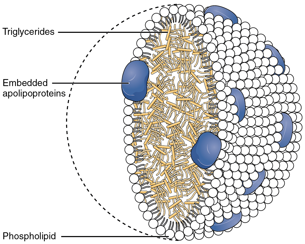
Chylomicrons (from the Greek χυλός, chylos, meaning ''juice'' (of plants or animals), and micron, meaning ''small particle''), also known as ultra low-density lipoproteins (ULDL), are lipoprotein particles that consist of triglycerides (85–92%), phospholipids (6–12%), cholesterol (1–3%), and proteins (1–2%). They transport dietary lipids from the intestines to other locations in the body. ULDLs are one of the five major groups of lipoproteins (sorted by density) that enable fats and cholesterol to move within the water-based solution of the bloodstream. A protein specific to chylomicrons is ApoB48. There is an inverse relationship in the density and size of lipoprotein particles: fats have a lower density than water or smaller protein molecules, and the larger particles have a higher ratio of internal fat molecules with respect to the outer emulsifying protein molecules in the shell. ULDLs, if in the region of 1,000 nm or more, are the only lipoprotein partic ... [...More Info...] [...Related Items...] OR: [Wikipedia] [Google] [Baidu] |
G0 Phase
The G0 phase describes a cellular state outside of the replicative cell cycle. Classically, cells were thought to enter G0 primarily due to environmental factors, like nutrient deprivation, that limited the resources necessary for proliferation. Thus it was thought of as a ''resting phase''. G0 is now known to take different forms and occur for multiple reasons. For example, most adult neuronal cells, among the most metabolically active cells in the body, are fully differentiated and reside in a terminal G0 phase. Neurons reside in this state, not because of stochastic or limited nutrient supply, but as a part of their developmental program. G0 was first suggested as a cell state based on early cell cycle studies. When the first studies defined the four phases of the cell cycle using radioactive labeling techniques, it was discovered that not all cells in a population proliferate at similar rates. A population's “growth fraction” – or the fraction of the population that w ... [...More Info...] [...Related Items...] OR: [Wikipedia] [Google] [Baidu] |
Hepatic Stellate Cell
Hepatic stellate cells (HSC), also known as perisinusoidal cells or Ito cells (earlier ''lipocytes'' or ''fat-storing cells''), are pericytes found in the perisinusoidal space of the liver, also known as the space of Disse (a small area between the sinusoids and hepatocytes). The stellate cell is the major cell type involved in liver fibrosis, which is the formation of scar tissue in response to liver damage. Structure Hepatic stellate cells can be selectively stained with gold chloride, but their distinguishing feature in routine histological preparations is the presence of multiple lipid droplets in their cytoplasm. Cytoglobin expression has been shown to be a specific marker with which hepatic stellate cells can be distinguished from portal myofibroblasts in the damaged human liver. In murine (rats, mice) liver, reelin expressed by Ito cells has been shown to be a reliable marker in discerning them from other myofibroblasts. The expression of reelin is increased after liver inju ... [...More Info...] [...Related Items...] OR: [Wikipedia] [Google] [Baidu] |
Basal Lamina
The basal lamina is a layer of extracellular matrix secreted by the epithelial cells, on which the epithelium sits. It is often incorrectly referred to as the basement membrane, though it does constitute a portion of the basement membrane. The basal lamina is visible only with the electron microscope, where it appears as an electron-dense layer that is 20–100 nm thick (with some exceptions that are thicker, such as basal lamina in lung alveoli and renal glomeruli). Structure The layers of the basal lamina ("BL") and those of the basement membrane ("BM") are described below: Anchoring fibrils composed of type VII collagen extend from the basal lamina into the underlying reticular lamina and loop around collagen bundles. Although found beneath all basal laminae, they are especially numerous in stratified squamous cells of the skin. These layers should not be confused with the lamina propria, which is found outside the basal lamina. Basement membrane The basement membra ... [...More Info...] [...Related Items...] OR: [Wikipedia] [Google] [Baidu] |
Liver Fibrosis
Cirrhosis, also known as liver cirrhosis or hepatic cirrhosis, and end-stage liver disease, is the impaired liver function caused by the formation of scar tissue known as fibrosis due to damage caused by liver disease. Damage causes tissue repair and subsequent formation of scar tissue, which over time can replace normal functioning tissue, leading to the impaired liver function of cirrhosis. The disease typically develops slowly over months or years. Early symptoms may include tiredness, weakness, loss of appetite, unexplained weight loss, nausea and vomiting, and discomfort in the right upper quadrant of the abdomen. As the disease worsens, symptoms may include itchiness, swelling in the lower legs, fluid build-up in the abdomen, jaundice, bruising easily, and the development of spider-like blood vessels in the skin. The fluid build-up in the abdomen may become spontaneously infected. More serious complications include hepatic encephalopathy, bleeding from dilated veins ... [...More Info...] [...Related Items...] OR: [Wikipedia] [Google] [Baidu] |


