|
Levator Scapulae Muscle
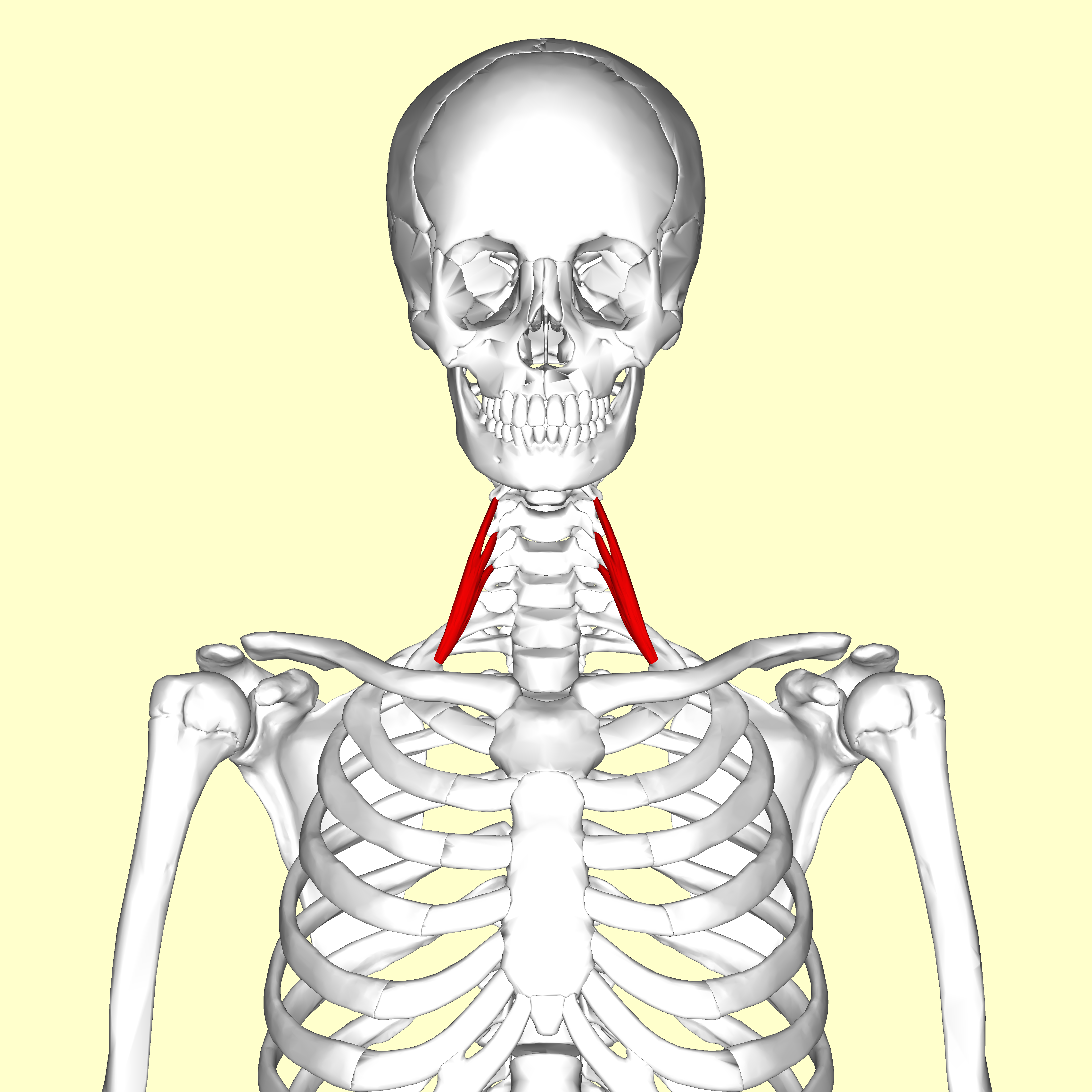
The levator scapulae is a slender skeletal muscle situated at the back and side of the neck. As the Latin name suggests, its main function is to lift the scapula. Anatomy Attachments The muscle descends diagonally from its origin to its insertion. Origin The levator scapulae originates from the posterior tubercles of the transverse processes of cervical vertebrae C1-4. At its origin, it attaches via tendinous slips. Insertion It inserts onto the medial border of the scapula (with its site of insertion extending between the superior angle of the scapula superiorly, and the junction of spine of scapula and medial border of scapula inferiorly). Relations One of the muscles within the floor of the posterior triangle of the neck, the superior part of levator scapulae is covered by sternocleidomastoid and its inferior part by the trapezius. It is bounded in front by the scalenus medius and behind by splenius cervicis. The spinal accessory nerve crosses laterally in the ... [...More Info...] [...Related Items...] OR: [Wikipedia] [Google] [Baidu] |
Vertebral Column
The vertebral column, also known as the backbone or spine, is part of the axial skeleton. The vertebral column is the defining characteristic of a vertebrate in which the notochord (a flexible rod of uniform composition) found in all chordates has been replaced by a segmented series of bone: vertebrae separated by intervertebral discs. Individual vertebrae are named according to their region and position, and can be used as anatomical landmarks in order to guide procedures such as lumbar punctures. The vertebral column houses the spinal canal, a cavity that encloses and protects the spinal cord. There are about 50,000 species of animals that have a vertebral column. The human vertebral column is one of the most-studied examples. Many different diseases in humans can affect the spine, with spina bifida and scoliosis being recognisable examples. The general structure of human vertebrae is fairly typical of that found in mammals, reptiles, and birds. The shape of the verte ... [...More Info...] [...Related Items...] OR: [Wikipedia] [Google] [Baidu] |
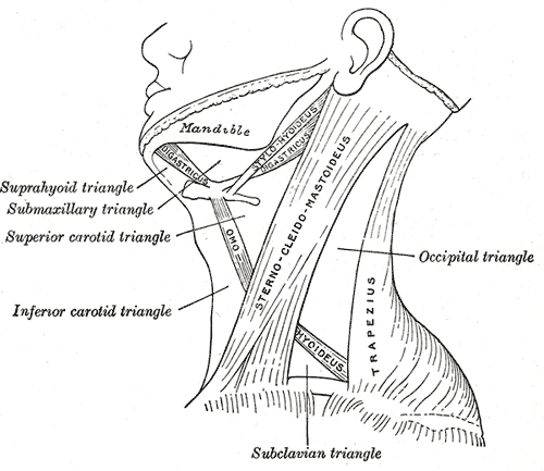
Posterior Triangle Of The Neck
Posterior may refer to: * Posterior (anatomy), the end of an organism opposite to its head ** Buttocks, as a euphemism * Posterior horn (other) * Posterior probability The posterior probability is a type of conditional probability that results from updating the prior probability with information summarized by the likelihood via an application of Bayes' rule. From an epistemological perspective, the posterior p ..., the conditional probability that is assigned when the relevant evidence is taken into account * Posterior tense, a relative future tense {{disambiguation ... [...More Info...] [...Related Items...] OR: [Wikipedia] [Google] [Baidu] |
Supraspinatous Fossa
The supraspinous fossa (supraspinatus fossa, supraspinatous fossa) of the posterior aspect of the scapula (the shoulder blade) is smaller than the infraspinous fossa, concave, smooth, and broader at its vertebral than at its humeral end. Its medial two-thirds give origin to the Supraspinatus. Structure The fossa can be exposed by the removal of skin and the superficial fascia of the back and the trapezius muscle. The supraspinous fossa is bounded by the spine of scapula on the inferior side, acromion process on the lateral side and the superior angle of scapula on the superior side. Supraspinatus muscle originates from the supraspinous fossa. Distal attachment of the levator scapulae muscle is also on the medial aspect of the fossa. Function The suprascapular artery and nerve are found within the fossa. The posterior branch of the suprascapular artery supplies the supraspinatous muscle. Dorsal scapular artery also gives off a collateral branch and anastomoses with the sup ... [...More Info...] [...Related Items...] OR: [Wikipedia] [Google] [Baidu] |
Cervical Nerves
A spinal nerve is a mixed nerve, which carries motor, sensory, and autonomic signals between the spinal cord and the body. In the human body there are 31 pairs of spinal nerves, one on each side of the vertebral column. These are grouped into the corresponding cervical, thoracic, lumbar, sacral and coccygeal regions of the spine. There are eight pairs of cervical nerves, twelve pairs of thoracic nerves, five pairs of lumbar nerves, five pairs of sacral nerves, and one pair of coccygeal nerves. The spinal nerves are part of the peripheral nervous system. Structure Each spinal nerve is a mixed nerve, formed from the combination of nerve fibers from its dorsal and ventral roots. The dorsal root is the afferent sensory root and carries sensory information to the brain. The ventral root is the efferent motor root and carries motor information from the brain. The spinal nerve emerges from the spinal column through an opening (intervertebral foramen) between adjacent verteb ... [...More Info...] [...Related Items...] OR: [Wikipedia] [Google] [Baidu] |
Gray's Anatomy
''Gray's Anatomy'' is a reference book of human anatomy written by Henry Gray, illustrated by Henry Vandyke Carter, and first published in London in 1858. It has gone through multiple revised editions and the current edition, the 42nd (October 2020), remains a standard reference, often considered "the doctors' bible". Earlier editions were called ''Anatomy: Descriptive and Surgical'', ''Anatomy of the Human Body'' and ''Gray's Anatomy: Descriptive and Applied'', but the book's name is commonly shortened to, and later editions are titled, ''Gray's Anatomy''. The book is widely regarded as an extremely influential work on the subject. Publication history Origins The English anatomist Henry Gray was born in 1827. He studied the development of the endocrine glands and spleen and in 1853 was appointed Lecturer on Anatomy at St George's Hospital Medical School in London. In 1855, he approached his colleague Henry Vandyke Carter with his idea to produce an inexpensive a ... [...More Info...] [...Related Items...] OR: [Wikipedia] [Google] [Baidu] |
Clavicle
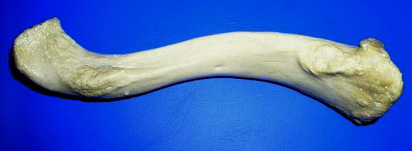
The clavicle, or collarbone, is a slender, S-shaped long bone approximately 6 inches (15 cm) long that serves as a strut between the shoulder blade and the sternum (breastbone). There are two clavicles, one on the left and one on the right. The clavicle is the only long bone in the body that lies horizontally. Together with the shoulder blade, it makes up the shoulder girdle. It is a palpable bone and, in people who have less fat in this region, the location of the bone is clearly visible. It receives its name from the Latin ''clavicula'' ("little key"), because the bone rotates along its axis like a key when the shoulder is abducted. The clavicle is the most commonly fractured bone. It can easily be fractured by impacts to the shoulder from the force of falling on outstretched arms or by a direct hit. Structure The collarbone is a thin doubly curved long bone that connects the arm to the trunk of the body. Located directly above the first rib, it acts as a strut t ... [...More Info...] [...Related Items...] OR: [Wikipedia] [Google] [Baidu] |
Serratus Anterior
The serratus anterior is a muscle that originates on the surface of the 1st to 8th ribs at the side of the chest and inserts along the entire anterior length of the medial border of the scapula. The serratus anterior acts to pull the scapula forward around the thorax. The muscle is named from Latin: ''serrare'' = to saw, referring to the shape, ''anterior'' = on the front side of the body. Structure Serratus anterior normally originates by nine or ten muscle slips – branches from either the first to ninth ribs or the first to eighth ribs. Because two slips usually arise from the second rib, the number of slips is greater than the number of ribs from which they originate. The muscle is inserted along the medial border of the scapula between the superior and inferior angles along with being inserted along the thoracic vertebrae. The muscle is divided into three named parts depending on their points of insertions: #the serratus anterior superior is inserted near the superior ... [...More Info...] [...Related Items...] OR: [Wikipedia] [Google] [Baidu] |
Scalene Muscles
The scalene muscles are a group of three pairs of muscles in the lateral neck, namely the anterior scalene, middle scalene, and posterior scalene. They are innervated by the third to the eight cervical spinal nerves (C3-C8). The anterior and middle scalene muscles lift the first rib and bend the neck to the same side; the posterior scalene lifts the second rib and tilts the neck to the same side. The muscles are named . Structure The scalene muscles originate from the transverse processes from the cervical vertebrae of C2 to C7 and insert onto the first and second ribs. Anterior scalene The anterior scalene muscle ( la, scalenus anterior), lies deeply at the side of the neck, behind the sternocleidomastoid muscle. It arises from the anterior tubercles of the transverse processes of the third, fourth, fifth, and sixth cervical vertebrae, and descending, almost vertically, is inserted by a narrow, flat tendon into the scalene tubercle on the inner border of the first rib, ... [...More Info...] [...Related Items...] OR: [Wikipedia] [Google] [Baidu] |
Trapezius
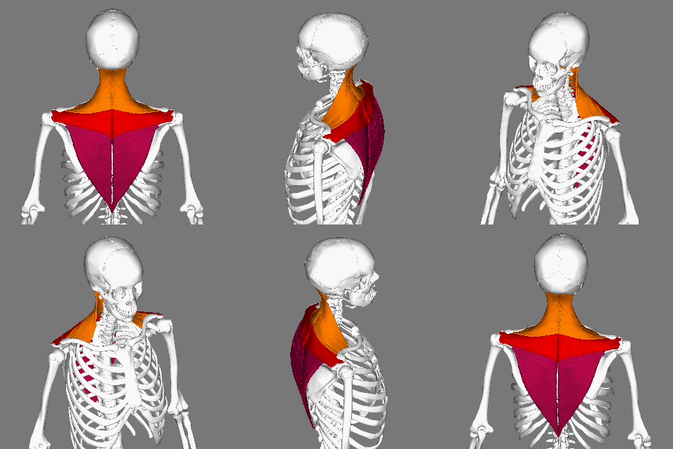
The trapezius is a large paired trapezoid-shaped surface muscle that extends longitudinally from the occipital bone to the lower thoracic vertebrae of the spine and laterally to the spine of the scapula. It moves the scapula and supports the arm. The trapezius has three functional parts: an upper (descending) part which supports the weight of the arm; a middle region (transverse), which retracts the scapula; and a lower (ascending) part which medially rotates and depresses the scapula. Name and history The trapezius muscle resembles a trapezium, also known as a trapezoid, or diamond-shaped quadrilateral. The word "spinotrapezius" refers to the human trapezius, although it is not commonly used in modern texts. In other mammals, it refers to a portion of the analogous muscle. Similarly, the term "tri-axle back plate" was historically used to describe the trapezius muscle. Structure The ''superior'' or ''upper'' (or descending) fibers of the trapezius originate from t ... [...More Info...] [...Related Items...] OR: [Wikipedia] [Google] [Baidu] |
Splenius Capitis
The splenius capitis () () is a broad, straplike muscle in the back of the neck. It pulls on the base of the skull from the vertebrae in the neck and upper thorax. It is involved in movements such as shaking the head. Structure It arises from the lower half of the nuchal ligament, from the spinous process of the seventh cervical vertebra, and from the spinous processes of the upper three or four thoracic vertebrae. The fibers of the muscle are directed upward and laterally and are inserted, under cover of the sternocleidomastoideus, into the mastoid process of the temporal bone, and into the rough surface on the occipital bone just below the lateral third of the superior nuchal line. The splenius capitis is deep to sternocleidomastoideus at the mastoid process, and to the trapezius for its lower portion. It is one of the muscles that forms the floor of the posterior triangle of the neck. The splenius capitis muscle is innervated by the posterior ramus of spinal nerves C3 and ... [...More Info...] [...Related Items...] OR: [Wikipedia] [Google] [Baidu] |
Sternocleidomastoid Muscle
The sternocleidomastoid muscle is one of the largest and most superficial cervical muscles. The primary actions of the muscle are rotation of the head to the opposite side and flexion of the neck. The sternocleidomastoid is innervated by the accessory nerve. Etymology and location It is given the name ''sternocleidomastoid'' because it originates at the manubrium of the sternum (''sterno-'') and the clavicle (''cleido-'') and has an insertion at the mastoid process of the temporal bone of the skull. Structure The sternocleidomastoid muscle originates from two locations: the manubrium of the sternum and the clavicle. It travels obliquely across the side of the neck and inserts at the mastoid process of the temporal bone of the skull by a thin aponeurosis. The sternocleidomastoid is thick and narrow at its centre, and broader and thinner at either end. The sternal head is a round fasciculus, tendinous in front, fleshy behind, arising from the upper part of the front of the manub ... [...More Info...] [...Related Items...] OR: [Wikipedia] [Google] [Baidu] |
Spinal Accessory Nerve
The accessory nerve, also known as the eleventh cranial nerve, cranial nerve XI, or simply CN XI, is a cranial nerve that supplies the sternocleidomastoid and trapezius muscles. It is classified as the eleventh of twelve pairs of cranial nerves because part of it was formerly believed to originate in the brain. The sternocleidomastoid muscle tilts and rotates the head, whereas the trapezius muscle, connecting to the scapula, acts to shrug the shoulder. Traditional descriptions of the accessory nerve divide it into a spinal part and a cranial part. The cranial component rapidly joins the vagus nerve, and there is ongoing debate about whether the cranial part should be considered part of the accessory nerve proper. Consequently, the term "accessory nerve" usually refers only to nerve supplying the sternocleidomastoid and trapezius muscles, also called the spinal accessory nerve. Strength testing of these muscles can be measured during a neurological examination to assess functio ... [...More Info...] [...Related Items...] OR: [Wikipedia] [Google] [Baidu] |


