|
Language Processing
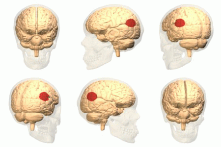
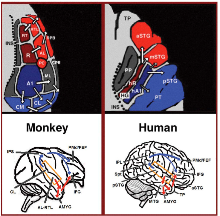
Language processing refers to the way humans use words to communicate ideas and feelings, and how such communications are processed and understood. Language processing is considered to be a uniquely human ability that is not produced with the same grammatical understanding or systematicity in even human's closest primate relatives. Throughout the 20th century the dominant model for language processing in the brain was the Geschwind-Lichteim-Wernicke model, which is based primarily on the analysis of brain damaged patients. However, due to improvements in intra-cortical electrophysiological recordings of monkey and human brains, as well non-invasive techniques such as fMRI, PET, MEG and EEG, a dual auditory pathway has been revealed and a two-streams model has been developed. In accordance with this model, there are two pathways that connect the auditory cortex to the frontal lobe, each pathway accounting for different linguistic roles. The auditory ventral stream pathway is re ... [...More Info...] [...Related Items...] OR: [Wikipedia] [Google] [Baidu] |
Neurolinguistics
Neurolinguistics is the study of neural mechanisms in the human brain that controls the comprehension, production, and acquisition of language. As an interdisciplinary field, neurolinguistics draws methods and theories from fields such as neuroscience, linguistics, cognitive science, communication disorders and neuropsychology. Researchers are drawn to the field from a variety of backgrounds, bringing along a variety of experimental techniques as well as widely varying theoretical perspectives. Much work in neurolinguistics is informed by models in psycholinguistics and theoretical linguistics, and is focused on investigating how the brain can implement the processes that theoretical and psycholinguistics propose are necessary in producing and comprehending language. Neurolinguists study the physiological mechanisms by which the brain processes information related to language, and evaluate linguistic and psycholinguistic theories, using aphasiology, brain imaging, electro ... [...More Info...] [...Related Items...] OR: [Wikipedia] [Google] [Baidu] |
Auditory Cortex
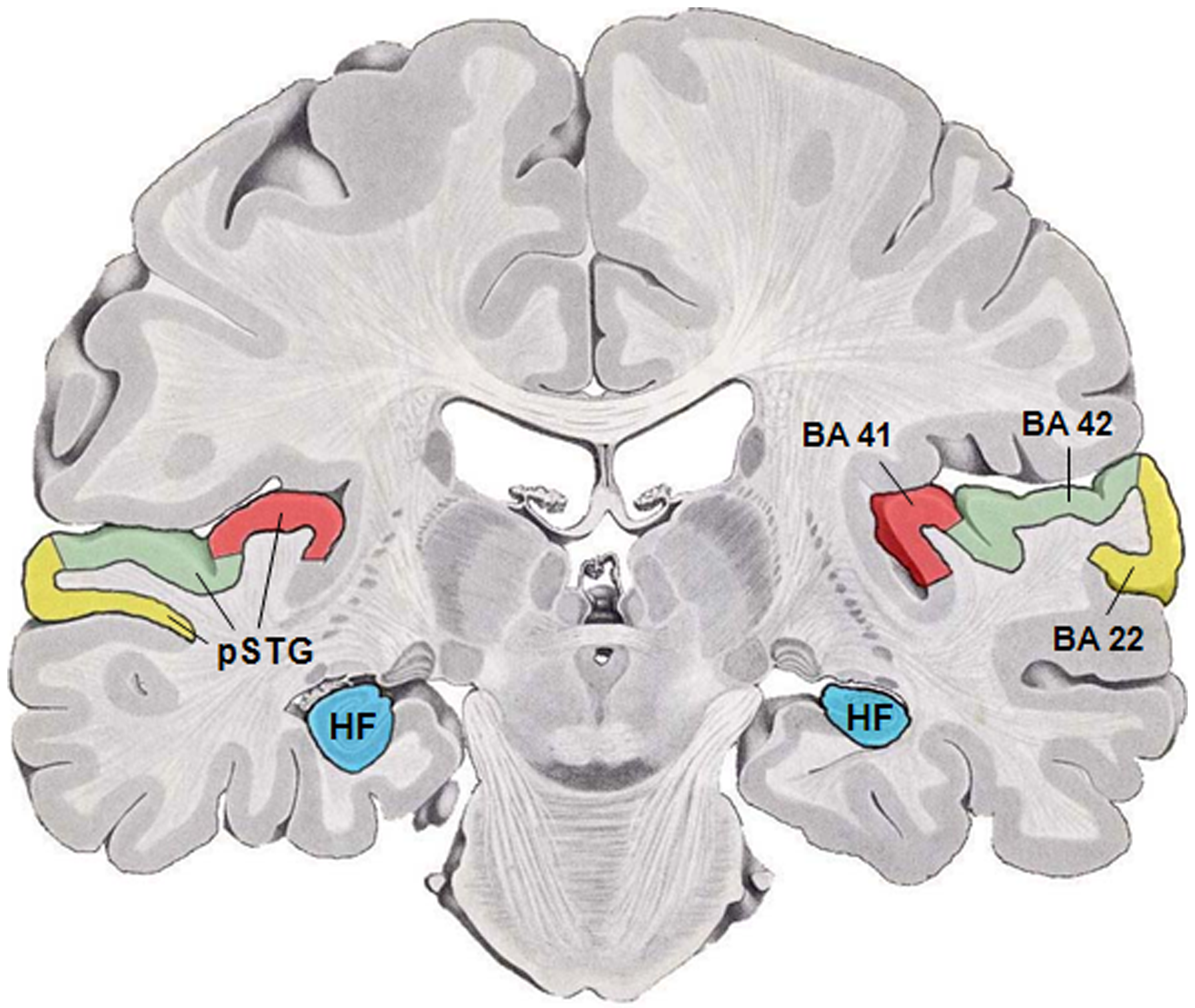
The auditory cortex is the part of the temporal lobe that processes auditory information in humans and many other vertebrates. It is a part of the auditory system, performing basic and higher functions in hearing, such as possible relations to language switching.Cf. Pickles, James O. (2012). ''An Introduction to the Physiology of Hearing'' (4th ed.). Bingley, UK: Emerald Group Publishing Limited, p. 238. It is located bilaterally, roughly at the upper sides of the temporal lobes – in humans, curving down and onto the medial surface, on the superior temporal plane, within the lateral sulcus and comprising parts of the transverse temporal gyri, and the superior temporal gyrus, including the planum polare and planum temporale (roughly Brodmann areas 41 and 42, and partially 22). The auditory cortex takes part in the spectrotemporal, meaning involving time and frequency, analysis of the inputs passed on from the ear. The cortex then filters and passes on the information to ... [...More Info...] [...Related Items...] OR: [Wikipedia] [Google] [Baidu] |
Brainstem
The brainstem (or brain stem) is the posterior stalk-like part of the brain that connects the cerebrum with the spinal cord. In the human brain the brainstem is composed of the midbrain, the pons, and the medulla oblongata. The midbrain is continuous with the thalamus of the diencephalon through the tentorial notch, and sometimes the diencephalon is included in the brainstem. The brainstem is very small, making up around only 2.6 percent of the brain's total weight. It has the critical roles of regulating cardiac, and respiratory function, helping to control heart rate and breathing rate. It also provides the main motor and sensory nerve supply to the face and neck via the cranial nerves. Ten pairs of cranial nerves come from the brainstem. Other roles include the regulation of the central nervous system and the body's sleep cycle. It is also of prime importance in the conveyance of motor and sensory pathways from the rest of the brain to the body, and from the bod ... [...More Info...] [...Related Items...] OR: [Wikipedia] [Google] [Baidu] |
Meta-analysis
A meta-analysis is a statistical analysis that combines the results of multiple scientific studies. Meta-analyses can be performed when there are multiple scientific studies addressing the same question, with each individual study reporting measurements that are expected to have some degree of error. The aim then is to use approaches from statistics to derive a pooled estimate closest to the unknown common truth based on how this error is perceived. Meta-analytic results are considered the most trustworthy source of evidence by the evidence-based medicine literature.Herrera Ortiz AF., Cadavid Camacho E, Cubillos Rojas J, Cadavid Camacho T, Zoe Guevara S, Tatiana Rincón Cuenca N, Vásquez Perdomo A, Del Castillo Herazo V, & Giraldo Malo R. A Practical Guide to Perform a Systematic Literature Review and Meta-analysis. Principles and Practice of Clinical Research. 2022;7(4):47–57. https://doi.org/10.21801/ppcrj.2021.74.6 Not only can meta-analyses provide an estimate of the u ... [...More Info...] [...Related Items...] OR: [Wikipedia] [Google] [Baidu] |
Temporal Pole
The vertebrate cerebrum (brain) is formed by two cerebral hemispheres that are separated by a groove, the longitudinal fissure. The brain can thus be described as being divided into left and right cerebral hemispheres. Each of these hemispheres has an outer layer of grey matter, the cerebral cortex, that is supported by an inner layer of white matter. In eutherian (placental) mammals, the hemispheres are linked by the corpus callosum, a very large bundle of nerve fibers. Smaller commissures, including the anterior commissure, the posterior commissure and the fornix, also join the hemispheres and these are also present in other vertebrates. These commissures transfer information between the two hemispheres to coordinate localized functions. There are three known poles of the cerebral hemispheres: the '' occipital pole'', the '' frontal pole'', and the '' temporal pole''. The central sulcus is a prominent fissure which separates the parietal lobe from the frontal lobe and the pr ... [...More Info...] [...Related Items...] OR: [Wikipedia] [Google] [Baidu] |
Middle Temporal Gyrus
Middle temporal gyrus is a gyrus in the brain on the temporal lobe. It is located between the superior temporal gyrus and inferior temporal gyrus. It corresponds largely to Brodmann area 21. The middle temporal gyrus is bounded by: * the superior temporal sulcus above; * the inferior temporal sulcus below; * an imaginary line drawn from the preoccipital notch to the lateral sulcus posteriorly. It has been connected with processes as different as contemplating distance, recognition of known faces, audio-visual emotional recognition, and accessing word meaning while reading. Some studies indicate that lesions of the posterior region of the middle temporal gyrus, in the left cerebral hemisphere, may result in alexia and agraphia for kanji are the logographic Chinese characters taken from the Chinese script and used in the writing of Japanese. They were made a major part of the Japanese writing system during the time of Old Japanese and are still used, along with ... [...More Info...] [...Related Items...] OR: [Wikipedia] [Google] [Baidu] |
Inferior Parietal Lobule
The inferior parietal lobule (subparietal district) lies below the horizontal portion of the intraparietal sulcus, and behind the lower part of the postcentral sulcus. Also known as Geschwind's territory after Norman Geschwind, an American neurologist, who in the early 1960s recognised its importance. It is a part of the parietal lobe. Structure It is divided from rostral to caudal into two gyri: * One, the supramarginal gyrus, arches over the upturned end of the lateral fissure; it is continuous in front with the postcentral gyrus, and behind with the superior temporal gyrus. * The second, the angular gyrus, arches over the posterior end of the superior temporal sulcus, behind which it is continuous with the middle temporal gyrus. In macaque neuroanatomy, this region is often divided into caudal and rostral portions, cIPL and rIPL, respectively. The cIPL is further divided into areas Opt and PG whereas rIPL is divided into PFG and PF areas. Function Inferior parietal lobule ... [...More Info...] [...Related Items...] OR: [Wikipedia] [Google] [Baidu] |
Temporoparietal Junction
The temporoparietal junction (TPJ) is an area of the brain where the temporal and parietal lobes meet, at the posterior end of the lateral sulcus (Sylvian fissure). The TPJ incorporates information from the thalamus and the limbic system as well as from the visual, auditory, and somatosensory systems. The TPJ also integrates information from both the external environment as well as from within the body. The TPJ is responsible for collecting all of this information and then processing it. This area is also known to play a crucial role in self–other distinctions processes and theory of mind (ToM). Furthermore, damage to the TPJ has been implicated in having adverse effects on an individual's ability to make moral decisions and has been known to produce out-of-body experiences (OBEs). Electromagnetic stimulation of the TPJ can also cause these effects. Apart from these diverse roles that the TPJ plays, it is also known for its involvement in a variety of widespread disorder ... [...More Info...] [...Related Items...] OR: [Wikipedia] [Google] [Baidu] |
Lateral Sulcus
In neuroanatomy, the lateral sulcus (also called Sylvian fissure, after Franciscus Sylvius, or lateral fissure) is one of the most prominent features of the human brain. The lateral sulcus is a deep fissure in each hemisphere that separates the frontal and parietal lobes from the temporal lobe. The insular cortex lies deep within the lateral sulcus. Anatomy The lateral sulcus divides both the frontal lobe and parietal lobe above from the temporal lobe below. It is in both hemispheres of the brain. The lateral sulcus is one of the earliest-developing sulci of the human brain. It first appears around the fourteenth gestational week. The insular cortex lies deep within the lateral sulcus. The lateral sulcus has a number of side branches. Two of the most prominent and most regularly found are the ascending (also called vertical) ramus and the horizontal ramus of the lateral fissure, which subdivide the inferior frontal gyrus. The lateral sulcus also contains the transverse te ... [...More Info...] [...Related Items...] OR: [Wikipedia] [Google] [Baidu] |
Diffusion MRI
Diffusion-weighted magnetic resonance imaging (DWI or DW-MRI) is the use of specific MRI sequences as well as software that generates images from the resulting data that uses the diffusion of water molecules to generate contrast in MR images. It allows the mapping of the diffusion process of molecules, mainly water, in biological tissues, in vivo and non-invasively. Molecular diffusion in tissues is not random, but reflects interactions with many obstacles, such as macromolecules, fibers, and membranes. Water molecule diffusion patterns can therefore reveal microscopic details about tissue architecture, either normal or in a diseased state. A special kind of DWI, diffusion tensor imaging (DTI), has been used extensively to map white matter tractography in the brain. Introduction In diffusion weighted imaging (DWI), the intensity of each image element ( voxel) reflects the best estimate of the rate of water diffusion at that location. Because the mobility of water is driven by ... [...More Info...] [...Related Items...] OR: [Wikipedia] [Google] [Baidu] |
Amygdala
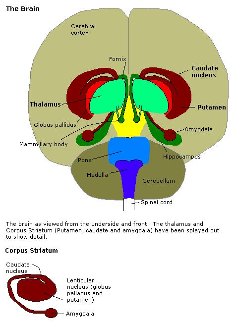
The amygdala (; plural: amygdalae or amygdalas; also '; Latin from Greek, , ', 'almond', 'tonsil') is one of two almond-shaped clusters of nuclei located deep and medially within the temporal lobes of the brain's cerebrum in complex vertebrates, including humans. Shown to perform a primary role in the processing of memory, decision making, and emotional responses (including fear, anxiety, and aggression), the amygdalae are considered part of the limbic system. The term "amygdala" was first introduced by Karl Friedrich Burdach in 1822. Structure The regions described as amygdala nuclei encompass several structures of the cerebrum with distinct connectional and functional characteristics in humans and other animals. Among these nuclei are the basolateral complex, the cortical nucleus, the medial nucleus, the central nucleus, and the intercalated cell clusters. The basolateral complex can be further subdivided into the lateral, the basal, and the accessory ba ... [...More Info...] [...Related Items...] OR: [Wikipedia] [Google] [Baidu] |



_-_inferiror_view.png)
