|
Lamina (anatomy)
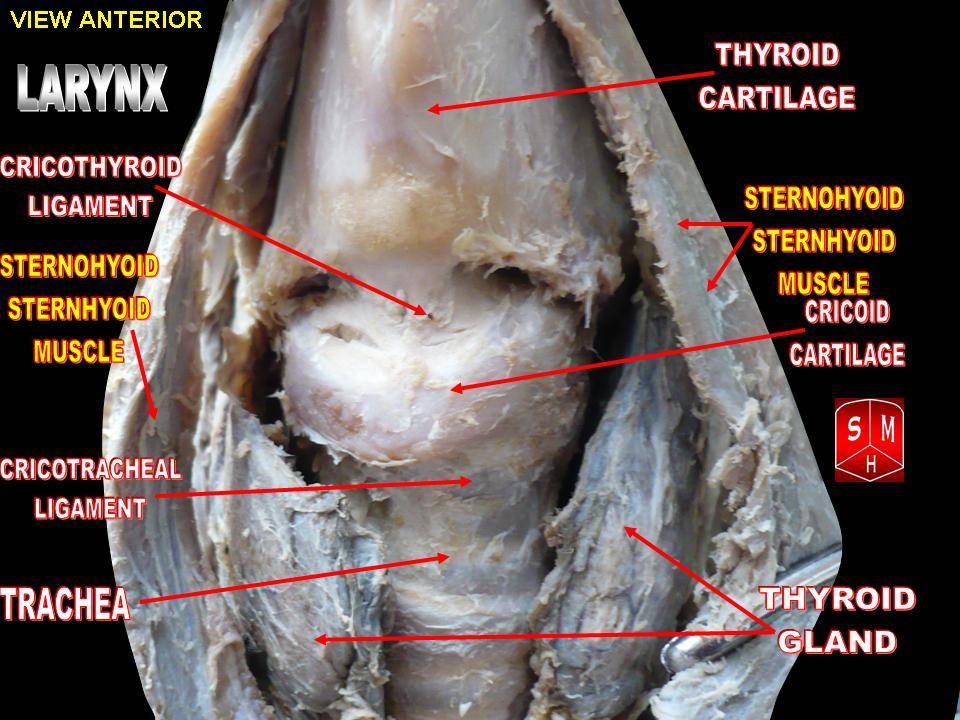
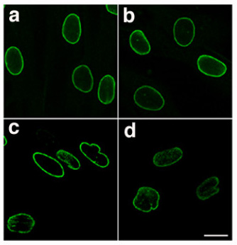
Lamina is a general anatomical term meaning "plate" or "layer". It is used in both gross anatomy and microscopic anatomy to describe structures. Some examples include: * The laminae of the thyroid cartilage: two leaf-like plates of cartilage that make up the walls of the structure. * The vertebral laminae: plates of bone that form the posterior walls of each vertebra, enclosing the spinal cord. * The laminae of the thalamus: the layers of thalamus tissue. * The lamina propria: a connective tissue layer under the epithelium of an organ. * The nuclear lamina: a dense fiber network inside the nucleus of cells. * The lamina affixa: a layer of epithelium growing on the surface of the thalamus The thalamus (from Greek θάλαμος, "chamber") is a large mass of gray matter located in the dorsal part of the diencephalon (a division of the forebrain). Nerve fibers project out of the thalamus to the cerebral cortex in all directions, .... * Lamina cribrosa with two different ... [...More Info...] [...Related Items...] OR: [Wikipedia] [Google] [Baidu] |
Anatomy
Anatomy () is the branch of biology concerned with the study of the structure of organisms and their parts. Anatomy is a branch of natural science that deals with the structural organization of living things. It is an old science, having its beginnings in prehistoric times. Anatomy is inherently tied to developmental biology, embryology, comparative anatomy, evolutionary biology, and phylogeny, as these are the processes by which anatomy is generated, both over immediate and long-term timescales. Anatomy and physiology, which study the structure and function (biology), function of organisms and their parts respectively, make a natural pair of related disciplines, and are often studied together. Human anatomy is one of the essential basic research, basic sciences that are applied in medicine. The discipline of anatomy is divided into macroscopic scale, macroscopic and microscopic scale, microscopic. Gross anatomy, Macroscopic anatomy, or gross anatomy, is the examination of an ... [...More Info...] [...Related Items...] OR: [Wikipedia] [Google] [Baidu] |
Gross Anatomy
Gross anatomy is the study of anatomy at the visible or macroscopic level. The counterpart to gross anatomy is the field of histology, which studies microscopic anatomy. Gross anatomy of the human body or other animals seeks to understand the relationship between components of an organism in order to gain a greater appreciation of the roles of those components and their relationships in maintaining the functions of life. The study of gross anatomy can be performed on deceased organisms using dissection or on living organisms using medical imaging. Education in the gross anatomy of humans is included training for most health professionals. Techniques of study Gross anatomy is studied using both invasive and noninvasive methods with the goal of obtaining information about the macroscopic structure and organization of organs and organ systems. Among the most common methods of study is dissection, in which the corpse of an animal or a human cadaver is surgically opened and its o ... [...More Info...] [...Related Items...] OR: [Wikipedia] [Google] [Baidu] |
Thyroid Cartilage
The thyroid cartilage is the largest of the nine cartilages that make up the ''laryngeal skeleton'', the cartilage structure in and around the trachea that contains the larynx. It does not completely encircle the larynx (only the cricoid cartilage encircles it). Structure The thyroid cartilage is a hyaline cartilage structure that sits in front of the larynx and above the thyroid gland. The cartilage is composed of two halves, which meet in the middle at a peak called the laryngeal prominence, also called the Adam's apple. In the midline above the prominence is the superior thyroid notch. A counterpart notch at the bottom of the cartilage is called the inferior thyroid notch. The two halves of the cartilage that make out the outer surfaces extend obliquely to cover the sides of the trachea. The posterior edge of each half articulates with the cricoid cartilage inferiorly at a joint called the cricothyroid joint. The most posterior part of the cartilage also has two projection ... [...More Info...] [...Related Items...] OR: [Wikipedia] [Google] [Baidu] |
Lamina Of The Vertebral Arch
The spinal column, a defining synapomorphy shared by nearly all vertebrates,Hagfish are believed to have secondarily lost their spinal column is a moderately flexible series of vertebrae (singular vertebra), each constituting a characteristic irregular bone whose complex structure is composed primarily of bone, and secondarily of hyaline cartilage. They show variation in the proportion contributed by these two tissue types; such variations correlate on one hand with the cerebral/caudal rank (i.e., location within the backbone), and on the other with phylogenetic differences among the vertebrate taxa. The basic configuration of a vertebra varies, but the bone is its ''body'', with the central part of the body constituting the ''centrum''. The upper (closer to) and lower (further from), respectively, the cranium and its central nervous system surfaces of the vertebra body support attachment to the intervertebral discs. The posterior part of a vertebra forms a vertebral arch (i ... [...More Info...] [...Related Items...] OR: [Wikipedia] [Google] [Baidu] |
Thalamus
The thalamus (from Greek θάλαμος, "chamber") is a large mass of gray matter located in the dorsal part of the diencephalon (a division of the forebrain). Nerve fibers project out of the thalamus to the cerebral cortex in all directions, allowing hub-like exchanges of information. It has several functions, such as the relaying of sensory signals, including motor signals to the cerebral cortex and the regulation of consciousness, sleep, and alertness. Anatomically, it is a paramedian symmetrical structure of two halves (left and right), within the vertebrate brain, situated between the cerebral cortex and the midbrain. It forms during embryonic development as the main product of the diencephalon, as first recognized by the Swiss embryologist and anatomist Wilhelm His Sr. in 1893. Anatomy The thalamus is a paired structure of gray matter located in the forebrain which is superior to the midbrain, near the center of the brain, with nerve fibers projecting out to the ... [...More Info...] [...Related Items...] OR: [Wikipedia] [Google] [Baidu] |
Lamina Propria
The lamina propria is a thin layer of connective tissue that forms part of the moist linings known as mucous membranes or mucosae, which line various tubes in the body, such as the respiratory tract, the gastrointestinal tract, and the urogenital tract. The lamina propria is a thin layer of loose (areolar) connective tissue, which lies beneath the epithelium, and together with the epithelium and basement membrane constitutes the mucosa. As its Latin name indicates, it is a characteristic component of the mucosa, or the mucosa's "own special layer." Thus, the term mucosa or mucous membrane refers to the combination of the epithelium and the lamina propria. The connective tissue of the lamina propria is loose and rich in cells. The cells of the lamina propria are variable and can include fibroblasts, lymphocytes, plasma cells, macrophages, eosinophilic leukocytes, and mast cells. It provides support and nutrition to the epithelium, as well as the means to bind to the underl ... [...More Info...] [...Related Items...] OR: [Wikipedia] [Google] [Baidu] |
Nuclear Lamina
The nuclear lamina is a dense (~30 to 100 nm thick) fibrillar network inside the nucleus of eukaryote cells. It is composed of intermediate filaments and membrane associated proteins. Besides providing mechanical support, the nuclear lamina regulates important cellular events such as DNA replication and cell division. Additionally, it participates in chromatin organization and it anchors the nuclear pore complexes embedded in the nuclear envelope. The nuclear lamina is associated with the inner face of the inner nuclear membrane of the nuclear envelope, whereas the outer face of the outer nuclear membrane is continuous with the endoplasmic reticulum. The nuclear lamina is similar in structure to the nuclear matrix, that extends throughout the nucleoplasm. Structure and composition The nuclear lamina consists of two components, lamins and nuclear lamin-associated membrane proteins. The lamins are type V intermediate filaments which can be categorized as either A-type (lam ... [...More Info...] [...Related Items...] OR: [Wikipedia] [Google] [Baidu] |
Lamina Affixa
Lamina affixa is a layer of epithelium growing on the surface of the thalamus and forming the floor of the central part of lateral ventricle, on whose medial margin is attached the choroid plexus of the lateral ventricle; it covers the superior thalamostriate vein and the superior choroid vein. The torn edge of this plexus is called the tela choroidea The tela choroidea (or tela chorioidea) is a region of meningeal pia mater that adheres to the underlying ependyma, and gives rise to the choroid plexus in each of the brain’s four ventricles. ''Tela'' is Latin for ''woven'' and is used to descr .... On the surface of the terminal vein is a narrow white band, named the lamina affixa. GDF-15/MIC-1 has been observed in lamina affixa cells. References External links * https://web.archive.org/web/20071024195305/http://www.univie.ac.at/anatomie2/plastinatedbrain/surfaceanatomy/surface-2-text.html Thalamus {{neuroanatomy-stub ... [...More Info...] [...Related Items...] OR: [Wikipedia] [Google] [Baidu] |
Epithelium
Epithelium or epithelial tissue is one of the four basic types of animal tissue, along with connective tissue, muscle tissue and nervous tissue. It is a thin, continuous, protective layer of compactly packed cells with a little intercellular matrix. Epithelial tissues line the outer surfaces of organs and blood vessels throughout the body, as well as the inner surfaces of cavities in many internal organs. An example is the epidermis, the outermost layer of the skin. There are three principal shapes of epithelial cell: squamous (scaly), columnar, and cuboidal. These can be arranged in a singular layer of cells as simple epithelium, either squamous, columnar, or cuboidal, or in layers of two or more cells deep as stratified (layered), or ''compound'', either squamous, columnar or cuboidal. In some tissues, a layer of columnar cells may appear to be stratified due to the placement of the nuclei. This sort of tissue is called pseudostratified. All glands are made up of epithe ... [...More Info...] [...Related Items...] OR: [Wikipedia] [Google] [Baidu] |
Lamina Cribrosa (other)
{{disambig ...
Lamina cribrosa may refer to: *Cribriform plate of the ethmoid bone (horizontal lamina or lamina cribrosa ossis ethmoidalis) *Lamina cribrosa sclerae, a mesh-like structures which allows nerve fibres of the optic nerve to pass through the sclera See also * Lamina (anatomy) Lamina is a general anatomical term meaning "plate" or "layer". It is used in both gross anatomy and microscopic anatomy to describe structures. Some examples include: * The laminae of the thyroid cartilage: two leaf-like plates of cartilage t ... [...More Info...] [...Related Items...] OR: [Wikipedia] [Google] [Baidu] |


