|
Ligaments
A ligament is a type of fibrous connective tissue in the body that connects bones to other bones. It also connects flight feathers to bones, in dinosaurs and birds. All 30,000 species of amniotes (land animals with internal bones) have ligaments. It is also known as ''articular ligament'', ''articular larua'', ''fibrous ligament'', or ''true ligament''. Comparative anatomy Ligaments are similar to tendons and fasciae as they are all made of connective tissue. The differences among them are in the connections that they make: ligaments connect one bone to another bone, tendons connect muscle to bone, and fasciae connect muscles to other muscles. These are all found in the skeletal system of the human body. Ligaments cannot usually be regenerated naturally; however, there are periodontal ligament stem cells located near the periodontal ligament which are involved in the adult regeneration of periodontist ligament. The study of ligaments is known as . Humans Other ligaments ... [...More Info...] [...Related Items...] OR: [Wikipedia] [Google] [Baidu] |
Ligament
A ligament is a type of fibrous connective tissue in the body that connects bones to other bones. It also connects flight feathers to bones, in dinosaurs and birds. All 30,000 species of amniotes (land animals with internal bones) have ligaments. It is also known as ''articular ligament'', ''articular larua'', ''fibrous ligament'', or ''true ligament''. Comparative anatomy Ligaments are similar to tendons and fasciae as they are all made of connective tissue. The differences among them are in the connections that they make: ligaments connect one bone to another bone, tendons connect muscle to bone, and fasciae connect muscles to other muscles. These are all found in the skeletal system of the human body. Ligaments cannot usually be regenerated naturally; however, there are periodontal ligament stem cells located near the periodontal ligament which are involved in the adult regeneration of periodontist ligament. The study of ligaments is known as . Humans Other ligame ... [...More Info...] [...Related Items...] OR: [Wikipedia] [Google] [Baidu] |
Musculoskeletal System
The human musculoskeletal system (also known as the human locomotor system, and previously the activity system) is an organ system that gives humans the ability to move using their Muscular system, muscular and Human skeleton, skeletal systems. The musculoskeletal system provides form, support, stability, and movement to the body. The human musculoskeletal system is made up of the bones of the human skeleton, skeleton, muscles, cartilage, tendons, ligaments, joints, and other connective tissue that supports and binds tissues and organs together. The musculoskeletal system's primary functions include supporting the body, allowing motion, and protecting vital organs. The skeletal portion of the system serves as the main storage system for calcium and phosphorus and contains critical components of the hematopoietic system. This system describes how bones are connected to other bones and muscle fibers via connective tissue such as tendons and ligaments. The bones provide stability t ... [...More Info...] [...Related Items...] OR: [Wikipedia] [Google] [Baidu] |
Cruciate Ligament
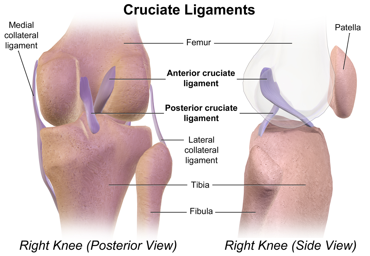
Cruciate ligaments (also cruciform ligaments) are pairs of ligaments arranged like a letter X. They occur in several joints of the body, such as the knee joint, wrist joint and the atlanto-axial joint. In a fashion similar to the cords in a toy Jacob's ladder, the crossed ligaments stabilize the joint while allowing a very large range of motion. Knee Structure Cruciate ligaments occur in the knee of humans and other bipedal animals and the corresponding stifle of quadrupedal animals, and in the neck, fingers, and foot. * The cruciate ligaments of the knee are the anterior cruciate ligament (ACL) and the posterior cruciate ligament (PCL). These ligaments are two strong, rounded bands that extend from the head of the tibia to the intercondyloid notch of the femur. The ACL is lateral and the PCL is medial. They cross each other like the limbs of an X. They are named for their insertion into the tibia: the ACL attaches to the anterior aspect of the intercondylar area, ... [...More Info...] [...Related Items...] OR: [Wikipedia] [Google] [Baidu] |
Dense Regular Connective Tissue
Dense regular connective tissue (DRCT) provides connection between different tissues in the human body. The collagen fibers in dense regular connective tissue are bundled in a parallel fashion. DRCT is divided into white fibrous connective tissue and yellow fibrous connective tissue, both of which occur in two forms: cord arrangement and sheath arrangement. In cord arrangement, bundles of collagen and matrix are distributed in regular alternate patterns. In sheath arrangement, collagen bundles and matrix are distributed in irregular patterns, sometimes in the form of a network. It is similar to areolar tissue, but in DRCT elastic fibers are completely absent. Structures formed * An example of their use is in tendon A tendon or sinew is a tough band of fibrous connective tissue, dense fibrous connective tissue that connects skeletal muscle, muscle to bone. It sends the mechanical forces of muscle contraction to the skeletal system, while withstanding tensi ...s, which connec ... [...More Info...] [...Related Items...] OR: [Wikipedia] [Google] [Baidu] |
Periodontal Ligament
The periodontal ligament, commonly abbreviated as the PDL, are a group of specialized connective tissue fibers that essentially attach a tooth to the alveolar bone within which they sit. It inserts into root cementum on one side and onto alveolar bone on the other. Structure The PDL consists of principal fibers, loose connective tissue, blast and clast cells, oxytalan fibers and cell rest of Malassez. Alveolodental ligament The main principal fiber group is the alveolodental ligament, which consists of five fiber subgroups: alveolar crest, horizontal, oblique, apical, and interradicular on multirooted teeth. Principal fibers other than the alveolodental ligament are the transseptal fibers. All these fibers help the tooth withstand the naturally substantial compressive forces that occur during chewing and remain embedded in the bone. The ends of the principal fibers that are within either cementum or alveolar bone proper are considered Sharpey fibers. * Alveolar cre ... [...More Info...] [...Related Items...] OR: [Wikipedia] [Google] [Baidu] |
Peritoneal Ligament
Peritoneal ligaments are folds of peritoneum that are used to connect viscera to viscera or the abdominal wall. There are multiple named ligaments that usually are named in accordance with what they are. * Gastrocolic ligament, connects the stomach and the colon. * Splenocolic ligament, connects the spleen and the colon. * Gastrosplenic ligament * Gastrophrenic ligament * Phrenicocolic ligament * Splenorenal ligament * Hepatic ligaments - Ligaments that are associated with the liver ** Coronary ligament ** Left triangular ligament ** Right triangular ligament The right triangular ligament is situated at the right extremity of the bare area, and is a small fold which passes to the Thoracic diaphragm, diaphragm, being formed by the apposition of the upper and lower layers of the coronary ligament. Addit ... ** Hepatoduodenal ligament ** Hepatogastric ligament ** Falciform ligament ** Round ligament of liver References {{reflist Abdomen ... [...More Info...] [...Related Items...] OR: [Wikipedia] [Google] [Baidu] |
Dislocation (medicine)
A joint dislocation, also called luxation, occurs when there is an abnormal separation in the joint, where two or more bones meet. A partial dislocation is referred to as a subluxation. Dislocations are commonly caused by sudden trauma to the joint like during a car accident or fall. A joint dislocation can damage the surrounding ligaments, tendons, muscles, and nerves. Dislocations can occur in any major joint (shoulder, knees, hips) or minor joint (toes, fingers). The most common joint dislocation is a shoulder dislocation. The treatment for joint dislocation is usually by closed reduction, that is, skilled manipulation to return the bones to their normal position. Only trained medical professionals should perform reductions since the manipulation can cause injury to the surrounding soft tissue, nerves, or vascular structures. Signs and Symptoms The following symptoms are common with any type of dislocation. * Intense pain * Joint instability * Deformity of the joint area ... [...More Info...] [...Related Items...] OR: [Wikipedia] [Google] [Baidu] |
Viscoelastic
In materials science and continuum mechanics, viscoelasticity is the property of materials that exhibit both Viscosity, viscous and Elasticity (physics), elastic characteristics when undergoing deformation (engineering), deformation. Viscous materials, like water, resist both shear flow and Strain (materials science), strain linearly with time when a Stress (physics), stress is applied. Elastic materials strain when stretched and immediately return to their original state once the stress is removed. Viscoelastic materials have elements of both of these properties and, as such, exhibit time-dependent strain. Whereas elasticity is usually the result of chemical bond, bond stretching along crystallographic planes in an ordered solid, viscosity is the result of the diffusion of atoms or molecules inside an amorphous material.Meyers and Chawla (1999): "Mechanical Behavior of Materials", 98-103. Background In the nineteenth century, physicists such as James Clerk Maxwell, Ludwig Boltzm ... [...More Info...] [...Related Items...] OR: [Wikipedia] [Google] [Baidu] |
Synovial Joint
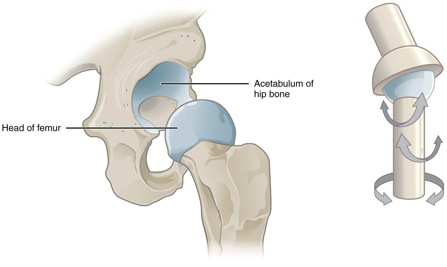
A synovial joint, also known as diarthrosis, joins bones or cartilage with a fibrous joint capsule that is continuous with the periosteum of the joined bones, constitutes the outer boundary of a synovial cavity, and surrounds the bones' articulating surfaces. This joint unites long bones and permits free bone movement and greater mobility. The synovial cavity/joint is filled with synovial fluid. The joint capsule is made up of an outer layer of fibrous membrane, which keeps the bones together structurally, and an inner layer, the synovial membrane, which seals in the synovial fluid. They are the most common and most movable type of joint in the body. As with most other joints, synovial joints achieve movement at the point of contact of the articulating bones. They originated 400 million years ago in the first jawed vertebrates. Structure Synovial joints contain the following structures: * Synovial cavity: all diarthroses have the characteristic space between the bones that is f ... [...More Info...] [...Related Items...] OR: [Wikipedia] [Google] [Baidu] |
Articular Capsule
In anatomy, a joint capsule or articular capsule is an envelope surrounding a synovial joint. Each joint capsule has two parts: an outer fibrous layer or membrane, and an inner synovial layer or membrane. Membranes Each capsule consists of two layers or membranes: * an outer (fibrous membrane, ''fibrous stratum'') composed of avascular white fibrous tissue * an inner ('''', ''synovial stratum'') which is a secreting layer On the inside of the capsule, articular cartilage covers the end surfaces of the bones that articulate within ...[...More Info...] [...Related Items...] OR: [Wikipedia] [Google] [Baidu] |
Muscle
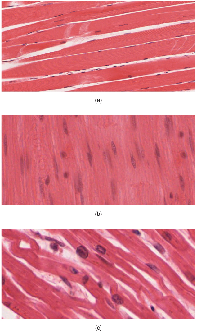
Muscle is a soft tissue, one of the four basic types of animal tissue. There are three types of muscle tissue in vertebrates: skeletal muscle, cardiac muscle, and smooth muscle. Muscle tissue gives skeletal muscles the ability to muscle contraction, contract. Muscle tissue contains special Muscle contraction, contractile proteins called actin and myosin which interact to cause movement. Among many other muscle proteins, present are two regulatory proteins, troponin and tropomyosin. Muscle is formed during embryonic development, in a process known as myogenesis. Skeletal muscle tissue is striated consisting of elongated, multinucleate muscle cells called muscle fibers, and is responsible for movements of the body. Other tissues in skeletal muscle include tendons and perimysium. Smooth and cardiac muscle contract involuntarily, without conscious intervention. These muscle types may be activated both through the interaction of the central nervous system as well as by innervation ... [...More Info...] [...Related Items...] OR: [Wikipedia] [Google] [Baidu] |
Joint
A joint or articulation (or articular surface) is the connection made between bones, ossicles, or other hard structures in the body which link an animal's skeletal system into a functional whole.Saladin, Ken. Anatomy & Physiology. 7th ed. McGraw-Hill Connect. Webp.274/ref> They are constructed to allow for different degrees and types of movement. Some joints, such as the knee, elbow, and shoulder, are self-lubricating, almost frictionless, and are able to withstand compression and maintain heavy loads while still executing smooth and precise movements. Other joints such as suture (joint), sutures between the bones of the skull permit very little movement (only during birth) in order to protect the brain and the sense organs. The connection between a tooth and the jawbone is also called a joint, and is described as a fibrous joint known as a gomphosis. Joints are classified both structurally and functionally. Joints play a vital role in the human body, contributing to movement, sta ... [...More Info...] [...Related Items...] OR: [Wikipedia] [Google] [Baidu] |


