|
Ischiopubic Ramus
The ischiopubic ramus is a compound structure consisting of the following two structures: * from the pubis, the inferior pubic ramus * from the ischium, the inferior ramus of the ischium It forms the inferior border of the obturator foramen and serves as part of the origin for the obturator internus and externus muscles. Also, most adductors originate at the ischiopubic ramus. The fascia of Colles The membranous layer of the superficial fascia of the perineum (Colles' fascia) is the deeper layer ( membranous layer) of the superficial perineal fascia. It is thin, aponeurotic in structure, and of considerable strength, serving to bind down th ... is attached to its margin. References External links * - "The Female Perineum" * * (, ) Bones of the pelvis {{musculoskeletal-stub ... [...More Info...] [...Related Items...] OR: [Wikipedia] [Google] [Baidu] |
Hip Bone
The hip bone (os coxae, innominate bone, pelvic bone or coxal bone) is a large flat bone, constricted in the center and expanded above and below. In some vertebrates (including humans before puberty) it is composed of three parts: the Ilium (bone), ilium, ischium, and the Pubis (bone), pubis. The two hip bones join at the pubic symphysis and together with the sacrum and coccyx (the pelvic part of the vertebral column, spine) comprise the human skeleton, skeletal component of the pelvis – the pelvic girdle which surrounds the pelvic cavity. They are connected to the sacrum, which is part of the axial skeleton, at the sacroiliac joint. Each hip bone is connected to the corresponding femur (thigh bone) (forming the primary connection between the bones of the lower limb and the axial skeleton) through the large ball and socket joint of the hip joint, hip. Structure The hip bone is formed by three parts: the Ilium (bone), ilium, ischium, and Pubis (bone), pubis. At birth, these three ... [...More Info...] [...Related Items...] OR: [Wikipedia] [Google] [Baidu] |
Pubis (bone)
In vertebrates, the pubic region ( la, pubis) is the most forward-facing (ventral and anterior) of the three main regions making up the coxal bone. The left and right pubic regions are each made up of three sections, a superior ramus, inferior ramus, and a body. Structure The pubic region is made up of a ''body'', ''superior ramus'', and ''inferior ramus'' (). The left and right coxal bones join at the pubic symphysis. It is covered by a layer of fat, which is covered by the mons pubis. The pubis is the lower limit of the suprapubic region. In the female, the pubic region is anterior to the urethral sponge. Body The body forms the wide, strong, middle and flat part of the pubic region. The bodies of the left and right pubic regions join at the pubic symphysis. The rough upper edge is the pubic crest, ending laterally in the pubic tubercle. This tubercle, found roughly 3 cm from the pubic symphysis, is a distinctive feature on the lower part of the abdominal wall; important ... [...More Info...] [...Related Items...] OR: [Wikipedia] [Google] [Baidu] |
Inferior Pubic Ramus
In vertebrates, the pubic region ( la, pubis) is the most forward-facing (ventral and anterior) of the three main regions making up the coxal bone. The left and right pubic regions are each made up of three sections, a superior ramus, inferior ramus, and a body. Structure The pubic region is made up of a ''body'', ''superior ramus'', and ''inferior ramus'' (). The left and right coxal bones join at the pubic symphysis. It is covered by a layer of fat, which is covered by the mons pubis. The pubis is the lower limit of the suprapubic region. In the female, the pubic region is anterior to the urethral sponge. Body The body forms the wide, strong, middle and flat part of the pubic region. The bodies of the left and right pubic regions join at the pubic symphysis. The rough upper edge is the pubic crest, ending laterally in the pubic tubercle. This tubercle, found roughly 3 cm from the pubic symphysis, is a distinctive feature on the lower part of the abdominal wall; important ... [...More Info...] [...Related Items...] OR: [Wikipedia] [Google] [Baidu] |
Ischium
The ischium () forms the lower and back region of the (''os coxae''). Situated below the ilium and behind the pubis, it is one of three regions whose fusion creates the . The superior portion of this region forms approximately one-third of the acetabulum. |
Inferior Ramus Of The Ischium
The ischium () forms the lower and back region of the (''os coxae''). Situated below the ilium and behind the pubis, it is one of three regions whose fusion creates the . The superior portion of this region forms approximately one-third of the |
Obturator Foramen
The obturator foramen (Latin foramen obturatum) is the large opening created by the ischium and pubis (bone), pubis bones of the pelvis through which nerves and blood vessels pass. Structure It is bounded by a thin, uneven margin, to which a strong membrane is attached, and presents, superiorly, a deep groove, the obturator groove, which runs from the pelvis obliquely medialward and downward. This groove is converted into the obturator canal by a ligamentous band, a specialized part of the obturator membrane, attached to two tubercles: * one, the posterior obturator tubercle, on the medial border of the ischium, just in front of the acetabular notch * the other, the anterior obturator tubercle, on the obturator crest of the superior pubic ramus, superior ramus of the pubis (bone), pubis Variation Reflecting the overall sex differences in human physiology, sex differences between male and female pelvises, the obturator foramina are oval in the male and wider and more triangular ... [...More Info...] [...Related Items...] OR: [Wikipedia] [Google] [Baidu] |
Internal Obturator Muscle
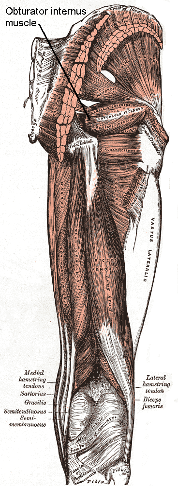
The internal obturator muscle or obturator internus muscle originates on the medial surface of the obturator membrane, the ischium near the membrane, and the rim of the pubis. It exits the pelvic cavity through the lesser sciatic foramen. The internal obturator is situated partly within the lesser pelvis, and partly at the back of the hip-joint. It functions to help laterally rotate femur with hip extension and abduct femur with hip flexion, as well as to steady the femoral head in the acetabulum. Structure Origin The internal obturator muscle arises from the inner surface of the antero-lateral wall of the pelvis. It surrounds the obturator foramen. It is attached to the inferior pubic ramus and ischium, and at the side to the inner surface of the hip bone below and behind the pelvic brim. It reaches from the upper part of the greater sciatic foramen above and behind to the obturator foramen below and in front. It also arises from the pelvic surface of the obturator membran ... [...More Info...] [...Related Items...] OR: [Wikipedia] [Google] [Baidu] |
External Obturator Muscle
The external obturator muscle, obturator externus muscle (; OE) is a flat, triangular muscle, which covers the outer surface of the anterior wall of the pelvis. It is sometimes considered part of the medial compartment of thigh, and sometimes considered part of the gluteal region. Structure It arises from the margin of bone immediately around the medial side of the obturator membrane and surrounding bone, viz., from the inferior pubic ramus, and the ramus of the ischium; it also arises from the medial two-thirds of the outer surface of the obturator membrane, and from the tendinous arch which completes the canal for the passage of the obturator vessels and nerves. The fibers springing from the pubic arch extend on to the inner surface of the bone, where they obtain a narrow origin between the margin of the foramen and the attachment of the obturator membrane. The fibers converge and pass posterolateral and upward, and end in a tendon which runs across the back of the neck o ... [...More Info...] [...Related Items...] OR: [Wikipedia] [Google] [Baidu] |
Adductor Muscles Of The Hip
The adductor muscles of the hip are a group of muscles mostly used for bringing the thighs together (called adduction). Structure The adductor group is made up of: *Adductor brevis *Adductor longus *Adductor magnus * Adductor minimus This is often considered to be a part of adductor magnus. * pectineus * gracilis *Obturator externusPlatzer, Werner (2004), Color Atlas of Human Anatomy, Vol. 1, Locomotor System', Thieme, 5th ed, p 240 and are also part of the medial compartment of thigh The adductors originate on the pubis and ischium bones and insert mainly on the medial posterior surface of the femur. Nerve supply The pectineus is the only adductor muscle that is innervated by the femoral nerve. The other adductor muscles are innervated by the obturator nerve with the exception of a small part of the adductor magnus which is innervated by the tibial nerve. Variation In 33% of people a supernumerary muscle is found between the adductor brevis and adductor minimus. When ... [...More Info...] [...Related Items...] OR: [Wikipedia] [Google] [Baidu] |
Fascia Of Colles
The membranous layer of the superficial fascia of the perineum (Colles' fascia) is the deeper layer (membranous layer) of the superficial perineal fascia. It is thin, aponeurotic in structure, and of considerable strength, serving to bind down the muscles of the root of the penis. Colles' fascia emerges from the perineal membrane, which divides the base of the penis from the prostate. Colles' fascia emerges from the inferior side of the perineal membrane and continues along the ventral (inferior) penis without covering the scrotum. It separates the skin and subcutaneous fat from the superficial perineal pouch. Relations ''In front'', it is continuous with the dartos fascia of the penis and Scarpa's fascia upon the anterior wall of the abdomen; ''On either side'' it is firmly attached to the margins of the rami of the pubis and ischium, lateral to the crus penis and as far back as the tuberosity of the ischium. ''Posteriorly'', it curves around the superficial transverse perine ... [...More Info...] [...Related Items...] OR: [Wikipedia] [Google] [Baidu] |

