|
Intestinal Lining
The intestinal epithelium is the single cell layer that form the luminal surface (lining) of both the small and large intestine (colon) of the gastrointestinal tract. Composed of simple columnar epithelial cells, it serves two main functions: absorbing useful substances into the body and restricting the entry of harmful substances. As part of its protective role, the intestinal epithelium forms an important component of the intestinal mucosal barrier. Certain diseases and conditions are caused by functional defects in the intestinal epithelium. On the other hand, various diseases and conditions can lead to its dysfunction which, in turn, can lead to further complications. Structure The intestinal epithelium is part of the intestinal mucosa. The epithelium is composed of a single layer of cells, while the other two layers of the mucosa, the lamina propria and the muscularis mucosae, support and articulate the epithelial layer. To securely contain the contents of the intest ... [...More Info...] [...Related Items...] OR: [Wikipedia] [Google] [Baidu] |
Lumen (anatomy)
In biology, a lumen (plural lumina) is the inside space of a tubular structure, such as an artery or intestine. It comes . It can refer to: *The interior of a vessel, such as the central space in an artery, vein or capillary through which blood flows. *The interior of the gastrointestinal tract *The pathways of the bronchi in the lungs *The interior of renal tubules and urinary collecting ducts *The pathways of the female genital tract, starting with a single pathway of the vagina, splitting up in two lumina in the uterus, both of which continue through the Fallopian tubes In cell biology, a lumen is a membrane-defined space that is found inside several organelles, cellular components, or structures: *thylakoid, endoplasmic reticulum, Golgi apparatus, lysosome, mitochondrion, or microtubule Transluminal procedures ''Transluminal procedures'' are procedures occurring through lumina, including: *Natural orifice transluminal endoscopic surgery in the lumina of, for example, the ... [...More Info...] [...Related Items...] OR: [Wikipedia] [Google] [Baidu] |
Circular Folds
The circular folds (also known as valves of Kerckring, valves of Kerchkring, plicae circulares, ''plicae circulae, and'' ''valvulae conniventes'') are large valvular flaps projecting into the lumen of the small intestine. Structure The entire small intestine has circular folds of mucous membrane. The majority extend transversely around the cylinder of the small intestine, for about one-half or two-thirds of its circumference. Some form complete circles. Others have a spiral direction. The latter usually extend a little more than once around the bowel, but occasionally two or three times. The larger folds are about 1 cm. in depth at their broadest part; but the greater number are smaller. The larger and smaller folds alternate with each other. These can increase the surface area of the small intestine threefold. These larger folds are called ''plica circularis''. Distribution They are not found at the commencement of the duodenum, but begin to appear about 2.5 or 5 cm b ... [...More Info...] [...Related Items...] OR: [Wikipedia] [Google] [Baidu] |
Enteroendocrine Cells
Enteroendocrine cells are specialized cells of the gastrointestinal tract and pancreas with endocrine function. They produce gastrointestinal hormones or peptides in response to various stimuli and release them into the bloodstream for systemic effect, diffuse them as local messengers, or transmit them to the enteric nervous system to activate nervous responses. Enteroendocrine cells of the intestine are the most numerous endocrine cells of the body. They constitute an enteric endocrine system as a subset of the endocrine system just as the enteric nervous system is a subset of the nervous system. In a sense they are known to act as chemoreceptors, initiating digestive actions and detecting harmful substances and initiating protective responses. Enteroendocrine cells are located in the stomach, in the intestine and in the pancreas. Microbiota plays key roles in the intestinal immune and metabolic responses in these enteroendocrine cells via their fermentation product (short chain f ... [...More Info...] [...Related Items...] OR: [Wikipedia] [Google] [Baidu] |
Goblet Cells
Goblet cells are simple columnar epithelial cells that secrete gel-forming mucins, like mucin 5AC. The goblet cells mainly use the merocrine method of secretion, secreting vesicles into a duct, but may use apocrine methods, budding off their secretions, when under stress. The term ''goblet'' refers to the cell's goblet-like shape. The apical portion is shaped like a cup, as it is distended by abundant mucus laden granules; its basal portion lacks these granules and is shaped like a stem. The goblet cell is highly polarized with the nucleus and other organelles concentrated at the base of the cell and secretory granules containing mucin, at the apical surface. The apical plasma membrane projects short microvilli to give an increased surface area for secretion. Goblet cells are typically found in the respiratory, reproductive and gastrointestinal tracts and are surrounded by other columnar cells. Biased differentiation of airway basal cells in the respiratory epithelium, into gob ... [...More Info...] [...Related Items...] OR: [Wikipedia] [Google] [Baidu] |
Enterocyte
Enterocytes, or intestinal absorptive cells, are simple columnar epithelial cells which line the inner surface of the small and large intestines. A glycocalyx surface coat contains digestive enzymes. Microvilli on the apical surface increase its surface area. This facilitates transport of numerous small molecules into the enterocyte from the intestinal lumen. These include broken down proteins, fats, and sugars, as well as water, electrolytes, vitamins, and bile salts. Enterocytes also have an endocrine role, secreting hormones such as leptin. Function The major functions of enterocytes include: *Ion uptake, including sodium, calcium, magnesium, iron, zinc, and copper. This typically occurs through active transport. *Water uptake. This follows the osmotic gradient established by Na+/K+ ATPase on the basolateral surface. This can occur transcellularly or paracellularly. *Sugar uptake. Polysaccharides and disaccharidases in the glycocalyx break down large sugar molecules, which ... [...More Info...] [...Related Items...] OR: [Wikipedia] [Google] [Baidu] |
List Of Intestinal Epithelial Differentiation Genes
Table of genes implicated in development and differentiation of the intestinal epithelium Epithelium or epithelial tissue is one of the four basic types of animal tissue, along with connective tissue, muscle tissue and nervous tissue. It is a thin, continuous, protective layer of compactly packed cells with a little intercellul ... The table listed below is a running comprehensive list of all intestinal differential genes that have been reported in the literature. The PMID is the pubmed identification number of the papers that support the summarized information in the table corresponding to each row. References {{Reflist Developmental genes and proteins ... [...More Info...] [...Related Items...] OR: [Wikipedia] [Google] [Baidu] |
Cellular Differentiation
Cellular differentiation is the process in which a stem cell alters from one type to a differentiated one. Usually, the cell changes to a more specialized type. Differentiation happens multiple times during the development of a multicellular organism as it changes from a simple zygote to a complex system of tissues and cell types. Differentiation continues in adulthood as adult stem cells divide and create fully differentiated daughter cells during tissue repair and during normal cell turnover. Some differentiation occurs in response to antigen exposure. Differentiation dramatically changes a cell's size, shape, membrane potential, metabolic activity, and responsiveness to signals. These changes are largely due to highly controlled modifications in gene expression and are the study of epigenetics. With a few exceptions, cellular differentiation almost never involves a change in the DNA sequence itself. Although metabolic composition does get altered quite dramaticall ... [...More Info...] [...Related Items...] OR: [Wikipedia] [Google] [Baidu] |
Microvilli
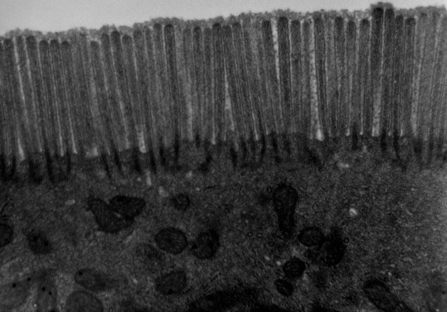
Microvilli (singular: microvillus) are microscopic cellular membrane protrusions that increase the surface area for diffusion and minimize any increase in volume, and are involved in a wide variety of functions, including absorption, secretion, cellular adhesion, and mechanotransduction. Structure Microvilli are covered in plasma membrane, which encloses cytoplasm and microfilaments. Though these are cellular extensions, there are little or no cellular organelles present in the microvilli. Each microvillus has a dense bundle of cross-linked actin filaments, which serves as its structural core. 20 to 30 tightly bundled actin filaments are cross-linked by bundling proteins fimbrin (or plastin-1), villin and espin to form the core of the microvilli. In the enterocyte microvillus, the structural core is attached to the plasma membrane along its length by lateral arms made of myosin 1a and Ca2+ binding protein calmodulin. Myosin 1a functions through a binding site for filamentous ... [...More Info...] [...Related Items...] OR: [Wikipedia] [Google] [Baidu] |
Glycolipid
Glycolipids are lipids with a carbohydrate attached by a glycosidic (covalent) bond. Their role is to maintain the stability of the cell membrane and to facilitate cellular recognition, which is crucial to the immune response and in the connections that allow cells to connect to one another to form tissues. Glycolipids are found on the surface of all eukaryotic cell membranes, where they extend from the phospholipid bilayer into the extracellular environment. Structure The essential feature of a glycolipid is the presence of a monosaccharide or oligosaccharide bound to a lipid moiety. The most common lipids in cellular membranes are glycerolipids and sphingolipids, which have glycerol or a sphingosine backbones, respectively. Fatty acids are connected to this backbone, so that the lipid as a whole has a polar head and a non-polar tail. The lipid bilayer of the cell membrane consists of two layers of lipids, with the inner and outer surfaces of the membrane made up of the pol ... [...More Info...] [...Related Items...] OR: [Wikipedia] [Google] [Baidu] |
Glycoprotein
Glycoproteins are proteins which contain oligosaccharide chains covalently attached to amino acid side-chains. The carbohydrate is attached to the protein in a cotranslational or posttranslational modification. This process is known as glycosylation. Secreted extracellular proteins are often glycosylated. In proteins that have segments extending extracellularly, the extracellular segments are also often glycosylated. Glycoproteins are also often important integral membrane proteins, where they play a role in cell–cell interactions. It is important to distinguish endoplasmic reticulum-based glycosylation of the secretory system from reversible cytosolic-nuclear glycosylation. Glycoproteins of the cytosol and nucleus can be modified through the reversible addition of a single GlcNAc residue that is considered reciprocal to phosphorylation and the functions of these are likely to be an additional regulatory mechanism that controls phosphorylation-based signalling. In contrast, ... [...More Info...] [...Related Items...] OR: [Wikipedia] [Google] [Baidu] |
Oligosaccharide
An oligosaccharide (/ˌɑlɪgoʊˈsækəˌɹaɪd/; from the Greek ὀλίγος ''olígos'', "a few", and σάκχαρ ''sácchar'', "sugar") is a saccharide polymer containing a small number (typically two to ten) of monosaccharides (simple sugars). Oligosaccharides can have many functions including cell recognition and cell adhesion. They are normally present as glycans: oligosaccharide chains are linked to lipids or to compatible amino acid side chains in proteins, by ''N''- or ''O''-glycosidic bonds. ''N''-Linked oligosaccharides are always pentasaccharides attached to asparagine via a beta linkage to the amine nitrogen of the side chain.. Alternately, ''O''-linked oligosaccharides are generally attached to threonine or serine on the alcohol group of the side chain. Not all natural oligosaccharides occur as components of glycoproteins or glycolipids. Some, such as the raffinose series, occur as storage or transport carbohydrates in plants. Others, such as maltodextrins or ... [...More Info...] [...Related Items...] OR: [Wikipedia] [Google] [Baidu] |
Glycocalyx
The glycocalyx, also known as the pericellular matrix, is a glycoprotein and glycolipid covering that surrounds the cell membranes of bacteria, epithelial cells, and other cells. In 1970, Martinez-Palomo discovered the cell coating in animal cells, which is known as the glycocalyx. Animal epithelial cells have a fuzz-like coating on the external surface of their plasma membranes. This viscous coating consists of several carbohydrate moieties of membrane glycolipids and glycoproteins, which serve as backbone molecules for support. Generally, the carbohydrate portion of the glycolipids found on the surface of plasma membranes helps these molecules contribute to cell–cell recognition, communication, and intercellular adhesion. The glycocalyx is a type of identifier that the body uses to distinguish between its own healthy cells and transplanted tissues, diseased cells, or invading organisms. Included in the glycocalyx are cell-adhesion molecules that enable cells to adhere to eac ... [...More Info...] [...Related Items...] OR: [Wikipedia] [Google] [Baidu] |

_and_foveolar_cells_in_incomplete_Barrett's_esophagus.jpg)

