|
Interventional Neurology
Endovascular therapy (EVT), also known as neurointerventional surgery (NIS), interventional neuroradiology (INR), endovascular neurosurgery, and interventional neurology is a medical subspecialty of radiology, neurosurgery, and neurology specializing in minimally invasive image-based technologies and procedures used in diagnosis and treatment of diseases of the head, neck, and spine. History Diagnostic angiography Cerebral angiography was developed by Portuguese neurologist Egas Moniz at the University of Lisbon, in order to identify central nervous system diseases such as tumors or arteriovenous malformations. He performed the first brain angiography in Lisbon in 1927 by injecting an iodinated contrast medium into the internal carotid artery and using the X-rays discovered 30 years earlier by Roentgen in order to visualize the cerebral vessels. In pre-CT and pre-MRI, it was the only tool to observe the structures within the skull and was also used to diagnose extravas ... [...More Info...] [...Related Items...] OR: [Wikipedia] [Google] [Baidu] |
Radiology
Radiology ( ) is the medical discipline that uses medical imaging to diagnose diseases and guide their treatment, within the bodies of humans and other animals. It began with radiography (which is why its name has a root referring to radiation), but today it includes all imaging modalities, including those that use no electromagnetic radiation (such as ultrasonography and magnetic resonance imaging), as well as others that do, such as computed tomography (CT), fluoroscopy, and nuclear medicine including positron emission tomography (PET). Interventional radiology is the performance of usually minimally invasive medical procedures with the guidance of imaging technologies such as those mentioned above. The modern practice of radiology involves several different healthcare professions working as a team. The radiologist is a medical doctor who has completed the appropriate post-graduate training and interprets medical images, communicates these findings to other physicians ... [...More Info...] [...Related Items...] OR: [Wikipedia] [Google] [Baidu] |
Seldinger Technique
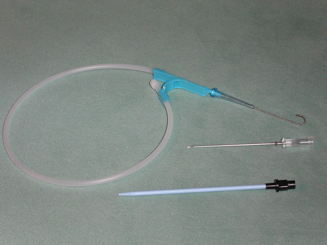
The Seldinger technique, also known as Seldinger wire technique, is a medical procedure to obtain safe access to blood vessels and other hollow organs. It is named after Sven Ivar Seldinger (1921–1998), a Swedish radiologist who introduced the procedure in 1953. Uses The Seldinger technique is used for angiography, insertion of chest drains and central venous catheters, insertion of PEG tubes using the push technique, insertion of the leads for an artificial pacemaker or implantable cardioverter-defibrillator, and numerous other interventional medical procedures. Complications The initial puncture is with a sharp instrument, and this may lead to hemorrhage or perforation of the organ in question. Infection is a possible complication, and hence asepsis is practiced during most Seldinger procedures. Loss of the guidewire into the cavity or blood vessel is a significant and generally preventable complication. Description The desired vessel or cavity is punctured with a shar ... [...More Info...] [...Related Items...] OR: [Wikipedia] [Google] [Baidu] |
Lumen (anatomy)
In biology, a lumen (plural lumina) is the inside space of a tubular structure, such as an artery or intestine. It comes . It can refer to: *The interior of a vessel, such as the central space in an artery, vein or capillary through which blood flows. *The interior of the gastrointestinal tract *The pathways of the bronchi in the lungs *The interior of renal tubules and urinary collecting ducts *The pathways of the female genital tract, starting with a single pathway of the vagina, splitting up in two lumina in the uterus, both of which continue through the Fallopian tubes In cell biology, a lumen is a membrane-defined space that is found inside several organelles, cellular components, or structures: *thylakoid, endoplasmic reticulum, Golgi apparatus, lysosome, mitochondrion, or microtubule Transluminal procedures ''Transluminal procedures'' are procedures occurring through lumina, including: *Natural orifice transluminal endoscopic surgery in the lumina of, for example, the ... [...More Info...] [...Related Items...] OR: [Wikipedia] [Google] [Baidu] |
Vascular Occlusion
Vascular occlusion is a blockage of a blood vessel, usually with a clot. It differs from thrombosis in that it can be used to describe any form of blockage, not just one formed by a clot. When it occurs in a major vein, it can, in some cases, cause deep vein thrombosis. The condition is also relatively common in the retina, and can cause partial or total loss of vision. An occlusion can often be diagnosed using Doppler sonography (a form of ultrasound). Some medical procedures, such as embolisation, involve occluding a blood vessel to treat a particular condition. This can be to reduce pressure on aneurysms (weakened blood vessels) or to restrict a haemorrhage. It can also be used to reduce blood supply to tumours or growths in the body, and therefore restrict their development. Occlusion can be carried out using a ligature; by implanting small coils which stimulate the formation of clots; or, particularly in the case of cerebral aneurysms, by clipping. See also * Central ... [...More Info...] [...Related Items...] OR: [Wikipedia] [Google] [Baidu] |
Intracranial Aneurysm
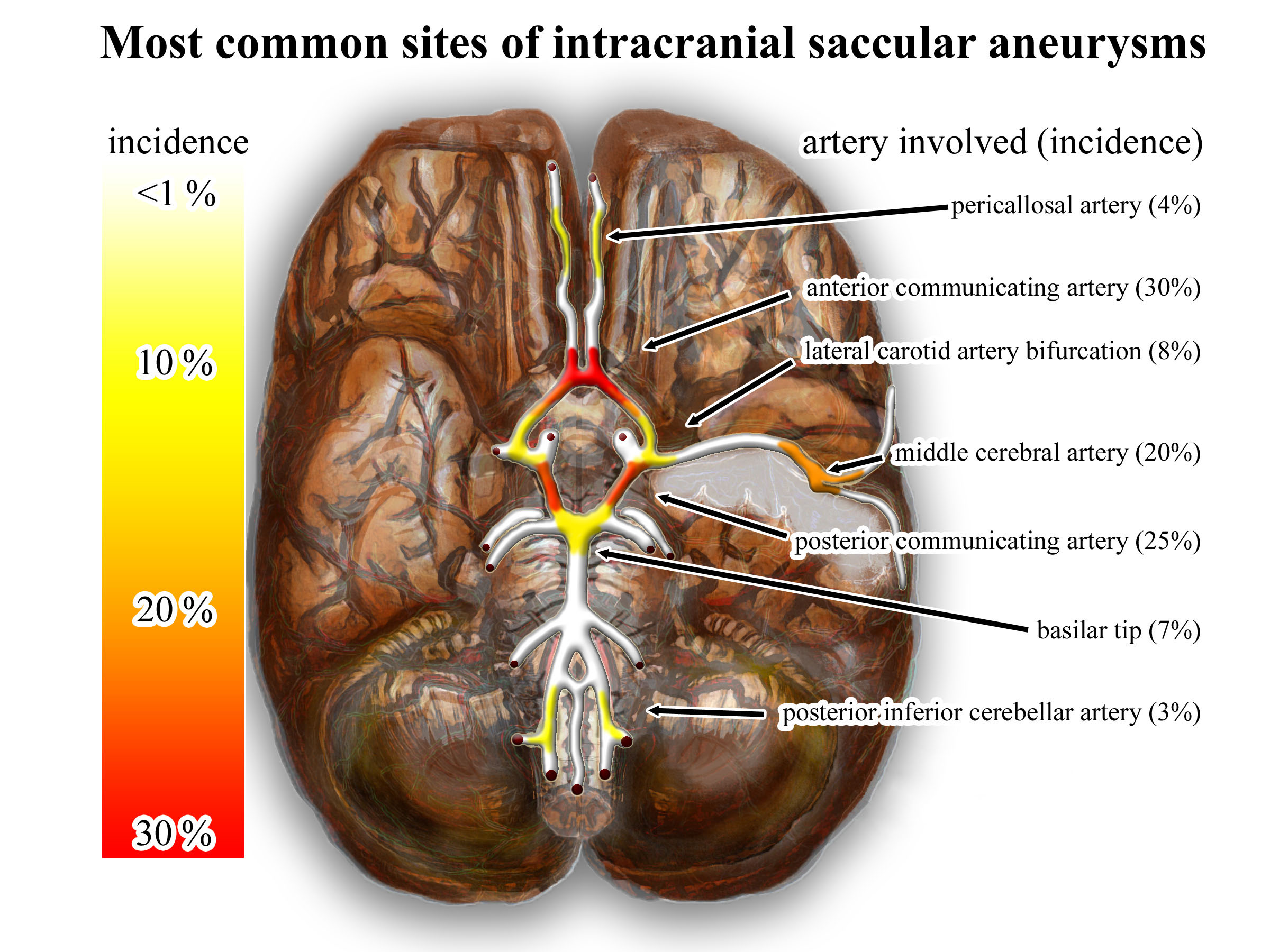
An intracranial aneurysm, also known as a brain aneurysm, is a cerebrovascular disorder in which weakness in the wall of a cerebral artery or vein causes a localized dilation or ballooning of the blood vessel. Aneurysms in the posterior circulation (basilar artery, vertebral arteries and posterior communicating artery) have a higher risk of rupture. Basilar artery aneurysms represent only 3–5% of all intracranial aneurysms but are the most common aneurysms in the posterior circulation. Classification Cerebral aneurysms are classified both by size and shape. Small aneurysms have a diameter of less than 15 mm. Larger aneurysms include those classified as large (15 to 25 mm), giant (25 to 50 mm), and super-giant (over 50 mm). Berry (saccular) aneurysms Saccular aneurysms, also known as berry aneurysms, appear as a round outpouching and are the most common form of cerebral aneurysm. Causes include connective tissue disorders, polycystic kidney disease, art ... [...More Info...] [...Related Items...] OR: [Wikipedia] [Google] [Baidu] |
Arterial Tortuosity Syndrome
Arterial tortuosity syndrome is a rare congenital connective tissue condition disorder characterized by elongation and generalized tortuosity of the major arteries including the aorta. It is associated with hyperextensible skin and hypermobility of joints, however symptoms vary depending on the person. Because ATS is so rare, not much is known about the disease. Signs and symptoms Among the signs and symptoms demonstrated, by this condition are the following: * Arachnodactyly * Congenital diaphragmatic hernia * Mental dysfunction * Keratoconus * Aortic regurgitation * Blepharophimosis Genetics Arterial tortuosity syndrome exhibits autosomal recessive inheritance, and the responsible gene is located at chromosome 20q13. The gene associated with arterial tortuosity syndrome is SLC2A10 and has no less than 23 mutations in those individuals found to have the aforementioned condition. Pathophysiology The mechanism of this condition is apparently controlled by(or due to) the SLC2 ... [...More Info...] [...Related Items...] OR: [Wikipedia] [Google] [Baidu] |
Lesion
A lesion is any damage or abnormal change in the tissue of an organism, usually caused by disease or trauma. ''Lesion'' is derived from the Latin "injury". Lesions may occur in plants as well as animals. Types There is no designated classification or naming convention for lesions. Since lesions can occur anywhere in the body and the definition of a lesion is so broad, the varieties of lesions are virtually endless. Generally, lesions may be classified by their patterns, their sizes, their locations, or their causes. They can also be named after the person who discovered them. For example, Ghon lesions, which are found in the lungs of those with tuberculosis, are named after the lesion's discoverer, Anton Ghon. The characteristic skin lesions of a varicella zoster virus infection are called '' chickenpox''. Lesions of the teeth are usually called dental caries. Location Lesions are often classified by their tissue types or locations. For example, a "skin lesion" or a " bra ... [...More Info...] [...Related Items...] OR: [Wikipedia] [Google] [Baidu] |
Cerebrovascular Disease
Cerebrovascular disease includes a variety of medical conditions that affect the blood vessels of the brain and the cerebral circulation. Arteries supplying oxygen and nutrients to the brain are often damaged or deformed in these disorders. The most common presentation of cerebrovascular disease is an ischemic stroke or mini-stroke and sometimes a hemorrhagic stroke. Hypertension (high blood pressure) is the most important contributing risk factor for stroke and cerebrovascular diseases as it can change the structure of blood vessels and result in atherosclerosis. Atherosclerosis narrows blood vessels in the brain, resulting in decreased cerebral perfusion. Other risk factors that contribute to stroke include smoking and diabetes. Narrowed cerebral arteries can lead to ischemic stroke, but continually elevated blood pressure can also cause tearing of vessels, leading to a hemorrhagic stroke. A stroke usually presents with an abrupt onset of a neurologic deficit – such as hemi ... [...More Info...] [...Related Items...] OR: [Wikipedia] [Google] [Baidu] |
Pneumonia
Pneumonia is an inflammatory condition of the lung primarily affecting the small air sacs known as alveoli. Symptoms typically include some combination of productive or dry cough, chest pain, fever, and difficulty breathing. The severity of the condition is variable. Pneumonia is usually caused by infection with viruses or bacteria, and less commonly by other microorganisms. Identifying the responsible pathogen can be difficult. Diagnosis is often based on symptoms and physical examination. Chest X-rays, blood tests, and culture of the sputum may help confirm the diagnosis. The disease may be classified by where it was acquired, such as community- or hospital-acquired or healthcare-associated pneumonia. Risk factors for pneumonia include cystic fibrosis, chronic obstructive pulmonary disease (COPD), sickle cell disease, asthma, diabetes, heart failure, a history of smoking, a poor ability to cough (such as following a stroke), and a weak immune system. Vaccines to ... [...More Info...] [...Related Items...] OR: [Wikipedia] [Google] [Baidu] |
Amputation
Amputation is the removal of a limb by trauma, medical illness, or surgery. As a surgical measure, it is used to control pain or a disease process in the affected limb, such as malignancy or gangrene. In some cases, it is carried out on individuals as a preventive surgery for such problems. A special case is that of congenital amputation, a congenital disorder, where fetal limbs have been cut off by constrictive bands. In some countries, amputation is currently used to punish people who commit crimes. Amputation has also been used as a tactic in war and acts of terrorism; it may also occur as a war injury. In some cultures and religions, minor amputations or mutilations are considered a ritual accomplishment. When done by a person, the person executing the amputation is an amputator. The oldest evidence of this practice comes from a skeleton found buried in Liang Tebo cave, East Kalimantan, Indonesian Borneo dating back to at least 31,000 years ago, where it was done when ... [...More Info...] [...Related Items...] OR: [Wikipedia] [Google] [Baidu] |
Ischemia
Ischemia or ischaemia is a restriction in blood supply to any tissue, muscle group, or organ of the body, causing a shortage of oxygen that is needed for cellular metabolism (to keep tissue alive). Ischemia is generally caused by problems with blood vessels, with resultant damage to or dysfunction of tissue i.e. hypoxia and microvascular dysfunction. It also implies local hypoxia in a part of a body resulting from constriction (such as vasoconstriction, thrombosis, or embolism). Ischemia causes not only insufficiency of oxygen, but also reduced availability of nutrients and inadequate removal of metabolic wastes. Ischemia can be partial (poor perfusion) or total blockage. The inadequate delivery of oxygenated blood to the organs must be resolved either by treating the cause of the inadequate delivery or reducing the oxygen demand of the system that needs it. For example, patients with myocardial ischemia have a decreased blood flow to the heart and are prescribed with medi ... [...More Info...] [...Related Items...] OR: [Wikipedia] [Google] [Baidu] |
Femoral Artery
The femoral artery is a large artery in the thigh and the main arterial supply to the thigh and leg. The femoral artery gives off the deep femoral artery or profunda femoris artery and descends along the anteromedial part of the thigh in the femoral triangle. It enters and passes through the adductor canal, and becomes the popliteal artery as it passes through the adductor hiatus in the adductor magnus near the junction of the middle and distal thirds of the thigh. Structure The femoral artery enters the thigh from behind the inguinal ligament as the continuation of the external iliac artery. Here, it lies midway between the anterior superior iliac spine and the symphysis pubis (Mid-inguinal point). Segments In clinical parlance, the femoral artery has the following segments: *The common femoral artery (CFA) is the segment of the femoral artery between the inferior margin of the inguinal ligament and the branching point of the deep femoral artery/profunda femoris artery. Its ... [...More Info...] [...Related Items...] OR: [Wikipedia] [Google] [Baidu] |


