|
Internal Thoracic Vein
In human anatomy, the internal thoracic vein (previously known as the internal mammary vein) is a vessel that drains the chest wall and breasts. Structure Bilaterally, the internal thoracic vein arises from the superior epigastric vein, and accompanies the internal thoracic artery along its course. It drains the intercostal veins, although the posterior drainage is often handled by the azygous veins. It terminates in the brachiocephalic vein. It has a width of 2-3 mm. There is either one or two internal thoracic veins accompanying the corresponding artery (internal thoracic artery). If internal thoracic vein is single, it usually runs medial to the artery. If there are double thoracic veins, they run on either side of the internal thoracic artery. Variations Bifurcation of each internal thoracic vein is common. The left internal thoracic vein may bifurcate between ribs 3-4 or remain as a single vein. The right internal thoracic vein may bifurcate between ribs 2-4 or rem ... [...More Info...] [...Related Items...] OR: [Wikipedia] [Google] [Baidu] |
Thorax
The thorax or chest is a part of the anatomy of humans, mammals, and other tetrapod animals located between the neck and the abdomen. In insects, crustaceans, and the extinct trilobites, the thorax is one of the three main divisions of the creature's body, each of which is in turn composed of multiple segments. The human thorax includes the thoracic cavity and the thoracic wall. It contains organs including the heart, lungs, and thymus gland, as well as muscles and various other internal structures. Many diseases may affect the chest, and one of the most common symptoms is chest pain. Etymology The word thorax comes from the Greek θώραξ ''thorax'' " breastplate, cuirass, corslet" via la, thorax. Plural: ''thoraces'' or ''thoraxes''. Human thorax Structure In humans and other hominids, the thorax is the chest region of the body between the neck and the abdomen, along with its internal organs and other contents. It is mostly protected and supported by the rib ... [...More Info...] [...Related Items...] OR: [Wikipedia] [Google] [Baidu] |
Millimetre
330px, Different lengths as in respect to the electromagnetic spectrum, measured by the metre and its derived scales. The microwave is between 1 meter to 1 millimeter. The millimetre (American and British English spelling differences#-re, -er, international spelling; International System of Units, SI unit symbol mm) or millimeter (American and British English spelling differences#-re, -er, American spelling) is a Units of measurement, unit of length in the International System of Units (SI), equal to one thousandth of a metre, which is the SI base unit of length. Therefore, there are one thousand millimetres in a metre. There are ten millimetres in a centimetre. One millimetre is equal to micrometres or nanometres. Since an inch is officially defined as exactly 25.4 millimetres, a millimetre is equal to exactly (≈ 0.03937) of an inch. Definition Since 1983, the metre has been defined as "the length of the path travelled by light in vacuum during a time interval of of a ... [...More Info...] [...Related Items...] OR: [Wikipedia] [Google] [Baidu] |
Inferior Vena Cava
The inferior vena cava is a large vein that carries the deoxygenated blood from the lower and middle body into the right atrium of the heart. It is formed by the joining of the right and the left common iliac veins, usually at the level of the fifth lumbar vertebra. The inferior vena cava is the lower ("inferior") of the two venae cavae, the two large veins that carry deoxygenated blood from the body to the right atrium of the heart: the inferior vena cava carries blood from the lower half of the body whilst the superior vena cava carries blood from the upper half of the body. Together, the venae cavae (in addition to the coronary sinus, which carries blood from the muscle of the heart itself) form the venous counterparts of the aorta. It is a large retroperitoneal vein that lies posterior to the abdominal cavity and runs along the right side of the vertebral column. It enters the right auricle at the lower right, back side of the heart. The name derives from la, ven ... [...More Info...] [...Related Items...] OR: [Wikipedia] [Google] [Baidu] |
Sternum
The sternum or breastbone is a long flat bone located in the central part of the chest. It connects to the ribs via cartilage and forms the front of the rib cage, thus helping to protect the heart, lungs, and major blood vessels from injury. Shaped roughly like a necktie, it is one of the largest and longest flat bones of the body. Its three regions are the manubrium, the body, and the xiphoid process. The word "sternum" originates from the Ancient Greek στέρνον (stérnon), meaning "chest". Structure The sternum is a narrow, flat bone, forming the middle portion of the front of the chest. The top of the sternum supports the clavicles (collarbones) and its edges join with the costal cartilages of the first two pairs of ribs. The inner surface of the sternum is also the attachment of the sternopericardial ligaments. Its top is also connected to the sternocleidomastoid muscle. The sternum consists of three main parts, listed from the top: * Manubrium * Body (gl ... [...More Info...] [...Related Items...] OR: [Wikipedia] [Google] [Baidu] |
Central Venous Catheter
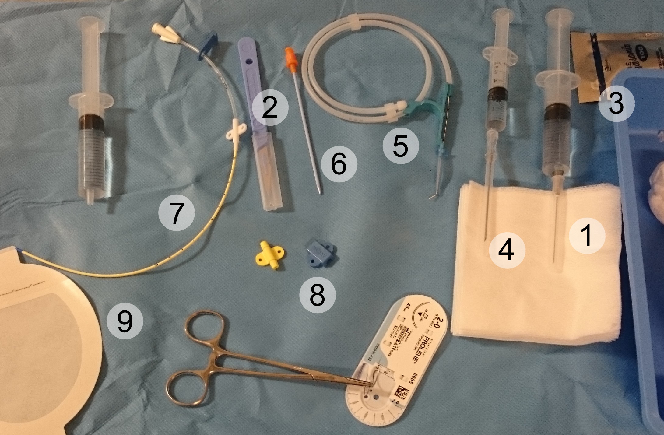
A central venous catheter (CVC), also known as a central line(c-line), central venous line, or central venous access catheter, is a catheter placed into a large vein. It is a form of venous access. Placement of larger catheters in more centrally located veins is often needed in critically ill patients, or in those requiring prolonged intravenous therapies, for more reliable vascular access. These catheters are commonly placed in veins in the neck (internal jugular vein), chest (subclavian vein or axillary vein), groin (femoral vein), or through veins in the arms (also known as a PICC line, or peripherally inserted central catheters). Central lines are used to administer medication or fluids that are unable to be taken by mouth or would harm a smaller peripheral vein, obtain blood tests (specifically the "central venous oxygen saturation"), administer fluid or blood products for large volume resuscitation, and measure central venous pressure. The catheters used are commonly 15� ... [...More Info...] [...Related Items...] OR: [Wikipedia] [Google] [Baidu] |
Empyema
An empyema () is a collection or gathering of pus within a naturally existing anatomical cavity. For example, pleural empyema is empyema of the pleural cavity. It must be differentiated from an abscess, which is a collection of pus in a newly formed cavity. The term is from Greek ἐμπύημα, "abscess". Classification Empyema occurs in: * the pleural cavity ( pleural empyema also known as pyothorax) * the thoracic cavity * the uterus (pyometra) * the appendix (appendicitis) * the meninges ( subdural empyema) * the joints (septic arthritis) * the gallbladder In vertebrates, the gallbladder, also known as the cholecyst, is a small hollow organ where bile is stored and concentrated before it is released into the small intestine. In humans, the pear-shaped gallbladder lies beneath the liver, althoug ... External links * {{Authority control Immune system Medical terminology ... [...More Info...] [...Related Items...] OR: [Wikipedia] [Google] [Baidu] |
Breast
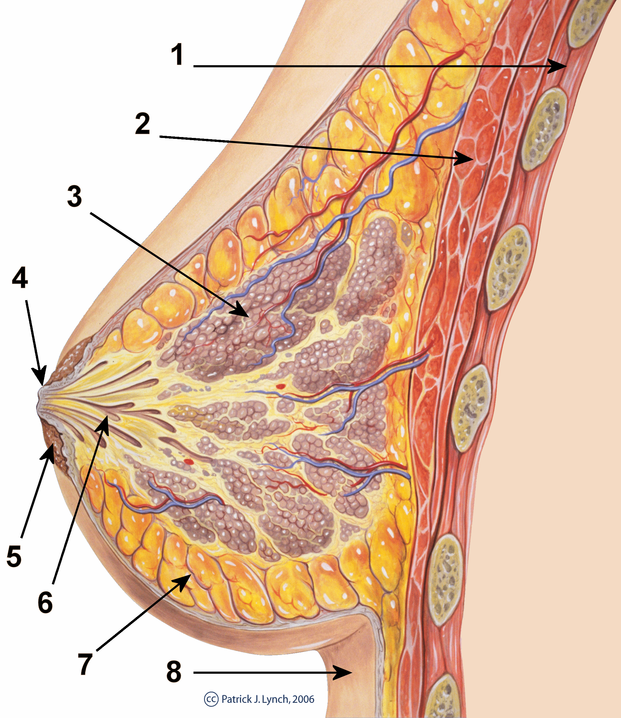
The breast is one of two prominences located on the upper ventral region of a primate's torso. Both females and males develop breasts from the same embryological tissues. In females, it serves as the mammary gland, which produces and secretes milk to feed infants. Subcutaneous fat covers and envelops a network of ducts that converge on the nipple, and these tissues give the breast its size and shape. At the ends of the ducts are lobules, or clusters of alveoli, where milk is produced and stored in response to hormonal signals. During pregnancy, the breast responds to a complex interaction of hormones, including estrogens, progesterone, and prolactin, that mediate the completion of its development, namely lobuloalveolar maturation, in preparation of lactation and breastfeeding. Humans are the only animals with permanent breasts. At puberty, estrogens, in conjunction with growth hormone, cause permanent breast growth in female humans. This happens only to ... [...More Info...] [...Related Items...] OR: [Wikipedia] [Google] [Baidu] |
Thoracic Wall
The thoracic wall or chest wall is the boundary of the thoracic cavity. Structure The bony skeletal part of the thoracic wall is the rib cage, and the rest is made up of muscle, skin, and fasciae. The chest wall has 10 layers, namely (from superficial to deep) skin (epidermis and dermis), superficial fascia, deep fascia and the invested extrinsic muscles (from the upper limbs), intrinsic muscles associated with the ribs (three layers of intercostal muscles), endothoracic fascia and parietal pleura. However, the extrinsic muscular layers vary according to the region of the chest wall. For example, the front and back sides may include attachments of large upper limb muscles like pectoralis major or latissimus dorsi, while the sides only have serratus anterior. Function Diving reflex When not breathing for long and dangerous periods of time in cold water, a person's body undergoes great temporary changes to try to prevent death. It achieves this through the activa ... [...More Info...] [...Related Items...] OR: [Wikipedia] [Google] [Baidu] |
Vena Comitans
Vena comitans is Latin for accompanying vein. It refers to a vein that is usually paired, with both veins lying on the sides of an artery. They are found in close proximity to arteries so that the pulsations of the artery aid venous return. Because they are generally found in pairs, they are often referred to by their plural form: venae comitantes. Venae comitantes are usually found with certain smaller arteries, especially those in the extremities. Larger arteries, on the other hand, generally do not have venae comitantes. They usually have a single, similarly sized vein which is not as intimately associated with the artery. Examples of arteries and their venae comitantes: * Radial artery and radial veins * Ulnar artery and ulnar veins * Brachial artery and brachial veins * Anterior tibial artery and anterior tibial veins * Posterior tibial artery and Posterior tibial veins * Fibular artery and Fibular veins Examples of arteries that do not have venae comitantes (i. ... [...More Info...] [...Related Items...] OR: [Wikipedia] [Google] [Baidu] |
Azygos Vein
The azygos vein is a vein running up the right side of the thoracic vertebral column draining itself towards the superior vena cava. It connects the systems of superior vena cava and inferior vena cava and can provide an alternative path for blood to the right atrium when either of the venae cavae is blocked. Structure The azygos vein transports deoxygenated blood from the posterior walls of the thorax and abdomen into the superior vena cava. It is formed by the union of the ascending lumbar veins with the right subcostal veins at the level of the 12th thoracic vertebra, ascending to the right of the descending aorta and thoracic duct, passing behind the right crus of diaphragm, anterior to the vertebral bodies of T12 to T5 and right posterior intercostal arteries. At the level of T4 vertebrae, it arches over the root of the right lung from behind to the front to join the superior vena cava. The trachea and oesophagus is located medially to the arch of the azygous vein. ... [...More Info...] [...Related Items...] OR: [Wikipedia] [Google] [Baidu] |
Abdomen
The abdomen (colloquially called the belly, tummy, midriff, tucky or stomach) is the part of the body between the thorax (chest) and pelvis, in humans and in other vertebrates. The abdomen is the front part of the abdominal segment of the torso. The area occupied by the abdomen is called the abdominal cavity. In arthropods it is the posterior tagma of the body; it follows the thorax or cephalothorax. In humans, the abdomen stretches from the thorax at the thoracic diaphragm to the pelvis at the pelvic brim. The pelvic brim stretches from the lumbosacral joint (the intervertebral disc between L5 and S1) to the pubic symphysis and is the edge of the pelvic inlet. The space above this inlet and under the thoracic diaphragm is termed the abdominal cavity. The boundary of the abdominal cavity is the abdominal wall in the front and the peritoneal surface at the rear. In vertebrates, the abdomen is a large body cavity enclosed by the abdominal muscles, at front and to ... [...More Info...] [...Related Items...] OR: [Wikipedia] [Google] [Baidu] |
Intercostal Veins
The intercostal veins are a group of veins which drain the area between the ribs ("costae"), called the intercostal space. They can be divided as follows: * Anterior intercostal veins * Posterior intercostal veins ** Posterior intercost vein that drain into the Supreme intercostal vein - 1st intercostal space ** Posterior intercost veins that drain into the Superior intercostal vein - 2nd, 3rd, and 4th intercostal spaces. The superior intercostal vein then drains into the Azygous vein. ** Posterior intercost veins that drain directly into the Azygous vein - in spaces 5-11. ** Subcostal vein -- below bottom (12th) rib and also drains into the Azygous vein. See also * Intercostal nerves The intercostal nerves are part of the somatic nervous system, and arise from the anterior rami of the thoracic spinal nerves from T1 to T11. The intercostal nerves are distributed chiefly to the thoracic pleura and abdominal peritoneum, and dif ... External links * * http://www.instantanatomy. ... [...More Info...] [...Related Items...] OR: [Wikipedia] [Google] [Baidu] |


