|
Intercostobrachial
The intercostobrachial nerves are cutaneous branches of the intercostal nerves. Second intercostal nerve The lateral cutaneous branch of the second intercostal nerve does not divide, like the others, into an anterior and a posterior branch; it is named the intercostobrachial nerve. It pierces the Intercostalis externus and the Serratus anterior, crosses the axilla to the medial side of the arm, and joins with a filament from the medial brachial cutaneous nerve. It then pierces the fascia, and supplies the skin of the upper half of the medial and posterior part of the arm, communicating with the posterior brachial cutaneous branch of the radial nerve. It is often the source of referred cardiac pain. The intercostobrachial nerve is also sometimes divided in axillary node clearance (ANC), such as that done for breast cancer surgery which requires the removal of the axillary nodes. Sensation to the cutaneous region supplied by the nerve is affected. The size of the intercostobrachial ... [...More Info...] [...Related Items...] OR: [Wikipedia] [Google] [Baidu] |
Cutaneous Innervation Of The Upper Limbs
Cutaneous innervation of the upper limbs is the nerve supply to areas of the skin of the upper limbs (including the arm, forearm, and hand) which are supplied by specific cutaneous nerves. Modern texts are in agreement about which areas of the skin are served by which cutaneous nerves, but there are minor variations in some of the details. The borders designated by the diagrams in the 1918 edition of Gray's Anatomy, provided below, are similar but not identical to those generally accepted today. Arm and shoulder * Supraclavicular nerves (yellow) * Axillary nerve (blue). Also Superior lateral cutaneous nerve of arm. * Inferior lateral cutaneous nerve of arm - Near blue "axillary" area, but actually branches from radial nerve. Most modern sources distinguish the superior and inferior, but some still include a single "lateral brachial cutaneous nerve". ) * Intercostobrachial nerve (brown) * Medial cutaneous nerve of arm (yellow) - labeled as "medial brachial cutaneous". * Posterior ... [...More Info...] [...Related Items...] OR: [Wikipedia] [Google] [Baidu] |
Intercostal Nerves
The intercostal nerves are part of the somatic nervous system, and arise from the anterior rami of the thoracic spinal nerves from T1 to T11. The intercostal nerves are distributed chiefly to the thoracic pleura and abdominal peritoneum, and differ from the anterior rami of the other spinal nerves in that each pursues an independent course without plexus formation. The first two nerves supply fibers to the upper limb and thorax; the next four distribute to the walls of the thorax; the lower five supply the walls of the thorax and abdomen. The 7th intercostal nerve end at the xyphoid process of the sternum. The 10th intercostal nerve terminates at the navel. The 12th ( subcostal) thoracic is distributed to the walls of the abdomen and groin. Each of these fibers contains around 1300 axons. Unlike the nerves from the autonomic nervous system that innervate the visceral pleura of the thoracic cavity, the intercostal nerves arise from the somatic nervous system. This enables them to c ... [...More Info...] [...Related Items...] OR: [Wikipedia] [Google] [Baidu] |
Cutaneous
Skin is the layer of usually soft, flexible outer tissue covering the body of a vertebrate animal, with three main functions: protection, regulation, and sensation. Other animal coverings, such as the arthropod exoskeleton, have different developmental origin, structure and chemical composition. The adjective cutaneous means "of the skin" (from Latin ''cutis'' 'skin'). In mammals, the skin is an organ of the integumentary system made up of multiple layers of ectodermal tissue and guards the underlying muscles, bones, ligaments, and internal organs. Skin of a different nature exists in amphibians, reptiles, and birds. Skin (including cutaneous and subcutaneous tissues) plays crucial roles in formation, structure, and function of extraskeletal apparatus such as horns of bovids (e.g., cattle) and rhinos, cervids' antlers, giraffids' ossicones, armadillos' osteoderm, and os penis/os clitoris. All mammals have some hair on their skin, even marine mammals like whales, dolphins, a ... [...More Info...] [...Related Items...] OR: [Wikipedia] [Google] [Baidu] |
Intercostal Nerve
The intercostal nerves are part of the somatic nervous system, and arise from the anterior rami of the thoracic spinal nerves from T1 to T11. The intercostal nerves are distributed chiefly to the thoracic pleura and abdominal peritoneum, and differ from the anterior rami of the other spinal nerves in that each pursues an independent course without plexus formation. The first two nerves supply fibers to the upper limb and thorax; the next four distribute to the walls of the thorax; the lower five supply the walls of the thorax and abdomen. The 7th intercostal nerve end at the xyphoid process of the sternum. The 10th intercostal nerve terminates at the navel. The 12th ( subcostal) thoracic is distributed to the walls of the abdomen and groin. Each of these fibers contains around 1300 axons. Unlike the nerves from the autonomic nervous system that innervate the visceral pleura of the thoracic cavity, the intercostal nerves arise from the somatic nervous system. This enables them to c ... [...More Info...] [...Related Items...] OR: [Wikipedia] [Google] [Baidu] |
Intercostal Nerves
The intercostal nerves are part of the somatic nervous system, and arise from the anterior rami of the thoracic spinal nerves from T1 to T11. The intercostal nerves are distributed chiefly to the thoracic pleura and abdominal peritoneum, and differ from the anterior rami of the other spinal nerves in that each pursues an independent course without plexus formation. The first two nerves supply fibers to the upper limb and thorax; the next four distribute to the walls of the thorax; the lower five supply the walls of the thorax and abdomen. The 7th intercostal nerve end at the xyphoid process of the sternum. The 10th intercostal nerve terminates at the navel. The 12th ( subcostal) thoracic is distributed to the walls of the abdomen and groin. Each of these fibers contains around 1300 axons. Unlike the nerves from the autonomic nervous system that innervate the visceral pleura of the thoracic cavity, the intercostal nerves arise from the somatic nervous system. This enables them to c ... [...More Info...] [...Related Items...] OR: [Wikipedia] [Google] [Baidu] |
Second Intercostal Nerve
The intercostal nerves are part of the somatic nervous system, and arise from the anterior rami of the thoracic spinal nerves from T1 to T11. The intercostal nerves are distributed chiefly to the thoracic pleura and abdominal peritoneum, and differ from the anterior rami of the other spinal nerves in that each pursues an independent course without plexus formation. The first two nerves supply fibers to the upper limb and thorax; the next four distribute to the walls of the thorax; the lower five supply the walls of the thorax and abdomen. The 7th intercostal nerve end at the xyphoid process of the sternum. The 10th intercostal nerve terminates at the navel. The 12th ( subcostal) thoracic is distributed to the walls of the abdomen and groin. Each of these fibers contains around 1300 axons. Unlike the nerves from the autonomic nervous system that innervate the visceral pleura of the thoracic cavity, the intercostal nerves arise from the somatic nervous system. This enables them to c ... [...More Info...] [...Related Items...] OR: [Wikipedia] [Google] [Baidu] |
Intercostalis Externus
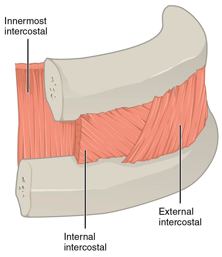
The external intercostal muscles, or external intercostals (Intercostales externi) are eleven in number on both sides. Structure The muscles extend from the tubercles of the ribs behind, to the cartilages of the ribs in front, where they end in thin membranes, the external intercostal membranes, which are continued forward to the sternum. These muscles work in unison when inhalation occurs. The internal intercostal muscles relax while the external muscles contract causing the expansion of the chest cavity and an influx of air into the lungs. Each arises from the lower border of a rib, and is inserted into the upper border of the rib below. In the two lower spaces they extend to the ends of the cartilages, and in the upper two or three spaces they do not quite reach the ends of the ribs. They are thicker than the internal intercostals, and their fibers are directed obliquely downward and laterally on the back of the thorax, and downward, forward, and medially on the front. ... [...More Info...] [...Related Items...] OR: [Wikipedia] [Google] [Baidu] |
Serratus Anterior
The serratus anterior is a muscle that originates on the surface of the 1st to 8th ribs at the side of the chest and inserts along the entire anterior length of the medial border of the scapula. The serratus anterior acts to pull the scapula forward around the thorax. The muscle is named from Latin: ''serrare'' = to saw, referring to the shape, ''anterior'' = on the front side of the body. Structure Serratus anterior normally originates by nine or ten muscle slips – branches from either the first to ninth ribs or the first to eighth ribs. Because two slips usually arise from the second rib, the number of slips is greater than the number of ribs from which they originate. The muscle is inserted along the medial border of the scapula between the superior and inferior angles along with being inserted along the thoracic vertebrae. The muscle is divided into three named parts depending on their points of insertions: #the serratus anterior superior is inserted near the superior a ... [...More Info...] [...Related Items...] OR: [Wikipedia] [Google] [Baidu] |
Axilla
The axilla (also, armpit, underarm or oxter) is the area on the human body directly under the shoulder joint. It includes the axillary space, an anatomical space within the shoulder girdle between the arm and the thoracic cage, bounded superiorly by the imaginary plane between the superior borders of the first rib, clavicle and scapula (above which are considered part of the neck), medially by the serratus anterior muscle and thoracolumbar fascia, anteriorly by the pectoral muscles and posteriorly by the subscapularis, teres major and latissimus dorsi muscle. The soft skin covering the lateral axilla contains many hair and sweat glands. In humans, the formation of body odor happens mostly in the axilla. These odorant substances have been suggested by some to serve as pheromones, which play a role related to mate selection, although this is a controversial topic within the scientific community. The underarms seem more important than the pubic area for emitting body odor, whi ... [...More Info...] [...Related Items...] OR: [Wikipedia] [Google] [Baidu] |
Medial Brachial Cutaneous Nerve
The medial brachial cutaneous nerve (lesser internal cutaneous nerve; medial cutaneous nerve of arm) is distributed to the skin on the medial brachial side of the arm. Anatomy It is the smallest branch of the brachial plexus, and arising from the medial cord receives its fibers from the eighth cervical and first thoracic nerves. It passes through the axilla, at first lying behind, and then medial to the axillary vein, and communicates with the intercostobrachial nerve. It descends along the medial side of the brachial artery to the middle of the arm, where it pierces the deep fascia, and is distributed to the skin of the back of the lower third of the arm, extending as far as the elbow, where some filaments are lost in the skin in front of the medial epicondyle, and others over the olecranon. It communicates with the ulnar branch of the medial antebrachial cutaneous nerve. Eponym The term nerve of Wrisberg (after Heinrich August Wrisberg) has been used to describe this nerve. ... [...More Info...] [...Related Items...] OR: [Wikipedia] [Google] [Baidu] |
Posterior Brachial Cutaneous Branch
The posterior cutaneous nerve of arm (internal cutaneous branch of musculospiral, posterior brachial cutaneous nerve) is a branch of the radial nerve that provides sensory innervation for much of the skin on the back of the arm. It arises in the axilla. It is of small size, and passes through the axilla to the medial side of the area supplying the skin on its dorsal surface nearly as far as the olecranon. In its course it crosses behind and communicates with the intercostobrachial. See also * Superior lateral cutaneous nerve of arm * Inferior lateral cutaneous nerve of arm * Medial cutaneous nerve of arm * Posterior cutaneous nerve of forearm The posterior cutaneous nerve of forearm is a nerve found in humans and other animals. It is also known as the dorsal antebrachial cutaneous nerve, the external cutaneous branch of the musculospiral nerve, and the posterior antebrachial cutaneous n ... Additional images File:Gray413_color.png, Cross-section through the middle of upper ar ... [...More Info...] [...Related Items...] OR: [Wikipedia] [Google] [Baidu] |
Radial Nerve
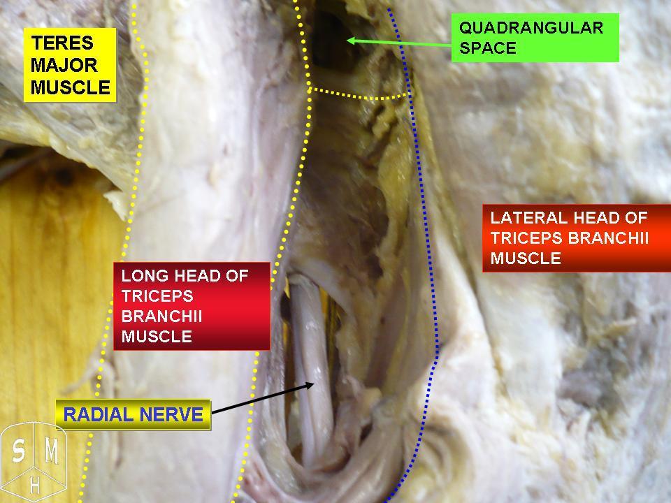
The radial nerve is a nerve in the human body that supplies the posterior portion of the upper limb. It innervates the medial and lateral heads of the triceps brachii muscle of the arm, as well as all 12 muscles in the posterior osteofascial compartment of the forearm and the associated joints and overlying skin. It originates from the brachial plexus, carrying fibers from the ventral roots of spinal nerves C5, C6, C7, C8 & T1. The radial nerve and its branches provide motor innervation to the dorsal arm muscles (the triceps brachii and the anconeus) and the extrinsic extensors of the wrists and hands; it also provides cutaneous sensory innervation to most of the back of the hand, except for the back of the little finger and adjacent half of the ring finger (which are innervated by the ulnar nerve). The radial nerve divides into a deep branch, which becomes the posterior interosseous nerve, and a superficial branch, which goes on to innervate the dorsum (back) of the hand. Th ... [...More Info...] [...Related Items...] OR: [Wikipedia] [Google] [Baidu] |


