|
Inferior Mesenteric Vein
In human anatomy, the inferior mesenteric vein (IMV) is a blood vessel that drains blood from the large intestine. It usually terminates when reaching the splenic vein, which goes on to form the portal vein with the superior mesenteric vein (SMV). Structure The inferior mesenteric vein merges with the splenic vein, posterior to the middle of the body of the pancreas. The splenic vein then merges with the superior mesenteric vein to form the portal vein. Tributaries Tributaries of the inferior mesenteric vein drain the large intestine, sigmoid colon and rectum. These include: * left colic vein * sigmoid veins * superior rectal vein * rectosigmoid veins Variation Anatomical variations include the inferior mesenteric vein draining into the confluence of the superior mesenteric vein ''and'' splenic vein and the inferior mesenteric vein draining in the superior mesenteric vein. Clinical significance The inferior mesenteric vein may be damaged during surgery on the body and ... [...More Info...] [...Related Items...] OR: [Wikipedia] [Google] [Baidu] |
Portal Vein
The portal vein or hepatic portal vein (HPV) is a blood vessel that carries blood from the gastrointestinal tract, gallbladder, pancreas and spleen to the liver. This blood contains nutrients and toxins extracted from digested contents. Approximately 75% of total liver blood flow is through the portal vein, with the remainder coming from the hepatic artery proper. The blood leaves the liver to the heart in the hepatic veins. The portal vein is not a true vein, because it conducts blood to capillary beds in the liver and not directly to the heart. It is a major component of the hepatic portal system, one of only two portal venous systems in the body – with the hypophyseal portal system being the other. The portal vein is usually formed by the confluence of the superior mesenteric, splenic veins, inferior mesenteric, left, right gastric veins and the pancreatic vein. Conditions involving the portal vein cause considerable illness and death. An important example of such a condi ... [...More Info...] [...Related Items...] OR: [Wikipedia] [Google] [Baidu] |
Sigmoid Colon
The sigmoid colon (or pelvic colon) is the part of the large intestine that is closest to the rectum and anus. It forms a loop that averages about in length. The loop is typically shaped like a Greek letter sigma (ς) or Latin letter S (thus ''sigma'' + ''-oid''). This part of the colon normally lies within the pelvis, but due to its freedom of movement it is liable to be displaced into the abdominal cavity. Structure The sigmoid colon begins at the superior aperture of the lesser pelvis, where it is continuous with the iliac colon, and passes transversely across the front of the sacrum to the right side of the pelvis. It then curves on itself and turns toward the left to reach the middle line at the level of the third piece of the sacrum, where it bends downward and ends in the rectum. Its function is to expel solid and gaseous waste from the gastrointestinal tract. The curving path it takes toward the anus allows it to store gas in the superior arched portion, enabling the co ... [...More Info...] [...Related Items...] OR: [Wikipedia] [Google] [Baidu] |
Ligature (medicine)
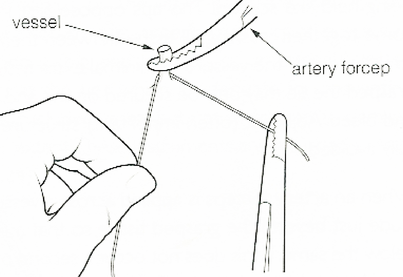
In surgery or medical procedure, a ligature consists of a piece of thread ( suture) tied around an anatomical structure, usually a blood vessel or another hollow structure (e.g. urethra) to shut it off. History The principle of ligation is attributed to Hippocrates and Galen. In ancient Rome, ligatures were used to treat hemorrhoids. The concept of a ligature was reintroduced some 1,500 years later by Ambroise Paré, and finally it found its modern use in 1870–80, made popular by Jules-Émile Péan. Procedure With a blood vessel the surgeon will clamp the vessel perpendicular to the axis of the artery or vein with a hemostat, then secure it by ligating it; i.e. using a piece of suture around it before dividing the structure and releasing the hemostat. It is different from a tourniquet in that the tourniquet will not be secured by knots and it can therefore be released/tightened at will. Ligature is one of the remedies to treat skin tag, or acrochorda. It is done by tying str ... [...More Info...] [...Related Items...] OR: [Wikipedia] [Google] [Baidu] |
Wound
A wound is a rapid onset of injury that involves laceration, lacerated or puncture wound, punctured skin (an ''open'' wound), or a bruise, contusion (a ''closed'' wound) from blunt force physical trauma, trauma or compression. In pathology, a ''wound'' is an acute injury that damages the epidermis of the skin. To healing, heal a wound, the body undertakes a series of actions collectively known as the wound healing process. Classification According to level of contamination, a wound can be classified as: * Clean wound – made under sterile conditions where there are no organisms present, and the skin is likely to heal without complications. * Contaminated wound – usually resulting from accidental injury; there are pathogenic organisms and foreign bodies in the wound. * Infected wound – the wound has pathogenic organisms present and multiplying, exhibiting clinical signs of infection (yellow appearance, soreness, redness, oozing pus). * Colonized wound – a chronic situation, ... [...More Info...] [...Related Items...] OR: [Wikipedia] [Google] [Baidu] |
Abdominal Surgery
The term abdominal surgery broadly covers Surgery, surgical procedures that involve opening the abdomen (laparotomy). Surgery of each abdominal organ is dealt with separately in connection with the description of that organ (see stomach, kidney, liver, etc.) Diseases affecting the abdominal cavity are dealt with generally under their own names (e.g. appendicitis). Types The most common abdominal surgeries are described below. *Appendectomy: surgical opening of the abdominal cavity and removal of the vermiform appendix, appendix. Typically performed as definitive treatment for appendicitis, although sometimes the appendix is Preventive healthcare, prophylactically removed incidental to another abdominal procedure. *Caesarean section (also known as C-section): a surgical procedure in which one or more incisions are made through a mother's abdomen (laparotomy) and uterus (hysterotomy) to deliver one or more babies, or, rarely, to remove a dead fetus. *Inguinal hernia surgery: the ... [...More Info...] [...Related Items...] OR: [Wikipedia] [Google] [Baidu] |
Superior Rectal Vein
The inferior mesenteric vein begins in the rectum as the superior rectal vein (superior hemorrhoidal vein), which has its origin in the hemorrhoidal plexus, and through this plexus communicates with the middle and inferior hemorrhoidal veins. The superior rectal vein leaves the lesser pelvis and crosses the left common iliac vessels with the superior rectal artery, and is continued upward as the inferior mesenteric vein In human anatomy, the inferior mesenteric vein (IMV) is a blood vessel that drains blood from the large intestine. It usually terminates when reaching the splenic vein, which goes on to form the portal vein with the superior mesenteric vein (SMV) .... References Veins of the torso Rectum {{circulatory-stub ... [...More Info...] [...Related Items...] OR: [Wikipedia] [Google] [Baidu] |
Sigmoid Vein , similar shape, term sometimes used for sigmoids
{{disambiguation ...
Sigmoid means resembling the lower-case Greek letter sigma (uppercase Σ, lowercase σ, lowercase in word-final position ς) or the Latin letter S. Specific uses include: * Sigmoid function, a mathematical function * Sigmoid colon, part of the large intestine or colon * Sigmoid sinus, two structures that drain blood from the bottom of the brain * Sigmoid arteries, a pair/trio of arteries in the lower abdomen See also * Ogee An ogee ( ) is the name given to objects, elements, and curves—often seen in architecture and building trades—that have been variously described as serpentine-, extended S-, or sigmoid-shaped. Ogees consist of a "double curve", the combinatio ... [...More Info...] [...Related Items...] OR: [Wikipedia] [Google] [Baidu] |
Left Colic Vein
The left colic vein drains the descending colon. It is a tributary of the inferior mesenteric vein, and follows the path of its corresponding artery, the left colic artery The left colic artery is a branch of the inferior mesenteric artery distributed to the descending colon, and left part of the transverse colon. It ends by dividing into an ascending branch and a descending branch; the terminal branches of the tw .... Branches * Ascending branch * Descending branch References * Veins of the torso {{Circulatory-stub ... [...More Info...] [...Related Items...] OR: [Wikipedia] [Google] [Baidu] |
Rectum
The rectum is the final straight portion of the large intestine in humans and some other mammals, and the Gastrointestinal tract, gut in others. The adult human rectum is about long, and begins at the rectosigmoid junction (the end of the sigmoid colon) at the level of the third sacral vertebra or the sacral promontory depending upon what definition is used. Its diameter is similar to that of the sigmoid colon at its commencement, but it is dilated near its termination, forming the rectal ampulla. It terminates at the level of the anorectal ring (the level of the puborectalis sling) or the dentate line, again depending upon which definition is used. In humans, the rectum is followed by the anal canal which is about long, before the gastrointestinal tract terminates at the anal verge. The word rectum comes from the Latin ''Wikt:rectum, rectum Wikt:intestinum, intestinum'', meaning ''straight intestine''. Structure The rectum is a part of the lower gastrointestinal tract ... [...More Info...] [...Related Items...] OR: [Wikipedia] [Google] [Baidu] |
Pancreas
The pancreas is an organ of the digestive system and endocrine system of vertebrates. In humans, it is located in the abdomen behind the stomach and functions as a gland. The pancreas is a mixed or heterocrine gland, i.e. it has both an endocrine and a digestive exocrine function. 99% of the pancreas is exocrine and 1% is endocrine. As an endocrine gland, it functions mostly to regulate blood sugar levels, secreting the hormones insulin, glucagon, somatostatin, and pancreatic polypeptide. As a part of the digestive system, it functions as an exocrine gland secreting pancreatic juice into the duodenum through the pancreatic duct. This juice contains bicarbonate, which neutralizes acid entering the duodenum from the stomach; and digestive enzymes, which break down carbohydrates, proteins, and fats in food entering the duodenum from the stomach. Inflammation of the pancreas is known as pancreatitis, with common causes including chronic alcohol use and gallstones. Becaus ... [...More Info...] [...Related Items...] OR: [Wikipedia] [Google] [Baidu] |
Superior Mesenteric Vein
In human anatomy, the superior mesenteric vein (SMV) is a blood vessel that drains blood from the small intestine (jejunum and ileum). Behind the neck of the pancreas, the superior mesenteric vein combines with the splenic vein to form the hepatic portal vein. The superior mesenteric vein lies to the right of the similarly named artery, the superior mesenteric artery, which originates from the abdominal aorta. Structure Tributaries of the superior mesenteric vein drain the small intestine, large intestine, stomach, pancreas and appendix and include: * Right gastro-omental vein (also known as the right gastro-epiploic vein) * inferior pancreaticoduodenal veins * veins from jejunum * veins from ileum * middle colic vein – drains the transverse colon * right colic vein – drains the ascending colon * ileocolic vein The superior mesenteric vein combines with the splenic vein to form the portal vein. Clinical significance Thrombosis of the superior mesenteric vein is quite rare ... [...More Info...] [...Related Items...] OR: [Wikipedia] [Google] [Baidu] |
Large Intestine
The large intestine, also known as the large bowel, is the last part of the gastrointestinal tract and of the digestive system in tetrapods. Water is absorbed here and the remaining waste material is stored in the rectum as feces before being removed by defecation. The colon is the longest portion of the large intestine, and the terms are often used interchangeably but most sources define the large intestine as the combination of the cecum, colon, rectum, and anal canal. Some other sources exclude the anal canal. In humans, the large intestine begins in the right iliac region of the pelvis, just at or below the waist, where it is joined to the end of the small intestine at the cecum, via the ileocecal valve. It then continues as the colon ascending the abdomen, across the width of the abdominal cavity as the transverse colon, and then descending to the rectum and its endpoint at the anal canal. Overall, in humans, the large intestine is about long, which is about one-fi ... [...More Info...] [...Related Items...] OR: [Wikipedia] [Google] [Baidu] |


