|
Inferior Medullary Velum
The inferior medullary velum (posterior medullary velum) is a thin layer of white substance, prolonged from the white center of the cerebellum, above and on either side of the nodule; it forms the infero-posterior part of the fourth ventricle. Somewhat semilunar in shape, its convex edge is continuous with the white substance of the cerebellum, while its thin concave margin is apparently free; in reality, however, it is continuous with the epithelium of the ventricle, which is prolonged downward from the posterior medullary velum to the taeniae. See also * Superior medullary velum The superior medullary velum (anterior medullary velum) is a thin, transparent lamina of white matter, which stretches between the superior cerebellar peduncles; on the dorsal surface of its lower half the folia and lingula are prolonged. It fo ... References Neuroanatomy {{Portal bar, Anatomy ... [...More Info...] [...Related Items...] OR: [Wikipedia] [Google] [Baidu] |
Fourth Ventricle
The fourth ventricle is one of the four connected fluid-filled cavities within the human brain. These cavities, known collectively as the ventricular system, consist of the left and right lateral ventricles, the third ventricle, and the fourth ventricle. The fourth ventricle extends from the cerebral aqueduct (''aqueduct of Sylvius'') to the obex, and is filled with cerebrospinal fluid (CSF). The fourth ventricle has a characteristic diamond shape in cross-sections of the human brain. It is located within the pons or in the upper part of the medulla oblongata. CSF entering the fourth ventricle through the cerebral aqueduct can exit to the subarachnoid space of the spinal cord through two lateral apertures and a single, midline median aperture. Boundaries The fourth ventricle has a roof at its ''upper'' (posterior) surface and a floor at its ''lower'' (anterior) surface, and side walls formed by the cerebellar peduncles (nerve bundles joining the structure on the posterior sid ... [...More Info...] [...Related Items...] OR: [Wikipedia] [Google] [Baidu] |
Foramen Of Majendie
In anatomy and osteology, a foramen (; in Merriam-Webster Online Dictionary '. plural foramina, or foramens ) is an open hole that is present in extant or extinct s. Foramina inside the of typically allow , |
Choroid Plexus
The choroid plexus, or plica choroidea, is a plexus of cells that arises from the tela choroidea in each of the ventricles of the brain. Regions of the choroid plexus produce and secrete most of the cerebrospinal fluid (CSF) of the central nervous system. The choroid plexus consists of modified ependymal cells surrounding a core of capillaries and loose connective tissue. Multiple cilia on the ependymal cells move to circulate the cerebrospinal fluid. Structure Location There is a choroid plexus in each of the four ventricles. In the lateral ventricles it is found in the body, and continued in an enlarged amount in the atrium. There is no choroid plexus in the anterior horn. In the third ventricle there is a small amount in the roof that is continuous with that in the body, via the interventricular foramina, the channels that connect the lateral ventricles with the third ventricle. A choroid plexus is in part of the roof of the fourth ventricle. Microanatomy The chor ... [...More Info...] [...Related Items...] OR: [Wikipedia] [Google] [Baidu] |
Cerebellomedullary Cistern
The cisterna magna (or cerebellomedullar cistern) is one of three principal openings in the subarachnoid space between the arachnoid and pia mater layers of the meninges surrounding the brain. The openings are collectively referred to as the subarachnoid cisterns. The cisterna magna is located between the cerebellum and the dorsal surface of the medulla oblongata. Cerebrospinal fluid produced in the fourth ventricle drains into the cisterna magna via the lateral apertures and median aperture. The two other principal cisterns are the ''pontine cistern'' located between the pons and the medulla and the ''interpeduncular cistern'' located between the cerebral peduncles. While the most commonly used clinical method for obtaining cerebrospinal fluid is a lumbar puncture Lumbar puncture (LP), also known as a spinal tap, is a medical procedure in which a needle is inserted into the spinal canal, most commonly to collect cerebrospinal fluid (CSF) for diagnostic testing. The main r ... [...More Info...] [...Related Items...] OR: [Wikipedia] [Google] [Baidu] |
Subarachnoid Cavity
In anatomy, the meninges (, ''singular:'' meninx ( or ), ) are the three membranes that envelop the brain and spinal cord. In mammals, the meninges are the dura mater, the arachnoid mater, and the pia mater. Cerebrospinal fluid is located in the subarachnoid space between the arachnoid mater and the pia mater. The primary function of the meninges is to protect the central nervous system. Structure Dura mater The dura mater ( la, tough mother) (also rarely called ''meninx fibrosa'' or ''pachymeninx'') is a thick, durable membrane, closest to the skull and vertebrae. The dura mater, the outermost part, is a loosely arranged, fibroelastic layer of cells, characterized by multiple interdigitating cell processes, no extracellular collagen, and significant extracellular spaces. The middle region is a mostly fibrous portion. It consists of two layers: the endosteal layer, which lies closest to the skull, and the inner meningeal layer, which lies closer to the brain. It contains large ... [...More Info...] [...Related Items...] OR: [Wikipedia] [Google] [Baidu] |
Central Canal
The central canal (also known as spinal foramen or ependymal canal) is the cerebrospinal fluid-filled space that runs through the spinal cord. The central canal lies below and is connected to the ventricular system of the brain, from which it receives cerebrospinal fluid, and shares the same ependymal lining. The central canal helps to transport nutrients to the spinal cord as well as protect it by cushioning the impact of a force when the spine is affected. The central canal represents the adult remainder of the central cavity of the neural tube. It generally occludes (closes off) with age. Structure The central canal below at the ventricular system of the brain, beginning at a region called the obex where the fourth ventricle, a cavity present in the brainstem, narrows. The central canal is located in the third of the spinal cord in the cervical and thoracic regions. In the lumbar spine it enlarges and is located more centrally. At the conus medullaris, where the spinal co ... [...More Info...] [...Related Items...] OR: [Wikipedia] [Google] [Baidu] |
Corpora Quadrigemina
Corpus is Latin for "body". It may refer to: Linguistics * Text corpus, in linguistics, a large and structured set of texts * Speech corpus, in linguistics, a large set of speech audio files * Corpus linguistics, a branch of linguistics Music * ''Corpus'' (album), by Sebastian Santa Maria * Corpus Delicti (band), also known simply as Corpus Medicine * Corpus callosum, a structure in the brain * Corpus cavernosum (other), a pair of structures in human genitals * Corpus luteum, a temporary endocrine structure in mammals * Corpus gastricum, the Latin term referring to the body of the stomach * Corpus alienum, a foreign object originating outside the body * Corpus albicans * Corpora amylacea * Corpora arenacea Other uses * ''Corpus'' (Bernini), a 1650 sculpture of Christ by Gian Lorenzo Bernini * Corpus (museum), a human body themed museum in the Netherlands * Corpus Clock, a large sculptural clock * Corpus (dance troupe), a Canadian dance troupe * Corpus (typography), ... [...More Info...] [...Related Items...] OR: [Wikipedia] [Google] [Baidu] |
Cerebral Peduncle
The cerebral peduncles are the two stalks that attach the cerebrum to the brainstem. They are structures at the front of the midbrain which arise from the ventral pons and contain the large ascending (sensory) and descending (motor) nerve tracts that run to and from the cerebrum from the pons. Mainly, the three common areas that give rise to the cerebral peduncles are the cerebral cortex, the spinal cord and the cerebellum. The region includes the tegmentum, crus cerebri and pretectum. By this definition, the cerebral peduncles are also known as the basis pedunculi, while the large ventral bundle of efferent fibers is referred to as the cerebral crus or the pes pedunculi. The cerebral peduncles are located on either side of the midbrain and are the frontmost part of the midbrain, and act as the connectors between the rest of the midbrain and the thalamic nuclei and thus the cerebrum. As a whole, the cerebral peduncles assist in refining motor movements, learning new motor ski ... [...More Info...] [...Related Items...] OR: [Wikipedia] [Google] [Baidu] |
Superior Medullary Velum
The superior medullary velum (anterior medullary velum) is a thin, transparent lamina of white matter, which stretches between the superior cerebellar peduncles; on the dorsal surface of its lower half the folia and lingula are prolonged. It forms, together with the superior cerebellar peduncle, the roof of the upper part of the fourth ventricle; it is narrow above, where it passes beneath the facial colliculi, and broader below, where it is continuous with the white substance of the superior vermis. A slightly elevated ridge, the frenulum veli, descends upon its upper part from between the inferior colliculi, and on either side of this the trochlear nerve emerges. Blood is supplied by branches from the superior cerebellar artery. Additional images File:Gray708.svg, Scheme of roof of fourth ventricle. 1. Posterior medullary velum 2. Choroid plexus 3. Cisterna cerebellomedullaris of subarachnoid cavity 4. Central canal 5. Corpora quadrigemina 6. Cerebral peduncle 7. Ant ... [...More Info...] [...Related Items...] OR: [Wikipedia] [Google] [Baidu] |
Ependymal
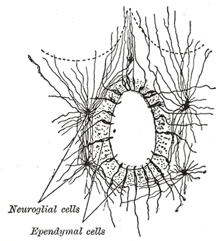
The ependyma is the thin neuroepithelial ( simple columnar ciliated epithelium) lining of the ventricular system of the brain and the central canal of the spinal cord. The ependyma is one of the four types of neuroglia in the central nervous system (CNS). It is involved in the production of cerebrospinal fluid (CSF), and is shown to serve as a reservoir for neuroregeneration. Structure The ependyma is made up of ependymal cells called ependymocytes, a type of glial cell. These cells line the ventricles in the brain and the central canal of the spinal cord, which become filled with cerebrospinal fluid. These are nervous tissue cells with simple columnar shape, much like that of some mucosal epithelial cells. Early monociliated ependymal cells are differentiated to multiciliated ependymal cells for their function in circulating cerebrospinal fluid. The basal membranes of these cells are characterized by tentacle-like extensions that attach to astrocytes. The apical side is cover ... [...More Info...] [...Related Items...] OR: [Wikipedia] [Google] [Baidu] |
Pontine Cistern
The pontine cistern, also cisterna pontis and cisterna pontocerebellaris is a notable subarachnoid cistern on the ventral aspect of the pons. It contains the basilar artery, and is continuous behind with the subarachnoid space of the spinal cord, and with the cisterna magna, and in front of the pons with the interpeduncular cistern The interpeduncular cistern of Sweeney is the subarachnoid cistern that encloses the cerebral peduncles and the structures contained in the interpeduncular fossa and contains the arterial circle of Willis The circle of Willis (also called Wil .... References Meninges {{Portal bar, Anatomy ... [...More Info...] [...Related Items...] OR: [Wikipedia] [Google] [Baidu] |
Cerebellum
The cerebellum (Latin for "little brain") is a major feature of the hindbrain of all vertebrates. Although usually smaller than the cerebrum, in some animals such as the mormyrid fishes it may be as large as or even larger. In humans, the cerebellum plays an important role in motor control. It may also be involved in some cognition, cognitive functions such as attention and language as well as emotion, emotional control such as regulating fear and pleasure responses, but its movement-related functions are the most solidly established. The human cerebellum does not initiate movement, but contributes to Motor coordination, coordination, precision, and accurate timing: it receives input from sensory systems of the spinal cord and from other parts of the brain, and integrates these inputs to fine-tune motor activity. Cerebellar damage produces disorders in Fine motor skill, fine movement, Equilibrioception, equilibrium, Human positions, posture, and motor learning in humans. Anatomica ... [...More Info...] [...Related Items...] OR: [Wikipedia] [Google] [Baidu] |


