|
Ischium
The ischium () forms the lower and back region of the (''os coxae''). Situated below the ilium and behind the pubis, it is one of three regions whose fusion creates the . The superior portion of this region forms approximately one-third of the acetabulum. |
Hip Bone
The hip bone (os coxae, innominate bone, pelvic bone or coxal bone) is a large flat bone, constricted in the center and expanded above and below. In some vertebrates (including humans before puberty) it is composed of three parts: the Ilium (bone), ilium, ischium, and the Pubis (bone), pubis. The two hip bones join at the pubic symphysis and together with the sacrum and coccyx (the pelvic part of the vertebral column, spine) comprise the human skeleton, skeletal component of the pelvis – the pelvic girdle which surrounds the pelvic cavity. They are connected to the sacrum, which is part of the axial skeleton, at the sacroiliac joint. Each hip bone is connected to the corresponding femur (thigh bone) (forming the primary connection between the bones of the lower limb and the axial skeleton) through the large ball and socket joint of the hip joint, hip. Structure The hip bone is formed by three parts: the Ilium (bone), ilium, ischium, and Pubis (bone), pubis. At birth, these three ... [...More Info...] [...Related Items...] OR: [Wikipedia] [Google] [Baidu] |
Coxal Bone
The hip bone (os coxae, innominate bone, pelvic bone or coxal bone) is a large flat bone, constricted in the center and expanded above and below. In some vertebrates (including humans before puberty) it is composed of three parts: the ilium, ischium, and the pubis. The two hip bones join at the pubic symphysis and together with the sacrum and coccyx (the pelvic part of the spine) comprise the skeletal component of the pelvis – the pelvic girdle which surrounds the pelvic cavity. They are connected to the sacrum, which is part of the axial skeleton, at the sacroiliac joint. Each hip bone is connected to the corresponding femur (thigh bone) (forming the primary connection between the bones of the lower limb and the axial skeleton) through the large ball and socket joint of the hip. Structure The hip bone is formed by three parts: the ilium, ischium, and pubis. At birth, these three components are separated by hyaline cartilage. They join each other in a Y-shaped portion of car ... [...More Info...] [...Related Items...] OR: [Wikipedia] [Google] [Baidu] |
Ischial Tuberosity
The ischial tuberosity (or tuberosity of the ischium, tuber ischiadicum), also known colloquially as the sit bones or sitz bones, or as a pair the sitting bones, is a large swelling posteriorly on the superior ramus of the ischium. It marks the lateral boundary of the pelvic outlet. When sitting, the weight is frequently placed upon the ischial tuberosity. The gluteus maximus provides cover in the upright posture, but leaves it free in the seated position.Platzer (2004), p 236 The distance between a cyclist's ischial tuberosities is one of the factors in the choice of a bicycle saddle. Divisions The tuberosity is divided into two portions: a lower, rough, somewhat triangular part, and an upper, smooth, quadrilateral portion. * The ''lower portion'' is subdivided by a prominent longitudinal ridge, passing from base to apex, into two parts: ** The outer gives attachment to the adductor magnus ** The inner to the sacrotuberous ligament * The ''upper portion'' is subdivided into ... [...More Info...] [...Related Items...] OR: [Wikipedia] [Google] [Baidu] |
Superficial Transverse Perineal Muscle
The transverse perineal muscles (transversus perinei) are the superficial and the deep transverse perineal muscles. Superficial transverse perineal The superficial transverse perineal muscle (transversus superficialis perinei or Lloyd-Beanie muscle) is a narrow muscular slip, which passes more or less transversely across the perineal space in front of the anus. It arises by tendinous fibers from the inner and forepart of the ischial tuberosity and, running medially, is inserted into the central tendinous point of the perineum (perineal body), joining in this situation with the muscle of the opposite side, with the external anal sphincter muscle behind, and with the bulbospongiosus muscle in front. In some cases, the fibers of the deeper layer of the external anal sphincter cross over in front of the anus and are continued into this mus ... [...More Info...] [...Related Items...] OR: [Wikipedia] [Google] [Baidu] |
Ischial Spine
The ischial spine is part of the posterior border of the body of the ischium bone of the pelvis. It is a thin and pointed triangular eminence, more or less elongated in different subjects. Structure The pudendal nerve travels close to the ischial spine. Clinical significance The ischial spine can serve as a landmark in pudendal anesthesia, as the pudendal nerve The pudendal nerve is the main nerve of the perineum. It carries sensation from the external genitalia of both sexes and the skin around the anus and perineum, as well as the motor supply to various pelvic muscles, including the male or fem ... lies close to the ischial spine. Additional images File:Sciatic notches.png, Right hip bone, external surface, showing the greater and lesser sciatic notches, separated by the ischial spine. File:Gray319.png, Articulations of pelvis. Anterior view. File:Slide3ADA.JPG, PELVIS. ANTERIOR VIEW. References External links * - "The Female Perineum: Osteology" * - "Th ... [...More Info...] [...Related Items...] OR: [Wikipedia] [Google] [Baidu] |
Tuberosity Of The Ischium
The ischial tuberosity (or tuberosity of the ischium, tuber ischiadicum), also known colloquially as the sit bones or sitz bones, or as a pair the sitting bones, is a large swelling posteriorly on the superior ramus of the ischium. It marks the lateral boundary of the pelvic outlet. When sitting, the weight is frequently placed upon the ischial tuberosity. The gluteus maximus provides cover in the upright posture, but leaves it free in the seated position.Platzer (2004), p 236 The distance between a cyclist's ischial tuberosities is one of the factors in the choice of a bicycle saddle. Divisions The tuberosity is divided into two portions: a lower, rough, somewhat triangular part, and an upper, smooth, quadrilateral portion. * The ''lower portion'' is subdivided by a prominent longitudinal ridge, passing from base to apex, into two parts: ** The outer gives attachment to the adductor magnus ** The inner to the sacrotuberous ligament * The ''upper portion'' is subdivided into ... [...More Info...] [...Related Items...] OR: [Wikipedia] [Google] [Baidu] |
Internal Obturator Muscle
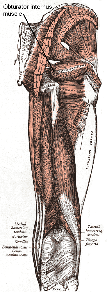
The internal obturator muscle or obturator internus muscle originates on the medial surface of the obturator membrane, the ischium near the membrane, and the rim of the pubis. It exits the pelvic cavity through the lesser sciatic foramen. The internal obturator is situated partly within the lesser pelvis, and partly at the back of the hip-joint. It functions to help laterally rotate femur with hip extension and abduct femur with hip flexion, as well as to steady the femoral head in the acetabulum. Structure Origin The internal obturator muscle arises from the inner surface of the antero-lateral wall of the pelvis. It surrounds the obturator foramen. It is attached to the inferior pubic ramus and ischium, and at the side to the inner surface of the hip bone below and behind the pelvic brim. It reaches from the upper part of the greater sciatic foramen above and behind to the obturator foramen below and in front. It also arises from the pelvic surface of the obturator membran ... [...More Info...] [...Related Items...] OR: [Wikipedia] [Google] [Baidu] |
Ilium (bone)
The ilium () (plural ilia) is the uppermost and largest part of the hip bone, and appears in most vertebrates including mammals and birds, but not bony fish. All reptiles have an ilium except snakes, although some snake species have a tiny bone which is considered to be an ilium. The ilium of the human is divisible into two parts, the body and the wing; the separation is indicated on the top surface by a curved line, the arcuate line, and on the external surface by the margin of the acetabulum. The name comes from the Latin (''ile'', ''ilis''), meaning "groin" or "flank". Structure The ilium consists of the body and wing. Together with the ischium and pubis, to which the ilium is connected, these form the pelvic bone, with only a faint line indicating the place of union. The body ( la, corpus) forms less than two-fifths of the acetabulum; and also forms part of the acetabular fossa. The internal surface of the body is part of the wall of the lesser pelvis and gives ... [...More Info...] [...Related Items...] OR: [Wikipedia] [Google] [Baidu] |
Pubis (bone)
In vertebrates, the pubic region ( la, pubis) is the most forward-facing (ventral and anterior) of the three main regions making up the coxal bone. The left and right pubic regions are each made up of three sections, a superior ramus, inferior ramus, and a body. Structure The pubic region is made up of a ''body'', ''superior ramus'', and ''inferior ramus'' (). The left and right coxal bones join at the pubic symphysis. It is covered by a layer of fat, which is covered by the mons pubis. The pubis is the lower limit of the suprapubic region. In the female, the pubic region is anterior to the urethral sponge. Body The body forms the wide, strong, middle and flat part of the pubic region. The bodies of the left and right pubic regions join at the pubic symphysis. The rough upper edge is the pubic crest, ending laterally in the pubic tubercle. This tubercle, found roughly 3 cm from the pubic symphysis, is a distinctive feature on the lower part of the abdominal wall; important ... [...More Info...] [...Related Items...] OR: [Wikipedia] [Google] [Baidu] |
Acetabulum
The acetabulum (), also called the cotyloid cavity, is a concave surface of the pelvis. The head of the femur meets with the pelvis at the acetabulum, forming the hip joint. Structure There are three bones of the ''os coxae'' (hip bone) that come together to form the ''acetabulum''. Contributing a little more than two-fifths of the structure is the ischium, which provides lower and side boundaries to the acetabulum. The ilium forms the upper boundary, providing a little less than two-fifths of the structure of the acetabulum. The rest is formed by the pubis, near the midline. It is bounded by a prominent uneven rim, which is thick and strong above, and serves for the attachment of the acetabular labrum, which reduces its opening, and deepens the surface for formation of the hip joint. At the lower part of the ''acetabulum'' is the acetabular notch, which is continuous with a circular depression, the acetabular fossa, at the bottom of the cavity of the ''acetabulum''. The re ... [...More Info...] [...Related Items...] OR: [Wikipedia] [Google] [Baidu] |
Obturator Foramen
The obturator foramen (Latin foramen obturatum) is the large opening created by the ischium and pubis (bone), pubis bones of the pelvis through which nerves and blood vessels pass. Structure It is bounded by a thin, uneven margin, to which a strong membrane is attached, and presents, superiorly, a deep groove, the obturator groove, which runs from the pelvis obliquely medialward and downward. This groove is converted into the obturator canal by a ligamentous band, a specialized part of the obturator membrane, attached to two tubercles: * one, the posterior obturator tubercle, on the medial border of the ischium, just in front of the acetabular notch * the other, the anterior obturator tubercle, on the obturator crest of the superior pubic ramus, superior ramus of the pubis (bone), pubis Variation Reflecting the overall sex differences in human physiology, sex differences between male and female pelvises, the obturator foramina are oval in the male and wider and more triangular ... [...More Info...] [...Related Items...] OR: [Wikipedia] [Google] [Baidu] |
Ischiocavernosus
The ischiocavernosus muscle (erectores penis ''or'' erector clitoridis in older texts) is a muscle just below the surface of the perineum, present in both men and women. Structure It arises by tendinous and fleshy fibers from the inner surface of the tuberosity of the ischium, behind the crus penis; and from the inferior pubic rami and ischium on either side of the crus. From these points fleshy fibers succeed, and end in an aponeurosis which is inserted into the sides and under surface of the crus penis. Function In females, the ischiocavernosus muscle assists with clitoral erection. In males, it helps to stabilize the erect penis by compressing the crus penis For their anterior three-fourths the corpora cavernosa penis lie in intimate apposition with one another, but behind they diverge in the form of two tapering processes, known as the crura, which are firmly connected to the ischial rami. Traced ... and retarding the return of blood through the veins. Additional i ... [...More Info...] [...Related Items...] OR: [Wikipedia] [Google] [Baidu] |

