|
Hyaline Cartilage
Hyaline cartilage is the glass-like (hyaline) and translucent cartilage found on many joint surfaces. It is also most commonly found in the ribs, nose, larynx, and trachea. Hyaline cartilage is pearl-gray in color, with a firm consistency and has a considerable amount of collagen. It contains no nerves or blood vessels, and its structure is relatively simple. Structure Hyaline cartilage is covered externally by a fibrous membrane known as the perichondrium or, when it's along articulating surfaces, the synovial membrane. This membrane contains vessels that provide the cartilage with nutrition through diffusion. Hyaline cartilage matrix is primarily made of type II collagen and chondroitin sulphate, both of which are also found in elastic cartilage. Hyaline cartilage exists on the sternal ends of the ribs, in the larynx, trachea, and bronchi, and on the articulating surfaces of bones. It gives the structures a definite but pliable form. The presence of collagen fibres makes suc ... [...More Info...] [...Related Items...] OR: [Wikipedia] [Google] [Baidu] |
Isogenous Group
An isogenous group (lat. "equal origin") is a cluster of up to eight chondrocytes found in hyaline and elastic cartilage.Wheater's Functional Histology, 6th ed. Young, O'Dowd and Woodford. Formation Chondrocytes develop in the embryo from mesenchymal progenitor cells through a process known as chondrogenesis. A chondrocyte can then undergo mitosis to form an isogenous group within its lacuna. Function Isogenous groups differentiate into individual chondrocytes where they continue to produce and deposit extracellular matrix (ECM), lengthening the cartilage and increasing its diameter. This is termed interstitial growth and is one of only two ways cartilage can grow. See also *Endochondral ossification * Hyaline A hyaline substance is one with a glassy appearance. The word is derived from el, ὑάλινος, translit=hyálinos, lit=transparent, and el, ὕαλος, translit=hýalos, lit=crystal, glass, label=none. Histopathology Hyaline cartilage is ... References Conn ... [...More Info...] [...Related Items...] OR: [Wikipedia] [Google] [Baidu] |
Light Micrograph
A micrograph or photomicrograph is a photograph or digital image taken through a microscope or similar device to show a magnify, magnified image of an object. This is opposed to a macrograph or photomacrograph, an image which is also taken on a microscope but is only slightly magnified, usually less than 10 times. Micrography is the practice or art of using microscopes to make photographs. A micrograph contains extensive details of microstructure. A wealth of information can be obtained from a simple micrograph like behavior of the material under different conditions, the phases found in the system, failure analysis, grain size estimation, elemental analysis and so on. Micrographs are widely used in all fields of microscopy. Types Photomicrograph A light micrograph or photomicrograph is a micrograph prepared using an optical microscope, a process referred to as ''photomicroscopy''. At a basic level, photomicroscopy may be performed simply by connecting a camera to a micros ... [...More Info...] [...Related Items...] OR: [Wikipedia] [Google] [Baidu] |
Elastic Cartilage
Elastic cartilage, fibroelastic cartilage or yellow fibrocartilage is a type of cartilage present in the pinnae (auricles) of the ear giving it shape, provides shape for the lateral region of the external auditory meatus, medial part of the auditory canal Eustachian tube, corniculate and cuneiform laryneal cartilages, and the epiglottis. It contains elastic fiber networks and collagen type II fibers. The principal protein is elastin. Structure Elastic cartilage is histologically similar to hyaline cartilage but contains many yellow elastic fibers lying in a solid matrix. These fibers form bundles that appear dark under a microscope. The elastic fibers require special staining since when it is stained using haematoxylin and eosin (H&E) stain it appears the same as hyaline cartilage. Verhoeff van Geison stains are used (giving the elastic fibers a black color), but aldehyde fuchsin stains, Weigert's elastic stains, and orcein stains also work. These fibers give elastic cartilage ... [...More Info...] [...Related Items...] OR: [Wikipedia] [Google] [Baidu] |
Synovial Joint
A synovial joint, also known as diarthrosis, joins bones or cartilage with a fibrous joint capsule that is continuous with the periosteum of the joined bones, constitutes the outer boundary of a synovial cavity, and surrounds the bones' articulating surfaces. This joint unites long bones and permits free bone movement and greater mobility. The synovial cavity/joint is filled with synovial fluid. The joint capsule is made up of an outer layer of fibrous membrane, which keeps the bones together structurally, and an inner layer, the synovial membrane, which seals in the synovial fluid. They are the most common and most movable type of joint in the body of a mammal. As with most other joints, synovial joints achieve movement at the point of contact of the articulating bones. Structure Synovial joints contain the following structures: * Synovial cavity: all diarthroses have the characteristic space between the bones that is filled with synovial fluid * Joint capsule: the fibrous ... [...More Info...] [...Related Items...] OR: [Wikipedia] [Google] [Baidu] |
Bone
A bone is a Stiffness, rigid Organ (biology), organ that constitutes part of the skeleton in most vertebrate animals. Bones protect the various other organs of the body, produce red blood cell, red and white blood cells, store minerals, provide structure and support for the body, and enable animal locomotion, mobility. Bones come in a variety of shapes and sizes and have complex internal and external structures. They are lightweight yet strong and hard and serve multiple Function (biology), functions. Bone tissue (osseous tissue), which is also called bone in the mass noun, uncountable sense of that word, is hard tissue, a type of specialized connective tissue. It has a honeycomb-like matrix (biology), matrix internally, which helps to give the bone rigidity. Bone tissue is made up of different types of bone cells. Osteoblasts and osteocytes are involved in the formation and mineralization (biology), mineralization of bone; osteoclasts are involved in the bone resorption, resor ... [...More Info...] [...Related Items...] OR: [Wikipedia] [Google] [Baidu] |
Articular
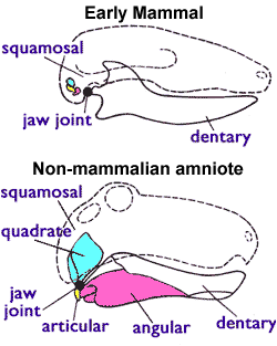
The articular bone is part of the lower jaw of most vertebrates, including most jawed fish, amphibians, birds and various kinds of reptiles, as well as ancestral mammals. Anatomy In most vertebrates, the articular bone is connected to two other lower jaw bones, the suprangular and the angular. Developmentally, it originates from the embryonic mandibular cartilage. The most caudal portion of the mandibular cartilage ossifies to form the articular bone, while the remainder of the mandibular cartilage either remains cartilaginous or disappears. In snakes In snakes, the articular, surangular, and prearticular bones have fused to form the compound bone. The mandible is suspended from the quadrate bone and articulates at this compound bone. Function In amphibians and reptiles In most tetrapods, the articular bone forms the lower portion of the jaw joint. The upper jaw articulates at the quadrate bone. In mammals In mammals, the articular bone evolves to form the malle ... [...More Info...] [...Related Items...] OR: [Wikipedia] [Google] [Baidu] |
Histology Of Articular Cartilage Zones
Histology, also known as microscopic anatomy or microanatomy, is the branch of biology which studies the microscopic anatomy of biological tissues. Histology is the microscopic counterpart to gross anatomy, which looks at larger structures visible without a microscope. Although one may divide microscopic anatomy into ''organology'', the study of organs, ''histology'', the study of tissues, and ''cytology'', the study of cells, modern usage places all of these topics under the field of histology. In medicine, histopathology is the branch of histology that includes the microscopic identification and study of diseased tissue. In the field of paleontology, the term paleohistology refers to the histology of fossil organisms. Biological tissues Animal tissue classification There are four basic types of animal tissues: muscle tissue, nervous tissue, connective tissue, and epithelial tissue. All animal tissues are considered to be subtypes of these four principal tissue types (for ... [...More Info...] [...Related Items...] OR: [Wikipedia] [Google] [Baidu] |
Mitosis
In cell biology, mitosis () is a part of the cell cycle in which replicated chromosomes are separated into two new nuclei. Cell division by mitosis gives rise to genetically identical cells in which the total number of chromosomes is maintained. Therefore, mitosis is also known as equational division. In general, mitosis is preceded by S phase of interphase (during which DNA replication occurs) and is often followed by telophase and cytokinesis; which divides the cytoplasm, organelles and cell membrane of one cell into two new cells containing roughly equal shares of these cellular components. The different stages of mitosis altogether define the mitotic (M) phase of an animal cell cycle—the division of the mother cell into two daughter cells genetically identical to each other. The process of mitosis is divided into stages corresponding to the completion of one set of activities and the start of the next. These stages are preprophase (specific to plant cells), prophase ... [...More Info...] [...Related Items...] OR: [Wikipedia] [Google] [Baidu] |
Cartilage Lacunae
In histology, a lacuna is a small space, containing an osteocyte in bone, or chondrocyte in cartilage. Bone The lacunae are situated between the lamellae, and consist of a number of oblong spaces. In an ordinary microscopic section, viewed by transmitted light, they appear as fusiform opaque spots. Each lacuna is occupied during life by a branched cell, termed an osteocyte, bone-cell or bone-corpuscle. Lacunae are connected to one another by small canals called canaliculi. A lacuna never contains more than one osteocyte. Sinuses are an example of lacuna. Cartilage The cartilage cells or chondrocytes are contained in cavities in the matrix, called cartilage lacunae; around these, the matrix is arranged in concentric lines as if it had been formed in successive portions around the cartilage cells. This constitutes the so-called capsule of the space. Each lacuna is generally occupied by a single cell, but during the division of the cells, it may contain two, four, or eight cells. La ... [...More Info...] [...Related Items...] OR: [Wikipedia] [Google] [Baidu] |
Cell Nucleus
The cell nucleus (pl. nuclei; from Latin or , meaning ''kernel'' or ''seed'') is a membrane-bound organelle found in eukaryotic cells. Eukaryotic cells usually have a single nucleus, but a few cell types, such as mammalian red blood cells, have no nuclei, and a few others including osteoclasts have many. The main structures making up the nucleus are the nuclear envelope, a double membrane that encloses the entire organelle and isolates its contents from the cellular cytoplasm; and the nuclear matrix, a network within the nucleus that adds mechanical support. The cell nucleus contains nearly all of the cell's genome. Nuclear DNA is often organized into multiple chromosomes – long stands of DNA dotted with various proteins, such as histones, that protect and organize the DNA. The genes within these chromosomes are structured in such a way to promote cell function. The nucleus maintains the integrity of genes and controls the activities of the cell by regulating gene expres ... [...More Info...] [...Related Items...] OR: [Wikipedia] [Google] [Baidu] |
Protoplasm
Protoplasm (; ) is the living part of a cell that is surrounded by a plasma membrane. It is a mixture of small molecules such as ions, monosaccharides, amino acid, and macromolecules such as proteins, polysaccharides, lipids, etc. In some definitions, it is a general term for the cytoplasm (e.g., Mohl, 1846), but for others, it also includes the nucleoplasm (e.g., Strasburger, 1882). For Sharp (1921), "According to the older usage the extra-nuclear portion of the protoplast 'the entire cell, excluding the cell wall''was called "protoplasm," but the nucleus also is composed of protoplasm, or living substance in its broader sense. The current consensus is to avoid this ambiguity by employing Strasburger's '(1882)''terms cytoplasm Albert_von_Kölliker.html"_;"title="'coined_by_Albert_von_Kölliker">Kölliker_(1863),_originally_as_synonym_for_protoplasm''and_nucleoplasm_([''term_coined_by_Edouard_Van_Beneden.html" ;"title="Albert von Kölliker">Kölliker (1863), originally as syn ... [...More Info...] [...Related Items...] OR: [Wikipedia] [Google] [Baidu] |
Matrix (biology)
In biology, matrix (plural: matrices) is the material (or tissue) in between a eukaryotic organism's cells. The structure of connective tissues is an extracellular matrix. Fingernails and toenails grow from matrices. It is found in various connective tissues. It is generally used as a jelly-like structure instead of cytoplasm in connective tissue. Tissue matrices Extracellular matrix (ECM) The main ingredients of the extracellular matrix are glycoproteins secreted by the cells. The most abundant glycoprotein in the ECM of most animal cells is collagen, which forms strong fibers outside the cells. In fact, collagen accounts for about 40% of the total protein in the human body. The collagen fibers are embedded in a network woven from proteoglycans. A proteoglycan molecule consists of a small core protein with many carbohydrate chains covalently attached, so that it may be up to 95% carbohydrate. Large proteoglycan complexes can form when hundreds of proteoglycans become noncoval ... [...More Info...] [...Related Items...] OR: [Wikipedia] [Google] [Baidu] |


