|
High Resolution Computed Tomography
High-resolution computed tomography (HRCT) is a type of computed tomography (CT) with specific techniques to enhance image resolution. It is used in the diagnosis of various health problems, though most commonly for lung disease, by assessing the lung parenchyma. On the other hand, HRCT of the temporal bone is used to diagnose various middle ear diseases such as otitis media, cholesteatoma, and evaluations after ear operations. Technique HRCT is performed using a conventional CT scanner. However, imaging parameters are chosen so as to maximize spatial resolution: a narrow slice width is used (usually 1–2 mm), a high spatial resolution image reconstruction algorithm is used, field of view is minimized, so as to minimize the size of each pixel, and other scan factors (e.g. focal spot) may be optimized for resolution at the expense of scan speed. Depending on the suspected diagnosis, the scan may be performed in both inspiration and expiration. In inspiration images ... [...More Info...] [...Related Items...] OR: [Wikipedia] [Google] [Baidu] |
CT Scan
A computed tomography scan (CT scan; formerly called computed axial tomography scan or CAT scan) is a medical imaging technique used to obtain detailed internal images of the body. The personnel that perform CT scans are called radiographers or radiology technologists. CT scanners use a rotating X-ray tube and a row of detectors placed in a gantry (medical), gantry to measure X-ray Attenuation#Radiography, attenuations by different tissues inside the body. The multiple X-ray measurements taken from different angles are then processed on a computer using tomographic reconstruction algorithms to produce Tomography, tomographic (cross-sectional) images (virtual "slices") of a body. CT scans can be used in patients with metallic implants or pacemakers, for whom magnetic resonance imaging (MRI) is Contraindication, contraindicated. Since its development in the 1970s, CT scanning has proven to be a versatile imaging technique. While CT is most prominently used in medical diagnosis, ... [...More Info...] [...Related Items...] OR: [Wikipedia] [Google] [Baidu] |
Pneumatosis
Pneumatosis is the abnormal presence of air or other gas within tissues. In the lungs, emphysema involves enlargement of the distal airspaces,page 64 in: and is a major feature of (COPD). Other pneumatoses in the lungs are focal (localized) blebs and bullae, pulmonary cysts and cavities. |
World Health Organization
The World Health Organization (WHO) is a specialized agency of the United Nations responsible for international public health. The WHO Constitution states its main objective as "the attainment by all peoples of the highest possible level of health". Headquartered in Geneva, Switzerland, it has six regional offices and 150 field offices worldwide. The WHO was established on 7 April 1948. The first meeting of the World Health Assembly (WHA), the agency's governing body, took place on 24 July of that year. The WHO incorporated the assets, personnel, and duties of the League of Nations' Health Organization and the , including the International Classification of Diseases (ICD). Its work began in earnest in 1951 after a significant infusion of financial and technical resources. The WHO's mandate seeks and includes: working worldwide to promote health, keeping the world safe, and serve the vulnerable. It advocates that a billion more people should have: universal health care coverag ... [...More Info...] [...Related Items...] OR: [Wikipedia] [Google] [Baidu] |
COVID-19
Coronavirus disease 2019 (COVID-19) is a contagious disease caused by a virus, the severe acute respiratory syndrome coronavirus 2 (SARS-CoV-2). The first known case was COVID-19 pandemic in Hubei, identified in Wuhan, China, in December 2019. The disease quickly spread worldwide, resulting in the COVID-19 pandemic. The symptoms of COVID‑19 are variable but often include fever, cough, headache, fatigue, breathing difficulties, Anosmia, loss of smell, and Ageusia, loss of taste. Symptoms may begin one to fourteen days incubation period, after exposure to the virus. At least a third of people who are infected Asymptomatic, do not develop noticeable symptoms. Of those who develop symptoms noticeable enough to be classified as patients, most (81%) develop mild to moderate symptoms (up to mild pneumonia), while 14% develop severe symptoms (dyspnea, Hypoxia (medical), hypoxia, or more than 50% lung involvement on imaging), and 5% develop critical symptoms (respiratory failure ... [...More Info...] [...Related Items...] OR: [Wikipedia] [Google] [Baidu] |
Immunosuppressive Drug
Immunosuppressive drugs, also known as immunosuppressive agents, immunosuppressants and antirejection medications, are drugs that inhibit or prevent activity of the immune system. Classification Immunosuppressive drugs can be classified into five groups: * glucocorticoids * cytostatics * antibodies * drugs acting on immunophilins * other drugs Glucocorticoids In pharmacologic (supraphysiologic) doses, glucocorticoids, such as prednisone, dexamethasone, and hydrocortisone are used to suppress various allergic, inflammatory, and autoimmune disorders. They are also administered as posttransplantory immunosuppressants to prevent the acute transplant rejection and graft-versus-host disease. Nevertheless, they do not prevent an infection and also inhibit later reparative processes. Immunosuppressive mechanism Glucocorticoids suppress cell-mediated immunity. They act by inhibiting genes that code for the cytokines Interleukin 1 (IL-1), IL-2, IL-3, IL-4, IL-5, IL-6, IL-8 ... [...More Info...] [...Related Items...] OR: [Wikipedia] [Google] [Baidu] |
Sarcoidosis
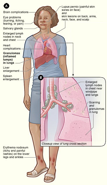
Sarcoidosis (also known as ''Besnier-Boeck-Schaumann disease'') is a disease involving abnormal collections of inflammatory cells that form lumps known as granulomata. The disease usually begins in the lungs, skin, or lymph nodes. Less commonly affected are the eyes, liver, heart, and brain. Any organ can be affected though. The signs and symptoms depend on the organ involved. Often, no, or only mild, symptoms are seen. When it affects the lungs, wheezing, coughing, shortness of breath, or chest pain may occur. Some may have Löfgren syndrome with fever, large lymph nodes, arthritis, and a rash known as erythema nodosum. The cause of sarcoidosis is unknown. Some believe it may be due to an immune reaction to a trigger such as an infection or chemicals in those who are genetically predisposed. Those with affected family members are at greater risk. Diagnosis is partly based on signs and symptoms, which may be supported by biopsy. Findings that make it likely include large lymph n ... [...More Info...] [...Related Items...] OR: [Wikipedia] [Google] [Baidu] |
Lymphangioleiomyomatosis
Lymphangioleiomyomatosis (LAM) is a rare, progressive and systemic disease that typically results in cystic lung destruction. It predominantly affects women, especially during childbearing years. The term sporadic LAM is used for patients with LAM not associated with tuberous sclerosis complex (TSC), while TSC-LAM refers to LAM that is associated with TSC. Signs and symptoms The average age of onset is the early to mid 30s. Exertional dyspnea (shortness of breath) and spontaneous pneumothorax (lung collapse) have been reported as the initial presentation of the disease in 49% and 46% of patients, respectively. Diagnosis is typically delayed 5 to 6 years. The condition is often misdiagnosed as asthma or chronic obstructive pulmonary disease. The first pneumothorax, or lung collapse, precedes the diagnosis of LAM in 82% of patients. The consensus clinical definition of LAM includes multiple symptoms: * Fatigue * Cough * Hemoptysis, Coughing up blood (rarely massive) * Chest pain * C ... [...More Info...] [...Related Items...] OR: [Wikipedia] [Google] [Baidu] |
Lymphangitis Carcinomatosa
Lymphangitis carcinomatosa is inflammation of the lymph vessels (lymphangitis) caused by a malignancy. Breast, lung, stomach, pancreas, and prostate cancers are the most common tumors that result in lymphangitis. Lymphangitis carcinomatosa was first described by pathologist Gabriel Andral in 1829 in a patient with uterine cancer. Lymphangitis carcinomatosa may show the presence of Kerley B lines on chest X-ray. Lymphangitis carcinomatosa most often affects people 40–49 years of age. Lymphangitis carcinomatosa may be caused by the following malignancies as suggested by the mnemonic: "''C''ertain ''C''ancers ''S''pread ''B''y ''P''lugging ''T''he ''L''ymphatics" (cervical cancer, colon cancer, stomach cancer, breast cancer/bronchiogenic carcinoma, pancreatic cancer, thyroid cancer, laryngeal cancer) Pathology In most cases, lymphangitis carcinomatosis is caused by the dissemination of a tumor with its cells along the lymphatics. However, in about 20 percent of cases, the in ... [...More Info...] [...Related Items...] OR: [Wikipedia] [Google] [Baidu] |
Biopsy
A biopsy is a medical test commonly performed by a surgeon, interventional radiologist, or an interventional cardiologist. The process involves extraction of sample cells or tissues for examination to determine the presence or extent of a disease. The tissue is then fixed, dehydrated, embedded, sectioned, stained and mounted before it is generally examined under a microscope by a pathologist; it may also be analyzed chemically. When an entire lump or suspicious area is removed, the procedure is called an excisional biopsy. An incisional biopsy or core biopsy samples a portion of the abnormal tissue without attempting to remove the entire lesion or tumor. When a sample of tissue or fluid is removed with a needle in such a way that cells are removed without preserving the histological architecture of the tissue cells, the procedure is called a needle aspiration biopsy. Biopsies are most commonly performed for insight into possible cancerous or inflammatory conditions. History T ... [...More Info...] [...Related Items...] OR: [Wikipedia] [Google] [Baidu] |
Non-specific Interstitial Pneumonitis
Signs and symptoms are the observed or detectable signs, and experienced symptoms of an illness, injury, or condition. A sign for example may be a higher or lower temperature than normal, raised or lowered blood pressure or an abnormality showing on a medical scan. A symptom is something out of the ordinary that is experienced by an individual such as feeling feverish, a headache or other pain or pains in the body. Signs and symptoms Signs A medical sign is an objective observable indication of a disease, injury, or abnormal physiological state that may be detected during a physical examination, examining the patient history, or diagnostic procedure. These signs are visible or otherwise detectable such as a rash or bruise. Medical signs, along with symptoms, assist in formulating diagnostic hypothesis. Examples of signs include elevated blood pressure, nail clubbing of the fingernails or toenails, staggering gait, and arcus senilis and arcus juvenilis of the eyes. Indic ... [...More Info...] [...Related Items...] OR: [Wikipedia] [Google] [Baidu] |
Air Trapping
Air trapping, also called gas trapping, is an abnormal retention of air in the lungs where it is difficult to exhale completely. It is observed in obstructive lung diseases such as asthma, bronchiolitis obliterans syndrome and chronic obstructive pulmonary diseases such as emphysema and chronic bronchitis. Air trapping is not a diagnosis but is a presentation of an illness, and can be a guide to the appropriate differential. __TOC__ Imaging On high resolution CT air trapping has a typical imaging appearance, although often evaluation with both maximum inhalation and exhalation, or inspiratory and expiratory views, are needed for a more specific diagnosis. One of its typical imaging patterns is mosaic attenuation. In the classic presentation, the lung will appear normal at inspiration, but on exhalation, the diseased portions of the lung which have lost connective tissue recoil will remain lucent while the healthy portions of the lung will become more dense due to atelectasis. Th ... [...More Info...] [...Related Items...] OR: [Wikipedia] [Google] [Baidu] |


