|
Hemorrhagic Infarct
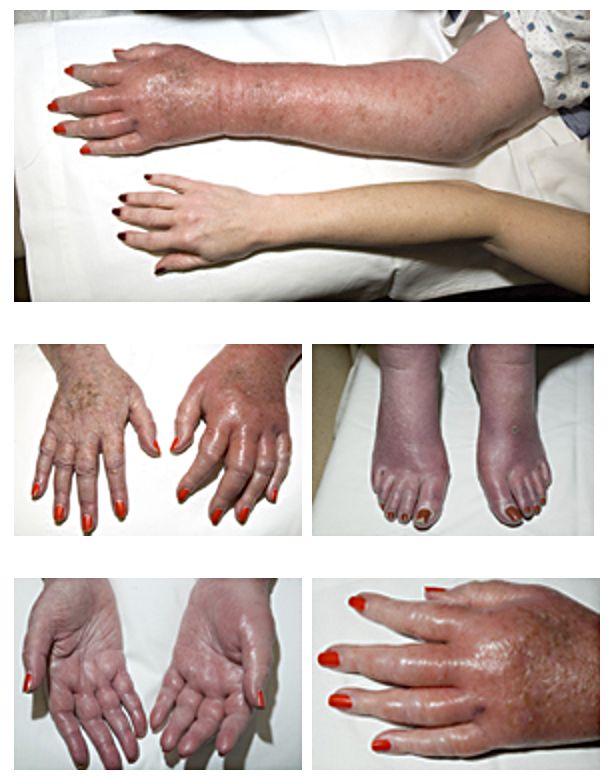
Hemorrhagic infarcts are infarcts commonly caused by occlusion of veins, with red blood cells entering the area of the infarct, or an artery occlusion of an organ with collaterals or dual circulation. These are typically seen in the brain, lungs, and the GI tract, areas referred to as having "loose tissue," or dual circulation. Loose-textured tissue allows red blood cells released from damaged vessels to diffuse through the necrotic tissue. A white infarct, also called an anemic infarct, can become hemorrhagic with reperfusion injury, reperfusion. Hemorrhagic infarction is also associated with testicular torsion. See also * Anemic infarct * Infarction References Vascular diseases Gross pathology {{circulatory-disease-stub ... [...More Info...] [...Related Items...] OR: [Wikipedia] [Google] [Baidu] |
Infarct
Infarction is tissue death (necrosis) due to Ischemia, inadequate blood supply to the affected area. It may be caused by Thrombosis, artery blockages, rupture, mechanical compression, or vasoconstriction. The resulting lesion is referred to as an infarct (from the Latin ''infarctus'', "stuffed into"). Causes Infarction occurs as a result of prolonged ischemia, which is the insufficient supply of oxygen and nutrition to an area of tissue due to a disruption in blood supply. The blood vessel supplying the affected area of tissue may be blocked due to an obstruction in the vessel (e.g., an arterial embolus, thrombus, or atherosclerotic plaque), compressed by something outside of the vessel causing it to narrow (e.g., tumor, volvulus, or hernia), ruptured by trauma causing a loss of blood pressure downstream of the rupture, or vasoconstricted, which is the narrowing of the blood vessel by contraction of the muscle wall rather than an external force (e.g., cocaine vaso ... [...More Info...] [...Related Items...] OR: [Wikipedia] [Google] [Baidu] |
Vein
Veins are blood vessels in humans and most other animals that carry blood towards the heart. Most veins carry deoxygenated blood from the tissues back to the heart; exceptions are the pulmonary and umbilical veins, both of which carry oxygenated blood to the heart. In contrast to veins, arteries carry blood away from the heart. Veins are less muscular than arteries and are often closer to the skin. There are valves (called ''pocket valves'') in most veins to prevent backflow. Structure Veins are present throughout the body as tubes that carry blood back to the heart. Veins are classified in a number of ways, including superficial vs. deep, pulmonary vs. systemic, and large vs. small. * Superficial veins are those closer to the surface of the body, and have no corresponding arteries. *Deep veins are deeper in the body and have corresponding arteries. *Perforator veins drain from the superficial to the deep veins. These are usually referred to in the lower limbs and feet. *Communic ... [...More Info...] [...Related Items...] OR: [Wikipedia] [Google] [Baidu] |
Red Blood Cell
Red blood cells (RBCs), also referred to as red cells, red blood corpuscles (in humans or other animals not having nucleus in red blood cells), haematids, erythroid cells or erythrocytes (from Greek ''erythros'' for "red" and ''kytos'' for "hollow vessel", with ''-cyte'' translated as "cell" in modern usage), are the most common type of blood cell and the vertebrate's principal means of delivering oxygen (O2) to the body tissues—via blood flow through the circulatory system. RBCs take up oxygen in the lungs, or in fish the gills, and release it into tissues while squeezing through the body's capillaries. The cytoplasm of a red blood cell is rich in hemoglobin, an iron-containing biomolecule that can bind oxygen and is responsible for the red color of the cells and the blood. Each human red blood cell contains approximately 270 million hemoglobin molecules. The cell membrane is composed of proteins and lipids, and this structure provides properties essential for physiolo ... [...More Info...] [...Related Items...] OR: [Wikipedia] [Google] [Baidu] |
Brain
A brain is an organ that serves as the center of the nervous system in all vertebrate and most invertebrate animals. It is located in the head, usually close to the sensory organs for senses such as vision. It is the most complex organ in a vertebrate's body. In a human, the cerebral cortex contains approximately 14–16 billion neurons, and the estimated number of neurons in the cerebellum is 55–70 billion. Each neuron is connected by synapses to several thousand other neurons. These neurons typically communicate with one another by means of long fibers called axons, which carry trains of signal pulses called action potentials to distant parts of the brain or body targeting specific recipient cells. Physiologically, brains exert centralized control over a body's other organs. They act on the rest of the body both by generating patterns of muscle activity and by driving the secretion of chemicals called hormones. This centralized control allows rapid and coordinated respon ... [...More Info...] [...Related Items...] OR: [Wikipedia] [Google] [Baidu] |
Lungs
The lungs are the primary organs of the respiratory system in humans and most other animals, including some snails and a small number of fish. In mammals and most other vertebrates, two lungs are located near the backbone on either side of the heart. Their function in the respiratory system is to extract oxygen from the air and transfer it into the bloodstream, and to release carbon dioxide from the bloodstream into the atmosphere, in a process of gas exchange. Respiration is driven by different muscular systems in different species. Mammals, reptiles and birds use their different muscles to support and foster breathing. In earlier tetrapods, air was driven into the lungs by the pharyngeal muscles via buccal pumping, a mechanism still seen in amphibians. In humans, the main muscle of respiration that drives breathing is the diaphragm. The lungs also provide airflow that makes vocal sounds including human speech possible. Humans have two lungs, one on the left and one on the ... [...More Info...] [...Related Items...] OR: [Wikipedia] [Google] [Baidu] |
GI Tract
The gastrointestinal tract (GI tract, digestive tract, alimentary canal) is the tract or passageway of the digestive system that leads from the mouth to the anus. The GI tract contains all the major organs of the digestive system, in humans and other animals, including the esophagus, stomach, and intestines. Food taken in through the mouth is digested to extract nutrients and absorb energy, and the waste expelled at the anus as feces. ''Gastrointestinal'' is an adjective meaning of or pertaining to the stomach and intestines. Most animals have a "through-gut" or complete digestive tract. Exceptions are more primitive ones: sponges have small pores ( ostia) throughout their body for digestion and a larger dorsal pore (osculum) for excretion, comb jellies have both a ventral mouth and dorsal anal pores, while cnidarians and acoels have a single pore for both digestion and excretion. The human gastrointestinal tract consists of the esophagus, stomach, and intestines, and is div ... [...More Info...] [...Related Items...] OR: [Wikipedia] [Google] [Baidu] |
Anemic Infarct
Anemic infarcts (also called white infarcts or pale infarcts) are white or pale infarcts caused by arterial occlusions, and are usually seen in the heart, kidney and spleen. These are referred to as "white" because of the lack of hemorrhaging and limited red blood cells accumulation, (compare to Hemorrhagic infarct). The tissues most likely to be affected are solid organs which limit the amount of hemorrhage that can seep into the area of ischemic necrosis from adjoining capillary beds. The organs typically include single blood supply (no dual arterial blood supply or anastomoses). The infarct generally results grossly in a wedge shaped area of necrosis with the apex closest to the occlusion and the base at the periphery of the organ. The margins will become better defined with time with a narrow rim of congestion attributable to inflammation at the edge of the lesion.Robbins Basic Pathology Relatively few extravasated red cells are lysed so the resulting hemosiderosis is limited an ... [...More Info...] [...Related Items...] OR: [Wikipedia] [Google] [Baidu] |
Reperfusion Injury
Reperfusion injury, sometimes called ischemia-reperfusion injury (IRI) or reoxygenation injury, is the tissue damage caused when blood supply returns to tissue ('' re-'' + '' perfusion'') after a period of ischemia or lack of oxygen (anoxia or hypoxia). The absence of oxygen and nutrients from blood during the ischemic period creates a condition in which the restoration of circulation results in inflammation and oxidative damage through the induction of oxidative stress rather than (or along with) restoration of normal function. Reperfusion injury is distinct from cerebral hyperperfusion syndrome (sometimes called "Reperfusion syndrome"), a state of abnormal cerebral vasodilation. Mechanisms Reperfusion of ischemic tissues is often associated with microvascular injury, particularly due to increased permeability of capillaries and arterioles that lead to an increase of diffusion and fluid filtration across the tissues. Activated endothelial cells produce more reactive oxygen sp ... [...More Info...] [...Related Items...] OR: [Wikipedia] [Google] [Baidu] |
Testicular Torsion
Testicular torsion occurs when the spermatic cord (from which the testicle is suspended) twists, cutting off the blood supply to the testicle. The most common symptom in children is sudden, severe testicular pain. The testicle may be higher than usual in the scrotum and vomiting may occur. In newborns, pain is often absent and instead the scrotum may become discolored or the testicle may disappear from its usual place. Most of those affected have no obvious prior underlying health problems. Testicular tumor or prior trauma may increase risk. Other risk factors include a congenital malformation known as a "bell-clapper deformity" wherein the testis is inadequately attached to the scrotum allowing it to move more freely and thus potentially twist. Cold temperatures may also be a risk factor. The diagnosis should usually be made based on the presenting symptoms, but requires timely diagnosis and treatment to avoid testicular loss. An ultrasound can be useful when the diagnosis is unc ... [...More Info...] [...Related Items...] OR: [Wikipedia] [Google] [Baidu] |
Anemic Infarct
Anemic infarcts (also called white infarcts or pale infarcts) are white or pale infarcts caused by arterial occlusions, and are usually seen in the heart, kidney and spleen. These are referred to as "white" because of the lack of hemorrhaging and limited red blood cells accumulation, (compare to Hemorrhagic infarct). The tissues most likely to be affected are solid organs which limit the amount of hemorrhage that can seep into the area of ischemic necrosis from adjoining capillary beds. The organs typically include single blood supply (no dual arterial blood supply or anastomoses). The infarct generally results grossly in a wedge shaped area of necrosis with the apex closest to the occlusion and the base at the periphery of the organ. The margins will become better defined with time with a narrow rim of congestion attributable to inflammation at the edge of the lesion.Robbins Basic Pathology Relatively few extravasated red cells are lysed so the resulting hemosiderosis is limited an ... [...More Info...] [...Related Items...] OR: [Wikipedia] [Google] [Baidu] |
Infarction
Infarction is tissue death (necrosis) due to inadequate blood supply to the affected area. It may be caused by artery blockages, rupture, mechanical compression, or vasoconstriction. The resulting lesion is referred to as an infarct (from the Latin ''infarctus'', "stuffed into"). Causes Infarction occurs as a result of prolonged ischemia, which is the insufficient supply of oxygen and nutrition to an area of tissue due to a disruption in blood supply. The blood vessel supplying the affected area of tissue may be blocked due to an obstruction in the vessel (e.g., an arterial embolus, thrombus, or atherosclerotic plaque), compressed by something outside of the vessel causing it to narrow (e.g., tumor, volvulus, or hernia), ruptured by trauma causing a loss of blood pressure downstream of the rupture, or vasoconstricted, which is the narrowing of the blood vessel by contraction of the muscle wall rather than an external force (e.g., cocaine vasoconstriction leading ... [...More Info...] [...Related Items...] OR: [Wikipedia] [Google] [Baidu] |
Vascular Diseases
Vascular disease is a class of diseases of the blood vessels – the arteries and veins of the circulatory system of the body. Vascular disease is a subgroup of cardiovascular disease. Disorders in this vast network of blood vessels can cause a range of health problems that can sometimes become severe. Types There are several types of vascular disease, and signs and symptoms can vary depending on the disease type. These types include: * Erythromelalgia - a rare peripheral vascular disease with syndromes that include burning pain, increased temperature, erythema and swelling that generally affect the hands and feet. * Peripheral artery disease – occurs when atheromatous plaques build up in the arteries that supply blood to the arms and legs, causing the arteries to narrow or become blocked. * Renal artery stenosis - the narrowing of renal arteries that carry blood to the kidneys from the aorta. * Buerger's disease – inflammation and swelling in small blood vessels, cau ... [...More Info...] [...Related Items...] OR: [Wikipedia] [Google] [Baidu] |


.jpg)
