|
Hemobilia
Haemobilia is a medical condition of bleeding into the biliary tree. Haemobilia occurs when there is a fistula between a vessel of the splanchnic circulation and the intrahepatic or extrahepatic biliary system. It can present as acute upper gastrointestinal (UGI) bleeding. It should be considered in upper abdominal pain presenting with UGI bleeding especially when there is a history of liver injury or instrumentation. First recorded in 1654 by Francis Glisson, a university of Cambridge, Cambridge professor. Presentation Quincke's triad of upper abdominal pain, Upper gastrointestinal bleeding, upper gastrointestinal haemorrhage and jaundice is classical but only present in 22% cases. It can be immediately life-threatening in major bleeding. However, in minor haemobilia, patient is haemodynamically stable despite significant blood loss being apparent. Causes The causes of haemobilia include Physical trauma, trauma (which can be accidental or iatrogenic due to procedures such as c ... [...More Info...] [...Related Items...] OR: [Wikipedia] [Google] [Baidu] |
Endoscopic Retrograde Cholangiopancreatography
Endoscopic retrograde cholangiopancreatography (ERCP) is a technique that combines the use of endoscopy and fluoroscopy to diagnose and treat certain problems of the biliary or pancreatic ductal systems. It is primarily performed by highly skilled and specialty trained gastroenterologists. Through the endoscope, the physician can see the inside of the stomach and duodenum, and inject a contrast medium into the ducts in the biliary tree and pancreas so they can be seen on radiographs. ERCP is used primarily to diagnose and treat conditions of the bile ducts and main pancreatic duct, including gallstones, inflammatory strictures (scars), leaks (from trauma and surgery), and cancer. ERCP can be performed for diagnostic and therapeutic reasons, although the development of safer and relatively non-invasive investigations such as magnetic resonance cholangiopancreatography (MRCP) and endoscopic ultrasound has meant that ERCP is now rarely performed without therapeutic intent. Medical u ... [...More Info...] [...Related Items...] OR: [Wikipedia] [Google] [Baidu] |
Biliary Tree
The biliary tract, (biliary tree or biliary system) refers to the liver, gallbladder and bile ducts, and how they work together to make, store and secrete bile. Bile consists of water, electrolytes, bile acids, cholesterol, phospholipids and conjugated bilirubin. Some components are synthesized by hepatocytes (liver cells), the rest are extracted from the blood by the liver. Bile is secreted by the liver into small ducts that join to form the common hepatic duct. Between meals, secreted bile is stored in the gallbladder. During a meal, the bile is secreted into the duodenum (part of the small intestine) to rid the body of waste stored in the bile as well as aid in the absorption of dietary fats and oils. Structure The biliary tract refers to the path by which bile is secreted by the liver then transported to the duodenum, the first part of the small intestine. A structure common to most members of the mammal family, the biliary tract is often referred to as a tree because ... [...More Info...] [...Related Items...] OR: [Wikipedia] [Google] [Baidu] |
Malformation
A birth defect, also known as a congenital disorder, is an abnormal condition that is present at birth regardless of its cause. Birth defects may result in disabilities that may be physical, intellectual, or developmental. The disabilities can range from mild to severe. Birth defects are divided into two main types: structural disorders in which problems are seen with the shape of a body part and functional disorders in which problems exist with how a body part works. Functional disorders include metabolic and degenerative disorders. Some birth defects include both structural and functional disorders. Birth defects may result from genetic or chromosomal disorders, exposure to certain medications or chemicals, or certain infections during pregnancy. Risk factors include folate deficiency, drinking alcohol or smoking during pregnancy, poorly controlled diabetes, and a mother over the age of 35 years old. Many are believed to involve multiple factors. Birth defects may be visib ... [...More Info...] [...Related Items...] OR: [Wikipedia] [Google] [Baidu] |
Biliary Tract Disorders
A bile duct is any of a number of long tube-like structures that carry bile, and is present in most vertebrates. Bile is required for the digestion of food and is secreted by the liver into passages that carry bile toward the hepatic duct. It joins the cystic duct (carrying bile to and from the gallbladder) to form the common bile duct which then opens into the intestine. Structure The top half of the common bile duct is associated with the liver, while the bottom half of the common bile duct is associated with the pancreas, through which it passes on its way to the intestine. It opens into the part of the intestine called the duodenum via the ampulla of Vater. Segments The biliary tree (see below) is the whole network of various sized ducts branching through the liver. The path is as follows: Bile canaliculi → Canals of Hering → interlobular bile ducts → intrahepatic bile ducts → left and right hepatic ducts ''merge to form'' → common hepatic duct ''exits liver a ... [...More Info...] [...Related Items...] OR: [Wikipedia] [Google] [Baidu] |
Embolisation
Embolization refers to the passage and lodging of an embolus within the bloodstream. It may be of natural origin (pathological), in which sense it is also called embolism, for example a pulmonary embolism; or it may be artificially induced (therapeutic), as a hemostatic treatment for bleeding or as a treatment for some types of cancer by deliberately blocking blood vessels to starve the tumor cells. In the cancer management application, the embolus, besides blocking the blood supply to the tumor, also often includes an ingredient to attack the tumor chemically or with irradiation. When it bears a chemotherapy drug, the process is called chemoembolization. Transcatheter arterial chemoembolization (TACE) is the usual form. When the embolus bears a radiopharmaceutical for unsealed source radiotherapy, the process is called radioembolization or selective internal radiation therapy (SIRT). Uses Embolization involves the selective occlusion of blood vessels by purposely introducin ... [...More Info...] [...Related Items...] OR: [Wikipedia] [Google] [Baidu] |
Endovascular
Interventional radiology (IR) is a medical specialty that performs various minimally-invasive procedures using medical imaging guidance, such as Fluoroscopy, x-ray fluoroscopy, CT scan, computed tomography, magnetic resonance imaging, or ultrasound. IR performs both diagnostic and therapeutic procedures Minimally invasive procedure, through very small incisions or body orifices. Diagnostic IR procedures are those intended to help make a diagnosis or guide further medical treatment, and include image-guided biopsy of a tumor or injection of an Radiocontrast agent, imaging contrast agent into a hollow structure, such as a blood vessel or a Bile duct, duct. By contrast, Therapy, therapeutic IR procedures provide direct treatment—they include catheter-based medicine delivery, medical device placement (e.g., stents), and angioplasty of narrowed structures. The main benefits of interventional radiology techniques are that they can reach the deep structures of the body through a body ... [...More Info...] [...Related Items...] OR: [Wikipedia] [Google] [Baidu] |
Surgical Ligation
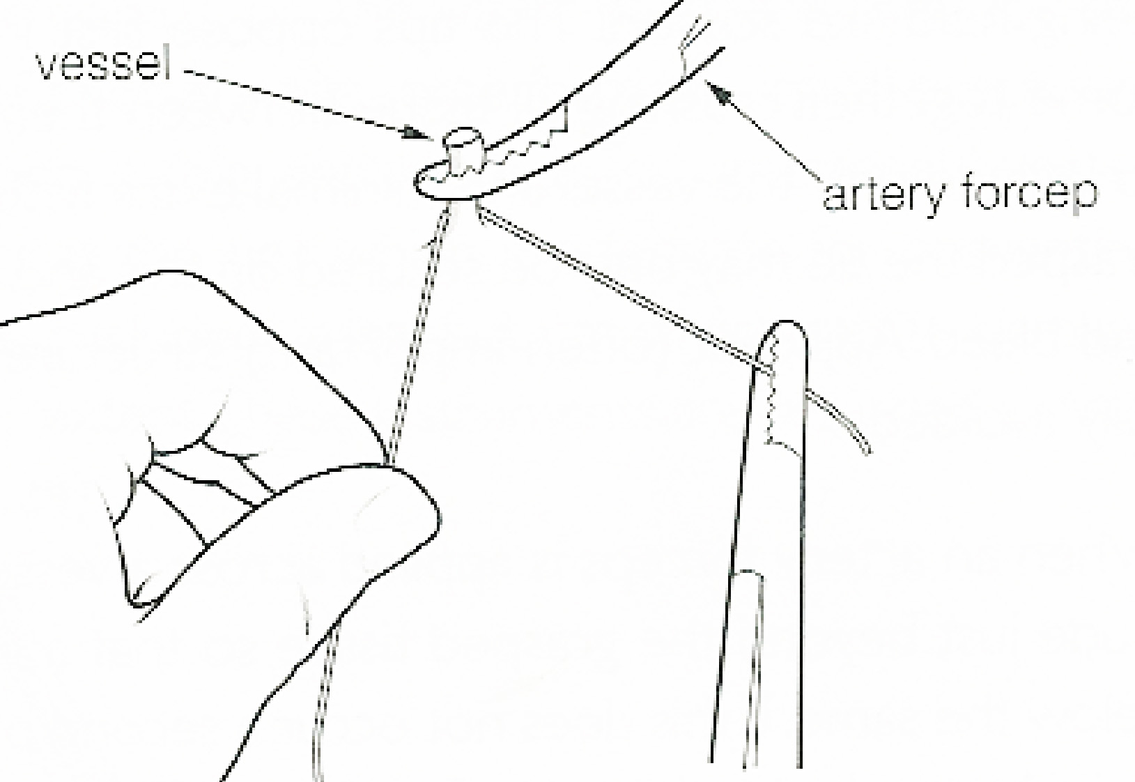
In surgery or medical procedure, a ligature consists of a piece of thread ( suture) tied around an anatomical structure, usually a blood vessel or another hollow structure (e.g. urethra) to shut it off. History The principle of ligation is attributed to Hippocrates and Galen. In ancient Rome, ligatures were used to treat hemorrhoids. The concept of a ligature was reintroduced some 1,500 years later by Ambroise Paré, and finally it found its modern use in 1870–80, made popular by Jules-Émile Péan. Procedure With a blood vessel the surgeon will clamp the vessel perpendicular to the axis of the artery or vein with a hemostat, then secure it by ligating it; i.e. using a piece of suture around it before dividing the structure and releasing the hemostat. It is different from a tourniquet in that the tourniquet will not be secured by knots and it can therefore be released/tightened at will. Ligature is one of the remedies to treat skin tag, or acrochorda. It is done by tying str ... [...More Info...] [...Related Items...] OR: [Wikipedia] [Google] [Baidu] |
Cholangiography
Cholangiography is the imaging of the bile duct (also known as the biliary tree) by x-rays and an injection of contrast medium. __TOC__ Types There are at least four types of cholangiography: # Percutaneous transhepatic cholangiography (PTC): Examination of liver and bile ducts by x-rays. This is accomplished by the insertion of a thin needle into the liver carrying a contrast medium to help to see blockage in liver and bile ducts. # Endoscopic retrograde cholangiopancreatography (ERCP). Although this is a form of imaging, it is both diagnostic and therapeutic, and is often classified with surgeries rather than with imaging. # Primary cholangiography (or ''perioperative''): Done in the operation room during a biliary drainage intervention. # Secondary cholangiography: Done after a biliary drainage intervention. In both cases fluorescent fluids are used to create contrasts that make the diagnosis possible. Cholangiography has largely replaced the previously used method of intravenou ... [...More Info...] [...Related Items...] OR: [Wikipedia] [Google] [Baidu] |
Angiography
Angiography or arteriography is a medical imaging technique used to visualize the inside, or lumen, of blood vessels and organs of the body, with particular interest in the arteries, veins, and the heart chambers. Modern angiography is performed by injecting a radio-opaque contrast agent into the blood vessel and imaging using X-ray based techniques such as fluoroscopy. The word itself comes from the Greek words ἀγγεῖον ''angeion'' 'vessel' and γράφειν ''graphein'' 'to write, record'. The film or image of the blood vessels is called an ''angiograph'', or more commonly an ''angiogram''. Though the word can describe both an arteriogram and a venogram, in everyday usage the terms angiogram and arteriogram are often used synonymously, whereas the term venogram is used more precisely. The term angiography has been applied to radionuclide angiography and newer vascular imaging techniques such as CO2 angiography, CT angiography and MR angiography. The term ''isotope a ... [...More Info...] [...Related Items...] OR: [Wikipedia] [Google] [Baidu] |
CT Scan
A computed tomography scan (CT scan; formerly called computed axial tomography scan or CAT scan) is a medical imaging technique used to obtain detailed internal images of the body. The personnel that perform CT scans are called radiographers or radiology technologists. CT scanners use a rotating X-ray tube and a row of detectors placed in a gantry (medical), gantry to measure X-ray Attenuation#Radiography, attenuations by different tissues inside the body. The multiple X-ray measurements taken from different angles are then processed on a computer using tomographic reconstruction algorithms to produce Tomography, tomographic (cross-sectional) images (virtual "slices") of a body. CT scans can be used in patients with metallic implants or pacemakers, for whom magnetic resonance imaging (MRI) is Contraindication, contraindicated. Since its development in the 1970s, CT scanning has proven to be a versatile imaging technique. While CT is most prominently used in medical diagnosis, ... [...More Info...] [...Related Items...] OR: [Wikipedia] [Google] [Baidu] |
Esophagogastroduodenoscopy
Esophagogastroduodenoscopy (EGD) or oesophagogastroduodenoscopy (OGD), also called by various other names, is a diagnostic endoscopic procedure that visualizes the upper part of the gastrointestinal tract down to the duodenum. It is considered a minimally invasive procedure since it does not require an incision into one of the major body cavities and does not require any significant recovery after the procedure (unless sedation or anesthesia has been used). However, a sore throat is common. Alternative names The words ''esophagogastroduodenoscopy'' (EGD; American English) and ''oesophagogastroduodenoscopy'' (OGD; British English; see spelling differences) are both pronounced . It is also called ''panendoscopy'' (PES) and ''upper GI endoscopy''. It is also often called just ''upper endoscopy'', ''upper GI'', or even just ''endoscopy''; because EGD is the most commonly performed type of endoscopy, the ambiguous term ''endoscopy'' is sometimes informally used to refer to EGD b ... [...More Info...] [...Related Items...] OR: [Wikipedia] [Google] [Baidu] |


