|
Hemianopia
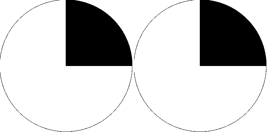
Hemianopsia, or hemianopia, is a loss of vision or blindness (anopsia) in half the visual field, usually on one side of the vertical midline. The most common causes of this damage are stroke, brain tumor, and trauma. This article deals only with permanent hemianopsia, and not with transitory or temporary hemianopsia, as identified by William Wollaston PRS in 1824. Temporary hemianopsia can occur in the aura phase of migraine. Etymology The word ''hemianopsia'' is from Greek origins, where: * ''hemi'' means "half", * ''an'' means "without", and * ''opsia'' means "seeing". Types When the pathology involves both eyes, it is either homonymous or heteronymous. Homonymous hemianopsia Paris as seen with left homonymous hemianopsia. A homonymous hemianopsia is the loss of half of the visual field on the same side in both eyes. The visual images that we see to the right side travel from both eyes to the left side of the brain, while the visual images we see to the left sid ... [...More Info...] [...Related Items...] OR: [Wikipedia] [Google] [Baidu] |
Homonymous Hemianopsia
Hemianopsia, or hemianopia, is a visual field loss on the left or right side of the vertical midline. It can affect one eye but usually affects both eyes. Homonymous hemianopsia (or homonymous hemianopia) is hemianopic visual field loss on the same side of both eyes. Homonymous hemianopsia occurs because the right half of the brain has visual pathways for the left hemifield of both eyes, and the left half of the brain has visual pathways for the right hemifield of both eyes. When one of these pathways is damaged, the corresponding visual field is lost. Signs and symptoms Paris as seen with right homonymous hemianopsia Mobility can be difficult for people with homonymous hemianopsia. "Patients frequently complain of bumping into obstacles on the side of the field loss, thereby bruising their arms and legs." People with homonymous hemianopsia often experience discomfort in crowds. "A patient with this condition may be unaware of what he or she cannot see and frequently bumps ... [...More Info...] [...Related Items...] OR: [Wikipedia] [Google] [Baidu] |
Visual Field
The visual field is the "spatial array of visual sensations available to observation in introspectionist psychological experiments". Or simply, visual field can be defined as the entire area that can be seen when an eye is fixed straight at a point. The equivalent concept for optical instruments and image sensors is the field of view (FOV). In optometry, ophthalmology, and neurology, a visual field test is used to determine whether the visual field is affected by diseases that cause local scotoma or a more extensive loss of vision or a reduction in sensitivity (increase in threshold). Normal limits The normal (monocular) human visual field extends to approximately 60 degrees nasally (toward the nose, or inward) from the vertical meridian in each eye, to 107 degrees temporally (away from the nose, or outwards) from the vertical meridian, and approximately 70 degrees above and 80 below the horizontal meridian. The binocular visual field is the superimposition of the two monocular ... [...More Info...] [...Related Items...] OR: [Wikipedia] [Google] [Baidu] |
Optic Radiation
In neuroanatomy, the optic radiation (also known as the geniculocalcarine tract, the geniculostriate pathway, and posterior thalamic radiation) are axons from the neurons in the lateral geniculate nucleus to the primary visual cortex. The optic radiation receives blood through deep branches of the middle cerebral artery and posterior cerebral artery. They carry visual information through two divisions (called upper and lower division) to the visual cortex (also called ''striate cortex'') along the calcarine fissure. There is one set of upper and lower divisions on each side of the brain. If a lesion only exists in one unilateral division of the optic radiation, the consequence is called quadrantanopia, which implies that only the respective superior or inferior quadrant of the visual field is affected. If both divisions on one side of the brain are affected, the result is a contralateral homonymous hemianopsia. Structure The upper division: :* Projects to the upper bank of th ... [...More Info...] [...Related Items...] OR: [Wikipedia] [Google] [Baidu] |
Meyer's Loop
In neuroanatomy, the optic radiation (also known as the geniculocalcarine tract, the geniculostriate pathway, and posterior thalamic radiation) are axons from the neurons in the lateral geniculate nucleus to the primary visual cortex. The optic radiation receives blood through deep branches of the middle cerebral artery and posterior cerebral artery. They carry visual information through two divisions (called upper and lower division) to the visual cortex (also called ''striate cortex'') along the calcarine fissure. There is one set of upper and lower divisions on each side of the brain. If a lesion only exists in one unilateral division of the optic radiation, the consequence is called quadrantanopia, which implies that only the respective superior or inferior quadrant of the visual field is affected. If both divisions on one side of the brain are affected, the result is a contralateral homonymous hemianopsia. Structure The upper division: :* Projects to the upper bank of th ... [...More Info...] [...Related Items...] OR: [Wikipedia] [Google] [Baidu] |
Quadrantanopia
Quadrantanopia, quadrantanopsia, refers to an anopia (loss of vision) affecting a quarter of the visual field. It can be associated with a lesion of an optic radiation. While quadrantanopia can be caused by lesions in the temporal and parietal lobes of the brain, it is most commonly associated with lesions in the occipital lobe.Kolb, B & Whishaw, I.Q. Human Neuropsychology, Sixth Edition, p.361; Worth Publishers (2008) Presentation An interesting aspect of quadrantanopia is that there exists a distinct and sharp border between the intact and damaged visual fields, due to an anatomical separation of the quadrants of the visual field. For example, information in the left half of visual field is processed in the right occipital lobe and information in the right half of the visual field is processed in the left occipital lobe. In a quadrantanopia that is partial, there also exists a distinct and sharp border between the intact and damaged field within the quadrant. The sufferer ... [...More Info...] [...Related Items...] OR: [Wikipedia] [Google] [Baidu] |
Bitemporal Hemianopsia
Bitemporal hemianopsia, is the medical description of a type of partial blindness where vision is missing in the outer half of both the right and left visual field. It is usually associated with lesions of the optic chiasm, the area where the optic nerves from the right and left eyes cross near the pituitary gland. Causes In bitemporal hemianopsia, vision is missing in the outer (temporal or lateral) half of both the right and left visual fields. Information from the temporal visual field falls on the nasal (medial) retina. The nasal retina is responsible for carrying the information along the optic nerve, and crosses to the other side at the optic chiasm. When there is compression at optic chiasm, the visual impulse from both nasal retina are affected, leading to inability to view the temporal, or peripheral, vision. This phenomenon is known as bitemporal hemianopsia. Knowing the neurocircuitry of visual signal flow through the optic tract is very important in understanding bitem ... [...More Info...] [...Related Items...] OR: [Wikipedia] [Google] [Baidu] |
Anopsia
An anopsia () is a defect in the visual field. If the defect is only partial, then the portion of the field with the defect can be used to isolate the underlying cause. Types of partial anopsia: *Hemianopsia **Homonymous hemianopsia **Heteronymous hemianopsia ***Binasal hemianopsia ***Bitemporal hemianopsia **Superior hemianopia **Inferior hemianopia *Quadrantanopia Quadrantanopia, quadrantanopsia, refers to an anopia (loss of vision) affecting a quarter of the visual field. It can be associated with a lesion of an optic radiation. While quadrantanopia can be caused by lesions in the temporal and parieta ... References External links Visual disturbances and blindness {{eye-stub ... [...More Info...] [...Related Items...] OR: [Wikipedia] [Google] [Baidu] |
Craniopharyngioma
A craniopharyngioma is a rare type of brain tumor derived from pituitary gland embryonic tissue that occurs most commonly in children, but also affects adults. It may present at any age, even in the prenatal and neonatal periods, but peak incidence rates are childhood-onset at 5–14 years and adult-onset at 50–74 years. People may present with bitemporal inferior quadrantanopia leading to bitemporal hemianopsia, as the tumor may compress the optic chiasm. It has a point prevalence around two per 1,000,000. Craniopharyngiomas are distinct from Rathke's cleft tumours and intrasellar arachnoid cysts. Symptoms and signs Craniopharyngiomas are almost always benign. However, as with many brain tumors, their treatment can be difficult, and significant morbidities are associated with both the tumor and treatment. * Headache (obstructive hydrocephalus) * Hypersomnia * Myxedema * Postsurgical weight gain * Polydipsia * Polyuria (diabetes insipidus) * Vision loss (bitemporal hemianopia ... [...More Info...] [...Related Items...] OR: [Wikipedia] [Google] [Baidu] |
Sprague Effect
The Sprague effect is the phenomenon where homonymous hemianopia, caused by damage to the visual cortex, gets slightly better when the contralesional superior colliculus is destroyed. The effect is named for its discoverer, James Sprague, who observed this phenomenon in 1966 using a cat model. Several reasons have been thought of for this happening, including mutual inhibition between the two brain hemispheres. For similar reasons of inhibiting an inhibitory structure, damaging the substantia nigra, for instance by using ibotenic acid Ibotenic acid or (''S'')-2-amino-2-(3-hydroxyisoxazol-5-yl)acetic acid, also referred to as ibotenate, is a chemical compound and psychoactive drug which occurs naturally in ''Amanita muscaria'' and related species of mushrooms typically found i ..., can also cause the same improvement. References {{reflist Visual disturbances and blindness Blindness ... [...More Info...] [...Related Items...] OR: [Wikipedia] [Google] [Baidu] |
Multisensory Integration
Multisensory integration, also known as multimodal integration, is the study of how information from the different sensory modalities (such as sight, sound, touch, smell, self-motion, and taste) may be integrated by the nervous system. A coherent representation of objects combining modalities enables animals to have meaningful perceptual experiences. Indeed, multisensory integration is central to adaptive behavior because it allows animals to perceive a world of coherent perceptual entities. Multisensory integration also deals with how different sensory modalities interact with one another and alter each other's processing. General introduction Multimodal perception is how animals form coherent, valid, and robust perception by processing sensory stimuli from various modalities. Surrounded by multiple objects and receiving multiple sensory stimulations, the brain is faced with the decision of how to categorize the stimuli resulting from different objects or events in the physical wo ... [...More Info...] [...Related Items...] OR: [Wikipedia] [Google] [Baidu] |
Binasal Hemianopsia
Binasal hemianopsia is the medical description of a type of partial blindness where vision is missing in the inner half of both the right and left visual field. It is associated with certain lesions of the eye and of the central nervous system, such as congenital hydrocephalus. Causes In binasal hemianopsia, vision is missing in the inner (nasal or medial) half of both the right and left visual fields. Information from the nasal visual field falls on the temporal (lateral) retina. Those lateral retinal nerve fibers do not cross in the optic chiasm. Calcification of the internal carotid arteries can impinge the uncrossed, lateral retinal fibers, leading to loss of vision in the nasal field. Clinical testing of visual fields (by confrontation) can produce a false positive result, particularly in inferior nasal quadrants. Management Etymology The absence of vision in half of a visual field is described as ''hemianopsia''. The absence of visual perception in one quarter of a vis ... [...More Info...] [...Related Items...] OR: [Wikipedia] [Google] [Baidu] |
Paris
Paris () is the capital and most populous city of France, with an estimated population of 2,165,423 residents in 2019 in an area of more than 105 km² (41 sq mi), making it the 30th most densely populated city in the world in 2020. Since the 17th century, Paris has been one of the world's major centres of finance, diplomacy, commerce, fashion, gastronomy, and science. For its leading role in the arts and sciences, as well as its very early system of street lighting, in the 19th century it became known as "the City of Light". Like London, prior to the Second World War, it was also sometimes called the capital of the world. The City of Paris is the centre of the Île-de-France region, or Paris Region, with an estimated population of 12,262,544 in 2019, or about 19% of the population of France, making the region France's primate city. The Paris Region had a GDP of €739 billion ($743 billion) in 2019, which is the highest in Europe. According to the Economist Intelli ... [...More Info...] [...Related Items...] OR: [Wikipedia] [Google] [Baidu] |


