|
Geniohyoid Muscle
The geniohyoid muscle is a narrow muscle situated superior to the medial border of the mylohyoid muscle. It is named for its passage from the chin ("genio-" is a standard prefix for "chin") to the hyoid bone. Structure It arises from the inferior mental spine, on the back of the mandibular symphysis, and runs backward and slightly downward, to be inserted into the anterior surface of the body of the hyoid bone. It lies in contact with its fellow of the opposite side. It thus belongs to the suprahyoid muscles. The muscle is supplied by branches of the lingual artery. Innervation The geniohyoid muscle is innervated by fibres from the first cervical spinal nerve travelling alongside the hypoglossal nerve. Although the first three cervical nerves give rise to the ansa cervicalis, the geniohyoid muscle is said to be innervated by the first cervical nerve, as some of its efferent fibers do not contribute to ansa cervicalis. Variations It may be blended with the one on opposite ... [...More Info...] [...Related Items...] OR: [Wikipedia] [Google] [Baidu] |
Muscles Of Tongue
The tongue is a muscular organ in the mouth of a typical tetrapod. It manipulates food for mastication and swallowing as part of the digestive process, and is the primary organ of taste. The tongue's upper surface (dorsum) is covered by taste buds housed in numerous lingual papillae. It is sensitive and kept moist by saliva and is richly supplied with nerves and blood vessels. The tongue also serves as a natural means of cleaning the teeth. A major function of the tongue is the enabling of speech in humans and vocalization in other animals. The human tongue is divided into two parts, an oral part at the front and a pharyngeal part at the back. The left and right sides are also separated along most of its length by a vertical section of fibrous tissue (the lingual septum) that results in a groove, the median sulcus, on the tongue's surface. There are two groups of muscles of the tongue. The four intrinsic muscles alter the shape of the tongue and are not attached to bone. The ... [...More Info...] [...Related Items...] OR: [Wikipedia] [Google] [Baidu] |
Chin
The chin is the forward pointed part of the anterior mandible (List_of_human_anatomical_regions#Regions, mental region) below the lower lip. A fully developed human skull has a chin of between 0.7 cm and 1.1 cm. Evolution The presence of a well-developed chin is considered to be one of the morphological characteristics of ''Homo sapiens'' that differentiates them from other human ancestors such as the closely related Neanderthal, Neanderthals. Early human ancestors have varied Mandibular symphysis, symphysial morphology, but none of them have a well-developed chin. The origin of the chin is traditionally associated with the anterior–posterior breadth shortening of the dental arch or tooth row; however, its general mechanical or functional advantage during feeding, developmental origin, and link with human speech, physiology, and social influence are highly debated. Functional perspectives Robinson (1913) suggests that the demand to resist Mastication, masticatory stresses ... [...More Info...] [...Related Items...] OR: [Wikipedia] [Google] [Baidu] |
Neanderthal
Neanderthals (, also ''Homo neanderthalensis'' and erroneously ''Homo sapiens neanderthalensis''), also written as Neandertals, are an extinct species or subspecies of archaic humans who lived in Eurasia until about 40,000 years ago. While the "causes of Neanderthal disappearance about 40,000 years ago remain highly contested," demographic factors such as small population size, inbreeding and genetic drift, are considered probable factors. Other scholars have proposed competitive replacement, assimilation into the modern human genome (bred into extinction), great climatic change, disease, or a combination of these factors. It is unclear when the line of Neanderthals split from that of modern humans; studies have produced various intervals ranging from 315,000 to more than 800,000 years ago. The date of divergence of Neanderthals from their ancestor ''H. heidelbergensis'' is also unclear. The oldest potential Neanderthal bones date to 430,000 years ago, but the classification ... [...More Info...] [...Related Items...] OR: [Wikipedia] [Google] [Baidu] |
Respiration (physiology)
In physiology, respiration is the movement of oxygen from the outside environment to the cells within Tissue (biology), tissues, and the transport, removal of carbon dioxide in the opposite direction that's to the environment. The physiological definition of respiration differs from the Cellular respiration, biochemical definition, which refers to a metabolic process by which an organism obtains energy (in the form of ATP and NADPH) by oxidizing nutrients and releasing waste products. Although physiologic respiration is necessary to sustain cellular respiration and thus life in animals, the processes are distinct: cellular respiration takes place in individual cells of the organism, while physiologic respiration concerns the Diffusion#Diffusion vs. bulk flow diffusion, diffusion and transport of metabolites between the organism and the external environment. Gas exchanges in the lung occurs by ventilation and perfusion. Ventilation refers to the in and out movement of air of the lu ... [...More Info...] [...Related Items...] OR: [Wikipedia] [Google] [Baidu] |
Larynx
The larynx (), commonly called the voice box, is an organ in the top of the neck involved in breathing, producing sound and protecting the trachea against food aspiration. The opening of larynx into pharynx known as the laryngeal inlet is about 4–5 centimeters in diameter. The larynx houses the vocal cords, and manipulates pitch and volume, which is essential for phonation. It is situated just below where the tract of the pharynx splits into the trachea and the esophagus. The word ʻlarynxʼ (plural ʻlaryngesʼ) comes from the Ancient Greek word ''lárunx'' ʻlarynx, gullet, throat.ʼ Structure The triangle-shaped larynx consists largely of cartilages that are attached to one another, and to surrounding structures, by muscles or by fibrous and elastic tissue components. The larynx is lined by a ciliated columnar epithelium except for the vocal folds. The cavity of the larynx extends from its triangle-shaped inlet, to the epiglottis, and to the circular outlet at the ... [...More Info...] [...Related Items...] OR: [Wikipedia] [Google] [Baidu] |
Genioglossus
The genioglossus is one of the paired extrinsic muscles of the tongue. The genioglossus is the major muscle responsible for protruding (or sticking out) the tongue. Structure Genioglossus is the fan-shaped extrinsic tongue muscle that forms the majority of the body of the tongue. It arises from the mental spine of the mandible and its insertions are the hyoid bone and the bottom of the tongue. The genioglossus is innervated by the hypoglossal nerve, as are all muscles of the tongue except for the palatoglossus. Blood is supplied to the sublingual branch of the lingual artery, a branch of the external carotid artery. The canine genioglossus muscle has been divided into horizontal and oblique compartments. Function The left and right genioglossus muscles protrude the tongue and deviate it towards the opposite side. When acting together, the muscles depress the center of the tongue at its back. Clinical significance Contraction of the genioglossus stabilizes and enlarges the porti ... [...More Info...] [...Related Items...] OR: [Wikipedia] [Google] [Baidu] |
Greater Cornu
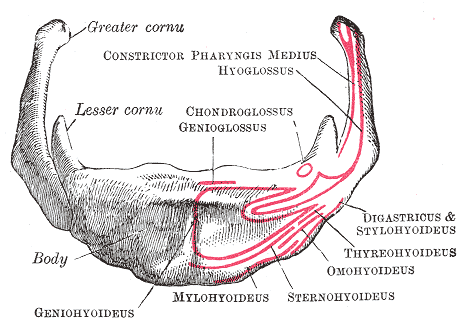
The hyoid bone (lingual bone or tongue-bone) () is a horseshoe-shaped bone situated in the anterior midline of the neck between the chin and the thyroid cartilage. At rest, it lies between the base of the mandible and the third cervical vertebra. Unlike other bones, the hyoid is only distantly articulated to other bones by muscles or ligaments. It is the only bone in the human body that is not connected to any other bones nearby. The hyoid is anchored by muscles from the anterior, posterior and inferior directions, and aids in tongue movement and swallowing. The hyoid bone provides attachment to the muscles of the floor of the mouth and the tongue above, the larynx below, and the epiglottis and pharynx behind. Its name is derived . Structure The hyoid bone is classed as an irregular bone and consists of a central part called the body, and two pairs of horns, the greater and lesser horns. Body The body of the hyoid bone is the central part of the hyoid bone. *At the front, ... [...More Info...] [...Related Items...] OR: [Wikipedia] [Google] [Baidu] |
Ansa Cervicalis
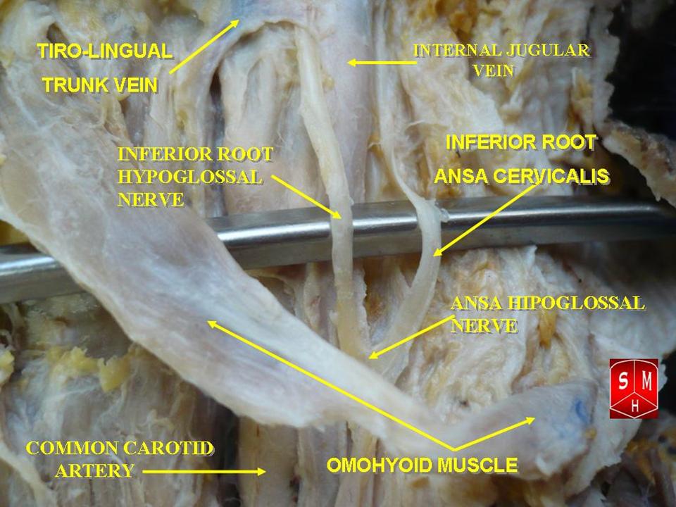
The ansa cervicalis (or ansa hypoglossi in older literature) is a loop of nerves that are part of the cervical plexus. It lies superficial to the internal jugular vein in the carotid triangle. Its name means "handle of the neck" in Latin. Branches from the ansa cervicalis innervate most of the infrahyoid muscles, including the sternothyroid muscle, sternohyoid muscle and the omohyoid muscle. Note that the thyrohyoid muscle, which is also an infrahyoid muscle and the geniohyoid muscle which is a suprahyoid muscle are innervated by cervical spinal nerve 1 via the hypoglossal nerve. Roots Two roots make up the ansa cervicalis, a superior root, and an inferior root. The superior root of the ansa cervicalis is formed from cervical spinal nerve 1 of the cervical plexus. These nerve fibers travel in the hypoglossal nerve before separating in the carotid triangle to form the superior root. The superior root goes around the occipital artery and then descends on the carotid sheath. I ... [...More Info...] [...Related Items...] OR: [Wikipedia] [Google] [Baidu] |
First Cervical Nerve
The cervical spinal nerve 1 (C1) is a spinal nerve of the cervical segment. from spinalcordinjuryzone.com. Published February 23, 2004 Archived Dec 23, 2011. Retrieved June 12, 2018. C1 carries predominantly motor fibres, but also a small meningeal branch that supplies sensation to parts of the dura around the foramen magnum (via dorsal rami). It originates from the spinal column from above the cervical vertebra 1 (C1). The dorsal root and ganglion of the first cervical nerve may be rudimentary or entirely absent. Muscles innervated by this nerve are: * Geniohyoid muscle- th ... [...More Info...] [...Related Items...] OR: [Wikipedia] [Google] [Baidu] |
Suprahyoid Muscles
The suprahyoid muscles are four muscles located above the hyoid bone in the neck. They are the digastric, stylohyoid, geniohyoid, and mylohyoid muscle, mylohyoid muscles. They are all pharyngeal muscles, with the exception of the geniohyoid muscle. The digastric is uniquely named for its two bellies. Its posterior belly rises from the mastoid process of the human skull, cranium and slopes downward and forward. The anterior belly arises from the digastric fossa on the inner surface of the mandibular body, which slopes downward and backward. The two bellies connect at the digastric muscle#intermediate tendon, intermediate tendon. The intermediate tendon passes through a connective tissue loop attached to the hyoid bone. The mylohyoid muscles are thin, flat muscles that form a sling inferior to the tongue supporting the floor of the mouth. The geniohyoids are short, narrow muscles that contact each other in the midline. The stylohyoids are long, thin muscles that are nearly parallel wit ... [...More Info...] [...Related Items...] OR: [Wikipedia] [Google] [Baidu] |
Mandibular Symphysis
In human anatomy, the facial skeleton of the skull the external surface of the mandible is marked in the median line by a faint ridge, indicating the mandibular symphysis (Latin: ''symphysis menti'') or line of junction where the two lateral halves of the mandible typically fuse at an early period of life (1-2 years). It is not a true symphysis as there is no cartilage between the two sides of the mandible. This ridge divides below and encloses a triangular eminence, the mental protuberance, the base of which is depressed in the center but raised on either side to form the mental tubercle. The lowest (most inferior) end of the mandibular symphysis — the point of the chin — is called the "menton". It serves as the origin for the geniohyoid and the genioglossus muscles. Other animals Solitary mammalian carnivores that rely on a powerful canine bite to subdue their prey have a strong mandibular symphysis, while pack hunters delivering shallow bites have a weaker one. When filter ... [...More Info...] [...Related Items...] OR: [Wikipedia] [Google] [Baidu] |
Mylohyoid Muscle
The mylohyoid muscle or diaphragma oris is a paired muscle of the neck. It runs from the mandible to the hyoid bone, forming the floor of the oral cavity of the mouth. It is named after its two attachments near the molar teeth. It forms the floor of the submental triangle. It elevates the hyoid bone and the tongue, important during swallowing and speaking. Structure The mylohyoid muscle is flat and triangular, and is situated immediately superior to the anterior belly of the digastric muscle. It is a pharyngeal muscle (derived from the first pharyngeal arch) and classified as one of the suprahyoid muscles. Together, the paired mylohyoid muscles form a muscular floor for the oral cavity of the mouth. The two mylohyoid muscles arise from the mandible at the mylohyoid line, which extends from the mandibular symphysis in front to the last molar tooth behind. The posterior fibers pass inferomedially and insert at anterior surface of the hyoid bone. The medial fibres of the two ... [...More Info...] [...Related Items...] OR: [Wikipedia] [Google] [Baidu] |


