|
Forehead
In human anatomy, the forehead is an area of the head bounded by three features, two of the skull and one of the scalp. The top of the forehead is marked by the hairline, the edge of the area where hair on the scalp grows. The bottom of the forehead is marked by the supraorbital ridge, the bone feature of the skull above the eyes. The two sides of the forehead are marked by the temporal ridge, a bone feature that links the supraorbital ridge to the coronal suture line and beyond. However, the eyebrows do not form part of the forehead. In ''Terminologia Anatomica'', ''sinciput'' is given as the Latin equivalent to "forehead". (Etymology of ''sinciput'': from ''semi-'' "half" + ''caput'' "head".) Structure The bone of the forehead is the squamous part of the frontal bone. The overlying muscles are the occipitofrontalis, procerus, and corrugator supercilii muscles, all of which are controlled by the temporal branch of the facial nerve. The sensory nerves of the forehead connect ... [...More Info...] [...Related Items...] OR: [Wikipedia] [Google] [Baidu] |
Supraorbital Ridge
The brow ridge, or supraorbital ridge known as superciliary arch in medicine, is a bony ridge located above the eye sockets of all primates. In humans, the eyebrows are located on their lower margin. Structure The brow ridge is a nodule or crest of bone situated on the frontal bone of the skull. It forms the separation between the forehead portion itself (the squama frontalis) and the roof of the eye sockets (the pars orbitalis). Normally, in humans, the ridges arch over each eye, offering mechanical protection. In other primates, the ridge is usually continuous and often straight rather than arched. The ridges are separated from the frontal eminences by a shallow groove. The ridges are most prominent medially, and are joined to one another by a smooth elevation named the glabella. Typically, the arches are more prominent in men than in women, and vary between different ethnic groups. Behind the ridges, deeper in the bone, are the frontal sinuses. Terminology The brow ridges, ... [...More Info...] [...Related Items...] OR: [Wikipedia] [Google] [Baidu] |
Corrugator Supercilii Muscle
The corrugator supercilii muscle is a small, narrow, pyramidal muscle close to the eye. It arises from the medial end of the superciliary arch, and inserts into the deep surface of the skin of the eyebrow. It draws the eyebrow downward and medially, producing the vertical wrinkles of the forehead. Structure The corrugator supercilii muscle is located at the medial end of the eyebrow, beneath the frontalis muscle and just above the orbicularis oculi muscle. It arises from the medial end of the superciliary arch. Its fibers pass upward and laterally, between the palpebral and orbital portions of the orbicularis oculi muscle. It inserts into the deep surface of the skin of the eyebrow, above the middle of the orbital arch. Relations The supratrochlear nerve passes by the corrugator supercilii muscle between it and the frontalis muscle. Function The corrugator supercilii muscle draws the eyebrow downward and medially, producing the vertical wrinkles of the forehead. It is th ... [...More Info...] [...Related Items...] OR: [Wikipedia] [Google] [Baidu] |
Supra-orbital Artery
The supraorbital artery is a branch of the ophthalmic artery. It passes anteriorly within the orbit to exit the orbit through the supraorbital foramen or notch alongside the supraorbital nerve, splitting into two terminal branches which go on to form anastomoses with arteries of the head. Structure Origin The supraorbital artery arises from the ophthalmic artery. Course and relations It travels anteriorly in the orbit by passing superior to the eye and medial to the superior rectus and levator palpebrae superioris. It then joins the supraorbital nerve to jointly pass between the periosteum of the roof of the orbit and the levator palpebrae superioris towards the supraorbital foramen or notch. After passing through the supraorbital foramen or notch, it often splits into a superficial branch and a deep branch. Distribution The supraorbital artery contributes arterial supply to: the superior rectus muscle, superior oblique muscle, levator palpebrae muscles, periorbita, the ... [...More Info...] [...Related Items...] OR: [Wikipedia] [Google] [Baidu] |
Physiognomica
''Physiognomonics'' ( el, Φυσιογνωμονικά; la, Physiognomonica) is an Ancient Greek pseudo-Aristotelian treatise on physiognomy attributed to Aristotle (and part of the Corpus Aristotelicum). Ancient physiognomy before the ''Physiognomonics'' Although ''Physiognomonics'' is the earliest work surviving in Greek devoted to the subject, texts preserved on clay tablets provide evidence of physiognomy manuals from the First Babylonian dynasty, containing divinatory case studies of the ominous significance of various bodily dispositions. At this point physiognomy is "a specific, already theorized, branch of knowledge" and the heir of a long-developed technical tradition.Raina, Introduction. While loosely physiognomic ways of thinking are present in Greek literature as early as Homer, physiognomy proper is not known before the classical period. The term ''physiognomonia'' first appears in the fifth-century BC Hippocratic treatise ''Epidemics'' (II.5.1). Physiognomy was ... [...More Info...] [...Related Items...] OR: [Wikipedia] [Google] [Baidu] |
Pseudo-Aristotle
Pseudo-Aristotle is a general cognomen for authors of philosophical or medical treatises who attributed their work to the Greek philosopher Aristotle, or whose work was later attributed to him by others. Such falsely attributed works are known as pseudepigrapha. The term Corpus Aristotelicum covers both the authentic and spurious works of Aristotle. History The first Pseudo-Aristotelian works were produced by the members of the Peripatetic school, which was founded by Aristotle. However, many more works were written much later, during the Middle Ages. Because Aristotle had produced so many works on such a variety of subjects, it was possible for writers in many different contexts—notably medieval Europeans, North Africans and Arabs—to write a work and ascribe it to Aristotle. Attaching his name to such a work guaranteed it a certain amount of respect and acceptance, since Aristotle was regarded as one of the most authoritative ancient writers for the learned men of both Chris ... [...More Info...] [...Related Items...] OR: [Wikipedia] [Google] [Baidu] |
Phrenology
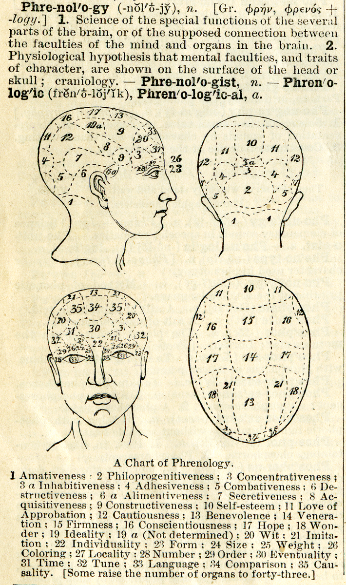
Phrenology () is a pseudoscience which involves the measurement of bumps on the skull to predict mental traits.Wihe, J. V. (2002). "Science and Pseudoscience: A Primer in Critical Thinking." In ''Encyclopedia of Pseudoscience'', pp. 195–203. California: Skeptics Society.Hines, T. (2002). ''Pseudoscience and the Paranormal''. New York: Prometheus Books. p. 200 It is based on the concept that the brain is the organ of the mind, and that certain brain areas have localized, specific functions or modules. It was said that the brain was composed of different muscles, so those that were used more often were bigger, resulting in the different skull shapes. This led to the reasoning behind why everyone had bumps on the skull in different locations. The brain "muscles" not being used as frequently remained small and were therefore not present on the exterior of the skull. Although both of those ideas have a basis in reality, phrenology generalized beyond empirical knowledge in a way that ... [...More Info...] [...Related Items...] OR: [Wikipedia] [Google] [Baidu] |
Physiognomy
Physiognomy (from the Greek , , meaning "nature", and , meaning "judge" or "interpreter") is the practice of assessing a person's character or personality from their outer appearance—especially the face. The term can also refer to the general appearance of a person, object, or terrain without reference to its implied characteristics—as in the physiognomy of an individual plant (see plant life-form) or of a plant community (see vegetation). Physiognomy as a practice meets the contemporary definition of pseudoscience and it is so regarded among academic circles because of its unsupported claims; popular belief in the practice of physiognomy is nonetheless still widespread. The practice was well-accepted by ancient Greek philosophers, but fell into disrepute in the Middle Ages while practised by vagabonds and mountebanks. It revived and was popularised by Johann Kaspar Lavater, before falling from favor in the late 19th century. [...More Info...] [...Related Items...] OR: [Wikipedia] [Google] [Baidu] |
Wrinkle
A wrinkle, also known as a rhytid, is a fold, ridge or crease in an otherwise smooth surface, such as on skin or fabric. Skin wrinkles typically appear as a result of ageing processes such as glycation, habitual sleeping positions, loss of body mass, sun damage, or temporarily, as the result of prolonged immersion in water. Age wrinkling in the skin is promoted by habitual facial expressions, aging, sun damage, smoking, poor hydration, and various other factors. In humans, it can also be prevented to some degree by avoiding excessive solar exposure and through diet (in particular through consumption of carotenoids, tocopherols and flavonoids, vitamins (A, C, D and E), essential omega-3-fatty acids, certain proteins and lactobacilli). Skin Causes for aging wrinkles Development of facial wrinkles is a kind of fibrosis of the skin. Misrepair-accumulation aging theory suggests that wrinkles develop from incorrect repairs of injured elastic fibers and collagen fibers. Repea ... [...More Info...] [...Related Items...] OR: [Wikipedia] [Google] [Baidu] |
Frown
A frown (also known as a scowl) is a facial expression in which the eyebrows are brought together, and the forehead is wrinkled, usually indicating displeasure, sadness or worry, or less often confusion or concentration. The appearance of a frown varies by culture. An alternative usage in North America is thought of as an expression of the mouth. In those cases when used iconically, as with an emoticon, it is entirely presented by the curve of the lips forming a down-open curve. The mouth expression is also commonly referred to in the colloquial English phrase, especially in the United States, to "turn that frown upside down" which indicates changing from sad to happy. Description Charles Darwin described the primary act of frowning as the furrowing of the brow which leads to a rise in the upper lip and a down-turning of the corners of the mouth. While the appearance of a frown varies from culture to culture, there appears to be some degree of universality to the recognition of t ... [...More Info...] [...Related Items...] OR: [Wikipedia] [Google] [Baidu] |
Aporia
In philosophy, an aporia ( grc, ᾰ̓πορῐ́ᾱ, aporíā, literally: "lacking passage", also: "impasse", "difficulty in passage", "puzzlement") is a conundrum or state of puzzlement. In rhetoric, it is a declaration of doubt, made for rhetorical purpose and often feigned. Definitions Definitions of the term ''aporia'' have varied throughout history. ''The Oxford English Dictionary'' includes two forms of the word: the adjective "aporetic", which it defines as "impassable", and "inclined to doubt, or to raise objections"; and the noun form "aporia", which it defines as the "state of the aporetic" and "a perplexity or difficulty". The dictionary entry also includes two early textual uses, which both refer to the term's rhetorical (rather than philosophical) usage. In George Puttenham's ''The Arte of English Poesie'' (1589), aporia is "the Doubtful, ocalled...because often we will seem to caste perils, and make doubts of things when by a plaine manner of speech we might a ... [...More Info...] [...Related Items...] OR: [Wikipedia] [Google] [Baidu] |
Surprise (emotion)
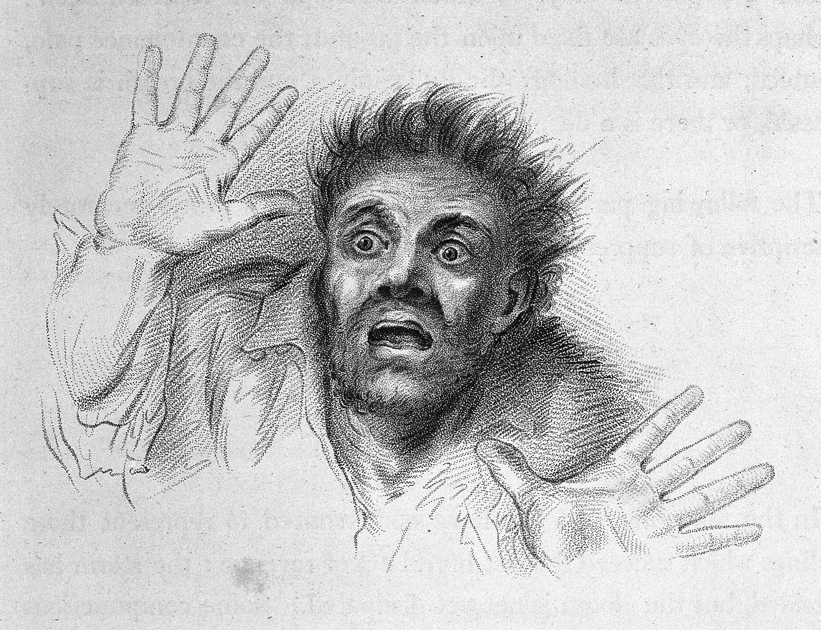
Surprise () is a brief mental and physiological state, a startle response experienced by animals and humans as the result of an unexpected event. Surprise can have any valence; that is, it can be neutral/moderate, pleasant, unpleasant, positive, or negative. Surprise can occur in varying levels of intensity ranging from very-surprised, which may induce the fight-or-flight response, or little-surprise that elicits a less intense response to the stimuli. Construct Surprise is intimately connected to the idea of acting in accordance with a set of rules. When the rules of reality generating events of daily life separate from the rule-of-thumb expectations, surprise is the outcome. Surprise represents the difference between expectations and reality, the gap between our assumptions and expectations about worldly events and the way that those events actually turn out. This gap can be deemed an important foundation on which new findings are based since surprises can make people awar ... [...More Info...] [...Related Items...] OR: [Wikipedia] [Google] [Baidu] |
Eyebrow
An eyebrow is an area of short hairs above each eye that follows the shape of the lower margin of the brow ridges of some mammals. In humans, eyebrows serve two main functions: first, communication through facial expression, and second, prevention of sweat, water, and other debris from falling down into the eye socket. It is common for people to modify their eyebrows by means of hair removal and makeup. Functions A number of theories have been proposed to explain the function of the eyebrow in humans. One approach suggests its main function is to prevent moisture (mostly sweat and rain) from flowing into the eye. Another theory holds that clearly visible eyebrows provided safety from predators when early hominid groups started sleeping on the ground. Recent research, however, suggests eyebrows in humans developed as a means of communication and that this is their primary function. Humans developed a smooth forehead with visible, hairy eyebrows capable of a wide range of movemen ... [...More Info...] [...Related Items...] OR: [Wikipedia] [Google] [Baidu] |


.jpg)

