|
Floor Plate
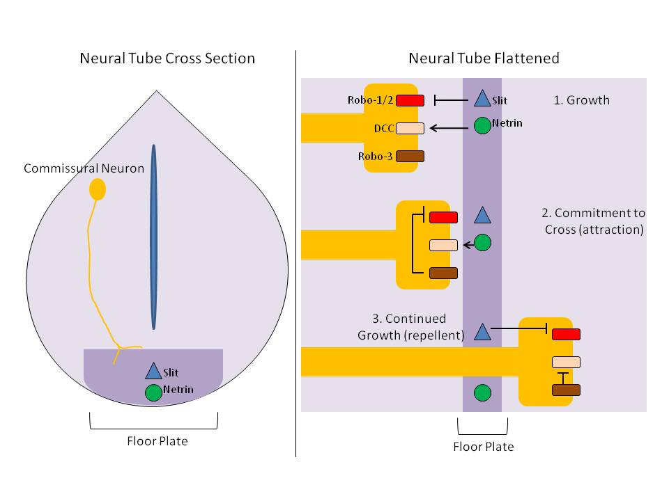
The floor plate is a structure integral to the developing nervous system of vertebrate organisms. Located on the ventral midline of the embryonic neural tube, the floor plate is a specialized glial structure that spans the anteroposterior axis from the midbrain to the tail regions. It has been shown that the floor plate is conserved among vertebrates, such as zebrafish and mice, with homologous structures in invertebrates such as the fruit fly ''Drosophila'' and the nematode ''C. elegans''. Functionally, the structure serves as an organizer to ventralize tissues in the embryo as well as to guide neuronal positioning and differentiation along the dorsoventral axis of the neural tube. Induction Induction of the floor plate during embryogenesis of vertebrate embryos has been studied extensively in chick and zebrafish and occurs as a result of a complex signaling network among tissues, the details of which have yet to be fully refined. Currently there are several competing lines o ... [...More Info...] [...Related Items...] OR: [Wikipedia] [Google] [Baidu] |
Basal Plate (neural Tube)
In the developing nervous system, the basal plate is the region of the neural tube ventral to the sulcus limitans. It extends from the rostral mesencephalon to the end of the spinal cord and contains primarily motor neurons, whereas neurons found in the alar plate are primarily associated with sensory functions. The cell types of the basal plate include lower motor neurons and four types of interneuron. Initially, the left and right sides of the basal plate are continuous, but during neurulation they become separated by the floor plate, and this process is directed by the notochord. Differentiation of neurons in the basal plate is under the influence of the protein Sonic hedgehog released by ventralizing structures, such as the notochord and floor plate. Gray642.png, The basal plate (basal lamina) is separated from the alar plate (alar lamina) by the sulcus limitans The sulcus limitans is found in the fourth ventricle of the brain. It separates the cranial nerve motor nuc ... [...More Info...] [...Related Items...] OR: [Wikipedia] [Google] [Baidu] |
Agnès Bernet
Agnès Bernet, (born 1968) is a French cell biologist and professor of cancer biology at the University Claude Bernard Lyon I. A co-founder of NETRIS Pharma, she has led within the Laboratory of Apoptosis, Cancer and Development, the research team that validated the use of interference ligand/dependence receptors as novel targeted therapies for cancer. Life and work Bernet earned her PhD at University Claude Bernard Lyon I in 1994 with the thesis ''Etude, par recombinaison homologue, de régions régulatrices de l'expression des gènes de globine alpha humains (Study, by homologous recombination, of regulatory regions of the expression of human alpha globin genes). Her'' research focused on the study of two regions that may be involved in the activation of human alpha globin genes during erythroid differentiation. In 2008, she co-founded the company Netris Pharma SAS, where she serves as scientific director and coordinates numerous research projects concerning clinical therapi ... [...More Info...] [...Related Items...] OR: [Wikipedia] [Google] [Baidu] |
AKIRIN2
Akirin-2 is a protein that in humans is encoded by the ''AKIRIN2'' gene In biology, the word gene (from , ; "... Wilhelm Johannsen coined the word gene to describe the Mendelian units of heredity..." meaning ''generation'' or ''birth'' or ''gender'') can have several different meanings. The Mendelian gene is a b .... References External links * Further reading * * * * {{gene-6-stub ... [...More Info...] [...Related Items...] OR: [Wikipedia] [Google] [Baidu] |
Embryogenesis
An embryo is an initial stage of development of a multicellular organism. In organisms that reproduce sexually, embryonic development is the part of the life cycle that begins just after fertilization of the female egg cell by the male sperm cell. The resulting fusion of these two cells produces a single-celled zygote that undergoes many cell divisions that produce cells known as blastomeres. The blastomeres are arranged as a solid ball that when reaching a certain size, called a morula, takes in fluid to create a cavity called a blastocoel. The structure is then termed a blastula, or a blastocyst in mammals. The mammalian blastocyst hatches before implantating into the endometrial lining of the womb. Once implanted the embryo will continue its development through the next stages of gastrulation, neurulation, and organogenesis. Gastrulation is the formation of the three germ layers that will form all of the different parts of the body. Neurulation forms the nervous sys ... [...More Info...] [...Related Items...] OR: [Wikipedia] [Google] [Baidu] |
DBX1
Homeobox protein DBX1, also known as developing brain homeobox protein 1, is a protein that in humans is encoded by the ''DBX1'' gene In biology, the word gene (from , ; "...Wilhelm Johannsen coined the word gene to describe the Mendelian units of heredity..." meaning ''generation'' or ''birth'' or ''gender'') can have several different meanings. The Mendelian gene is a ba .... The DBX1 gene is a transcription factor gene that is pivotal in interneuron differentiation in the ventral spinal cord. The spinal interneurons V0 and V1 are derived from progenitor domains that are differentiated by the expression of homeodomain proteins DBX1 and DBX2. DBX1 is spatially restricted and has a critical role in establishing the distinction of V0 and V1 neuronal fate. In DBX1 mutant mice, neural progenitors fail to generate V0 interneurons and instead gave rise to interneurons expressing V1 characteristics, such as their transcription factor profile, neurotransmitter phenotype, migrator ... [...More Info...] [...Related Items...] OR: [Wikipedia] [Google] [Baidu] |
SUMO Protein
In molecular biology, SUMO (Small Ubiquitin-like Modifier) proteins are a family of small proteins that are covalently attached to and detached from other proteins in cells to modify their function. This process is called SUMOylation (sometimes written sumoylation). SUMOylation is a post-translational modification involved in various cellular processes, such as nuclear-cytosolic transport, transcriptional regulation, apoptosis, protein stability, response to stress, and progression through the cell cycle. SUMO proteins are similar to ubiquitin and are considered members of the ubiquitin-like protein family. SUMOylation is directed by an enzymatic cascade analogous to that involved in ubiquitination. In contrast to ubiquitin, SUMO is not used to tag proteins for degradation. Mature SUMO is produced when the last four amino acids of the C-terminus have been cleaved off to allow formation of an isopeptide bond between the C-terminal glycine residue of SUMO and an acceptor lysine on ... [...More Info...] [...Related Items...] OR: [Wikipedia] [Google] [Baidu] |
PTCH1
Protein patched homolog 1 is a protein that is the member of the patched family and in humans is encoded by the ''PTCH1'' gene. Function PTCH1 is a member of the patched gene family and is the receptor for sonic hedgehog, a secreted molecule implicated in the formation of embryonic structures and in tumorigenesis. This gene functions as a tumor suppressor. The PTCH1 gene product, is a transmembrane protein that suppresses the release of another protein called smoothened, and when sonic hedgehog binds PTCH1, smoothened is released and signals cell proliferation. Clinical significance Mutations of this gene have been associated with nevoid basal cell carcinoma syndrome (AKA Gorlin's Syndrome), esophageal squamous cell carcinoma, trichoepitheliomas, transitional cell carcinomas of the bladder, as well as holoprosencephaly. Alternative splicing results in multiple transcript variants encoding different isoforms. Additional splice variants have been described, but their full leng ... [...More Info...] [...Related Items...] OR: [Wikipedia] [Google] [Baidu] |
GLI3
Zinc finger protein GLI3 is a protein that in humans is encoded by the ''GLI3'' gene. This gene encodes a protein that belongs to the C2H2-type zinc finger proteins subclass of the Gli family. They are characterized as DNA-binding transcription factors and are mediators of Sonic hedgehog (Shh) signaling. The protein encoded by this gene localizes in the cytoplasm and activates patched Drosophila homolog (PTCH1) gene expression. It is also thought to play a role during embryogenesis. Role in development Gli3 is a known transcriptional repressor but may also have a positive transcriptional function. Gli3 represses dHand and Gremlin, which are involved in developing digits. There is evidence that Shh-controlled processing (e.g., cleavage) regulates transcriptional activity of Gli3 similarly to that of Ci. Gli3 mutant mice have many abnormalities including CNS and lung defects and limb polydactyly. In the developing mouse limb bud, Gli3 derepression predominantly regulate ... [...More Info...] [...Related Items...] OR: [Wikipedia] [Google] [Baidu] |
Gliogenesis
Gliogenesis is the generation of non-neuronal glia populations derived from multipotent neural stem cells. Overview Gliogenesis results in the formation of non-neuronal glia populations from neuronal cells. In this capacity, glial cells provide multiple functions to both the central nervous system (CNS) and the peripheral nervous system (PNS). Subsequent differentiation of glial cell populations results in function-specialized glial lineages. Glial cell-derived astrocytes are specialized lineages responsible for modulating the chemical environment by altering ion gradients and neurotransmitter transduction. Similarly derived, oligodendrocytes produce myelin, which insulates axons to facilitate electric signal transduction. Finally, microglial cells are derived from glial precursors and carry out macrophage-like properties to remove cellular and foreign debris within the central nervous system ref. Functions of glial-derived cell lineages are reviewed by Baumann and Hauw. Glioge ... [...More Info...] [...Related Items...] OR: [Wikipedia] [Google] [Baidu] |
Astrocyte
Astrocytes (from Ancient Greek , , "star" + , , "cavity", "cell"), also known collectively as astroglia, are characteristic star-shaped glial cells in the brain and spinal cord. They perform many functions, including biochemical control of endothelial cells that form the blood–brain barrier, provision of nutrients to the nervous tissue, maintenance of extracellular ion balance, regulation of cerebral blood flow, and a role in the repair and scarring process of the brain and spinal cord following infection and traumatic injuries. The proportion of astrocytes in the brain is not well defined; depending on the counting technique used, studies have found that the astrocyte proportion varies by region and ranges from 20% to 40% of all glia. Another study reports that astrocytes are the most numerous cell type in the brain. Astrocytes are the major source of cholesterol in the central nervous system. Apolipoprotein E transports cholesterol from astrocytes to neurons and other glial ... [...More Info...] [...Related Items...] OR: [Wikipedia] [Google] [Baidu] |
Microglia
Microglia are a type of neuroglia (glial cell) located throughout the brain and spinal cord. Microglia account for about 7% of cells found within the brain. As the resident macrophage cells, they act as the first and main form of active immune defense in the central nervous system (CNS). Microglia (and other neuroglia including astrocytes) are distributed in large non-overlapping regions throughout the CNS. Microglia are key cells in overall brain maintenance—they are constantly scavenging the CNS for plaques, damaged or unnecessary neurons and synapses, and infectious agents. Since these processes must be efficient to prevent potentially fatal damage, microglia are extremely sensitive to even small pathological changes in the CNS. This sensitivity is achieved in part by the presence of unique potassium channels that respond to even small changes in extracellular potassium. Recent evidence shows that microglia are also key players in the sustainment of normal brain functions und ... [...More Info...] [...Related Items...] OR: [Wikipedia] [Google] [Baidu] |
Oligodendrocyte
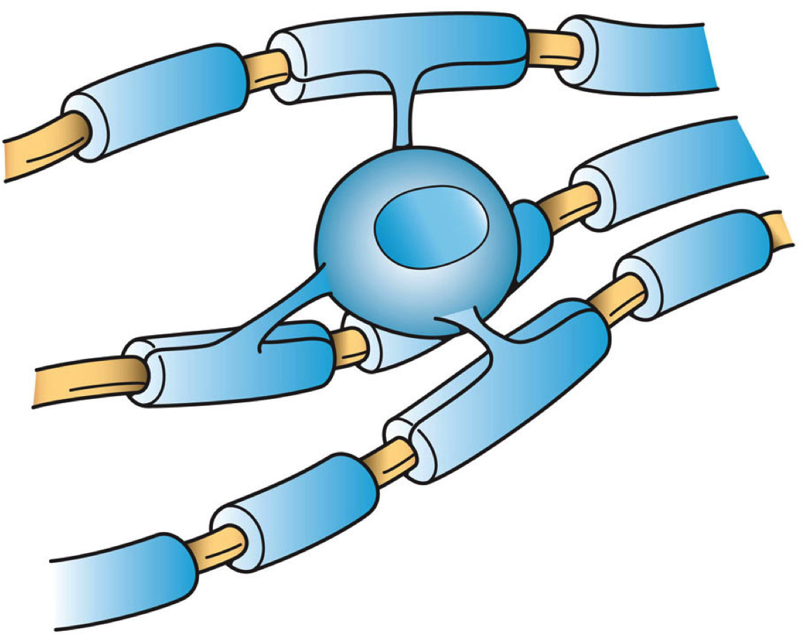
Oligodendrocytes (), or oligodendroglia, are a type of neuroglia whose main functions are to provide support and insulation to axons in the central nervous system of jawed vertebrates, equivalent to the function performed by Schwann cells in the peripheral nervous system. Oligodendrocytes do this by creating the myelin sheath. A single oligodendrocyte can extend its processes to 50 axons, wrapping approximately 1 μm of myelin sheath around each axon; Schwann cells, on the other hand, can wrap around only one axon. Each oligodendrocyte forms one segment of myelin for several adjacent axons. Oligodendrocytes are found only in the central nervous system, which comprises the brain and spinal cord. These cells were originally thought to have been produced in the ventral neural tube; however, research now shows oligodendrocytes originate from the ventral ventricular zone of the embryonic spinal cord and possibly have some concentrations in the forebrain. They are the last cell ... [...More Info...] [...Related Items...] OR: [Wikipedia] [Google] [Baidu] |


