|
Facial Bones
The facial skeleton comprises the ''facial bones'' that may attach to build a portion of the skull. The remainder of the skull is the braincase. In human anatomy and development, the facial skeleton is sometimes called the ''membranous viscerocranium'', which comprises the mandible and dermatocranial elements that are not part of the braincase. Structure In the human skull, the facial skeleton consists of fourteen bones in the face: * Inferior turbinal (2) * Lacrimal bones (2) * Mandible * Maxilla (2) * Nasal bones (2) * Palatine bones (2) * Vomer * Zygomatic bones (2) Variations Elements of the ''cartilaginous viscerocranium'' (i.e., splanchnocranial elements), such as the hyoid bone, are sometimes considered part of the facial skeleton. The ethmoid bone (or a part of it) and also the sphenoid bone are sometimes included, but otherwise considered part of the neurocranium. Because the maxillary bones are fused, they are often collectively listed as only one bone. The mandibl ... [...More Info...] [...Related Items...] OR: [Wikipedia] [Google] [Baidu] |
Face
The face is the front of an animal's head that features the eyes, nose and mouth, and through which animals express many of their emotions. The face is crucial for human identity, and damage such as scarring or developmental deformities may affect the psyche adversely. Structure The front of the human head is called the face. It includes several distinct areas, of which the main features are: *The forehead, comprising the skin beneath the hairline, bordered laterally by the temples and inferiorly by eyebrows and ears *The eyes, sitting in the orbit and protected by eyelids and eyelashes * The distinctive human nose shape, nostrils, and nasal septum *The cheeks, covering the maxilla and mandibula (or jaw), the extremity of which is the chin *The mouth, with the upper lip divided by the philtrum, sometimes revealing the teeth Facial appearance is vital for human recognition and communication. Facial muscles in humans allow expression of emotions. The face is itself a hi ... [...More Info...] [...Related Items...] OR: [Wikipedia] [Google] [Baidu] |
Inferior Turbinal
The inferior nasal concha (inferior turbinated bone or inferior turbinal/turbinate) is one of the three paired nasal conchae in the nose. It extends horizontally along the lateral wall of the nasal cavity and consists of a lamina of spongy bone, curled upon itself like a scroll, (''turbinate'' meaning inverted cone). The inferior nasal conchae are considered a pair of facial bones. As the air passes through the turbinates, the air is churned against these mucosa-lined bones in order to receive warmth, moisture and cleansing. Superior to inferior nasal concha are the middle nasal concha and superior nasal concha which both arise from the ethmoid bone, of the cranial portion of the skull. Hence, these two are considered as a part of the cranial bones. It has two surfaces, two borders, and two extremities. Structure Surfaces The medial surface is convex, perforated by numerous apertures, and traversed by longitudinal grooves for the lodgement of vessels. The lateral surface is con ... [...More Info...] [...Related Items...] OR: [Wikipedia] [Google] [Baidu] |
Mesoderm
The mesoderm is the middle layer of the three germ layers that develops during gastrulation in the very early development of the embryo of most animals. The outer layer is the ectoderm, and the inner layer is the endoderm.Langman's Medical Embryology, 11th edition. 2010. The mesoderm forms mesenchyme, mesothelium, non-epithelial blood cells and coelomocytes. Mesothelium lines coeloms. Mesoderm forms the muscles in a process known as myogenesis, septa (cross-wise partitions) and mesenteries (length-wise partitions); and forms part of the gonads (the rest being the gametes). Myogenesis is specifically a function of mesenchyme. The mesoderm differentiates from the rest of the embryo through intercellular signaling, after which the mesoderm is polarized by an organizing center. The position of the organizing center is in turn determined by the regions in which beta-catenin is protected from degradation by GSK-3. Beta-catenin acts as a co-factor that alters the activity of ... [...More Info...] [...Related Items...] OR: [Wikipedia] [Google] [Baidu] |
Somite
The somites (outdated term: primitive segments) are a set of bilaterally paired blocks of paraxial mesoderm that form in the embryonic stage of somitogenesis, along the head-to-tail axis in segmented animals. In vertebrates, somites subdivide into the dermatomes, myotomes, sclerotomes and syndetomes that give rise to the vertebrae of the vertebral column, rib cage, part of the occipital bone, skeletal muscle, cartilage, tendons, and skin (of the back). The word ''somite'' is sometimes also used in place of the word '' metamere''. In this definition, the somite is a homologously-paired structure in an animal body plan, such as is visible in annelids and arthropods. Development The mesoderm forms at the same time as the other two germ layers, the ectoderm and endoderm. The mesoderm at either side of the neural tube is called paraxial mesoderm. It is distinct from the mesoderm underneath the neural tube which is called the chordamesoderm that becomes the notochord. The ... [...More Info...] [...Related Items...] OR: [Wikipedia] [Google] [Baidu] |
Sclerotome
The somites (outdated term: primitive segments) are a set of bilaterally paired blocks of paraxial mesoderm that form in the embryonic stage of somitogenesis, along the head-to-tail axis in segmented animals. In vertebrates, somites subdivide into the dermatomes, myotomes, sclerotomes and syndetomes that give rise to the vertebrae of the vertebral column, rib cage, part of the occipital bone, skeletal muscle, cartilage, tendons, and skin (of the back). The word ''somite'' is sometimes also used in place of the word '' metamere''. In this definition, the somite is a homologously-paired structure in an animal body plan, such as is visible in annelids and arthropods. Development The mesoderm forms at the same time as the other two germ layers, the ectoderm and endoderm. The mesoderm at either side of the neural tube is called paraxial mesoderm. It is distinct from the mesoderm underneath the neural tube which is called the chordamesoderm that becomes the notochord. The p ... [...More Info...] [...Related Items...] OR: [Wikipedia] [Google] [Baidu] |
Adrenal Medulla
The adrenal medulla ( la, medulla glandulae suprarenalis) is part of the adrenal gland. It is located at the center of the gland, being surrounded by the adrenal cortex. It is the innermost part of the adrenal gland, consisting of chromaffin cells that secrete catecholamines, including epinephrine (adrenaline), norepinephrine (noradrenaline), and a small amount of dopamine, in response to stimulation by sympathetic preganglionic neurons. Structure The adrenal medulla consists of irregularly shaped cells grouped around blood vessels. These cells are intimately connected with the sympathetic division of the autonomic nervous system (ANS). These adrenal medullary cells are modified postganglionic neurons, and preganglionic autonomic nerve fibers lead to them directly from the central nervous system. The adrenal medulla affects energy availability, heart rate, and basal metabolic rate. Recent research indicates that the adrenal medulla may receive input from higher-order cogni ... [...More Info...] [...Related Items...] OR: [Wikipedia] [Google] [Baidu] |
Human Teeth
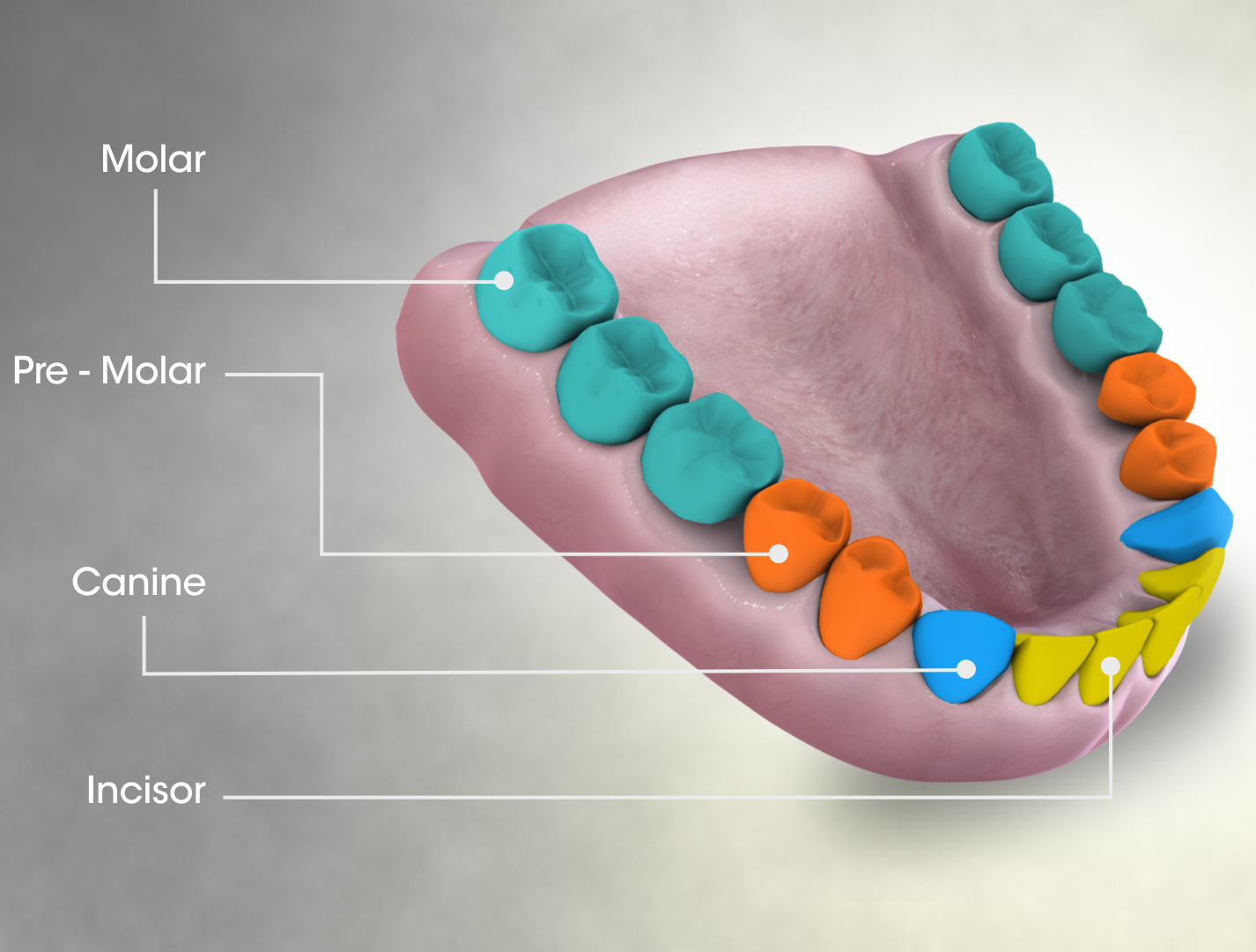
The human teeth function to mechanically break down items of food by cutting and crushing them in preparation for swallowing and digesting. As such, they are considered part of the human digestive system. Humans have four types of teeth: incisors, canines, premolars, and molars, which each have a specific function. The incisors cut the food, the canines tear the food and the molars and premolars crush the food. The roots of teeth are embedded in the maxilla (upper jaw) or the mandible (lower jaw) and are covered by gums. Teeth are made of multiple tissues of varying density and hardness. Humans, like most other mammals, are diphyodont, meaning that they develop two sets of teeth. The first set, deciduous teeth, also called "primary teeth", "baby teeth", or "milk teeth", normally eventually contains 20 teeth. Primary teeth typically start to appear (" erupt") around six months of age and this may be distracting and/or painful for the infant. However, some babies are born with ... [...More Info...] [...Related Items...] OR: [Wikipedia] [Google] [Baidu] |
Neural Crest
Neural crest cells are a temporary group of cells unique to vertebrates that arise from the embryonic ectoderm germ layer, and in turn give rise to a diverse cell lineage—including melanocytes, craniofacial cartilage and bone, smooth muscle, peripheral and enteric neurons and glia. After gastrulation, neural crest cells are specified at the border of the neural plate and the non-neural ectoderm. During neurulation, the borders of the neural plate, also known as the neural folds, converge at the dorsal midline to form the neural tube. Subsequently, neural crest cells from the roof plate of the neural tube undergo an epithelial to mesenchymal transition, delaminating from the neuroepithelium and migrating through the periphery where they differentiate into varied cell types. The emergence of neural crest was important in vertebrate evolution because many of its structural derivatives are defining features of the vertebrate clade. Underlying the development of neura ... [...More Info...] [...Related Items...] OR: [Wikipedia] [Google] [Baidu] |
Dermal Bone
A dermal bone or investing bone or membrane bone is a bony structure derived from intramembranous ossification forming components of the vertebrate skeleton including much of the skull, jaws, gill covers, shoulder girdle and fin spines rays ( lepidotrichia), and the shell (of tortoises and turtles). In contrast to endochondral bone, dermal bone does not form from cartilage that then calcifies, and it is often ornamented. Dermal bone is formed within the dermis and grows by accretion only – the outer portion of the bone is deposited by osteoblasts. The function of some dermal bone is conserved throughout vertebrates, although there is variation in shape and in the number of bones in the skull roof and postcranial structures. In bony fish, dermal bone is found in the fin rays and scales. A special example of dermal bone is the clavicle The clavicle, or collarbone, is a slender, S-shaped long bone approximately 6 inches (15 cm) long that serves as a strut between ... [...More Info...] [...Related Items...] OR: [Wikipedia] [Google] [Baidu] |
Neurocranium
In human anatomy, the neurocranium, also known as the braincase, brainpan, or brain-pan is the upper and back part of the skull, which forms a protective case around the brain. In the human skull, the neurocranium includes the calvaria or skullcap. The remainder of the skull is the facial skeleton. In comparative anatomy, neurocranium is sometimes used synonymously with endocranium or chondrocranium. Structure The neurocranium is divided into two portions: * the membranous part, consisting of flat bones, which surround the brain; and * the cartilaginous part, or chondrocranium, which forms bones of the base of the skull. In humans, the neurocranium is usually considered to include the following eight bones: * 1 ethmoid bone * 1 frontal bone * 1 occipital bone * 2 parietal bones * 1 sphenoid bone * 2 temporal bones The ossicles (three on each side) are usually not included as bones of the neurocranium. There may variably also be extra sutural bones present. Below the ... [...More Info...] [...Related Items...] OR: [Wikipedia] [Google] [Baidu] |
Sphenoid Bone
The sphenoid bone is an unpaired bone of the neurocranium. It is situated in the middle of the skull towards the front, in front of the basilar part of the occipital bone. The sphenoid bone is one of the seven bones that articulate to form the orbit. Its shape somewhat resembles that of a butterfly or bat with its wings extended. Structure It is divided into the following parts: * a median portion, known as the body of sphenoid bone, containing the sella turcica, which houses the pituitary gland as well as the paired paranasal sinuses, the sphenoidal sinuses * two greater wings on the lateral side of the body and two lesser wings from the anterior side. * Pterygoid processes of the sphenoides, directed downwards from the junction of the body and the greater wings. Two sphenoidal conchae are situated at the anterior and inferior part of the body. Intrinsic ligaments of the sphenoid The more important of these are: * the pterygospinous, stretching between the spin ... [...More Info...] [...Related Items...] OR: [Wikipedia] [Google] [Baidu] |
Ethmoid Bone
The ethmoid bone (; from grc, ἡθμός, hēthmós, sieve) is an unpaired bone in the skull that separates the nasal cavity from the brain. It is located at the roof of the nose, between the two orbits. The cubical bone is lightweight due to a spongy construction. The ethmoid bone is one of the bones that make up the orbit of the eye. Structure The ethmoid bone is an anterior cranial bone located between the eyes. It contributes to the medial wall of the orbit, the nasal cavity, and the nasal septum. The ethmoid has three parts: cribriform plate, ethmoidal labyrinth, and perpendicular plate. The cribriform plate forms the roof of the nasal cavity and also contributes to formation of the anterior cranial fossa, the ethmoidal labyrinth consists of a large mass on either side of the perpendicular plate, and the perpendicular plate forms the superior two-thirds of the nasal septum. Between the orbital plate and the nasal conchae are the ethmoidal sinuses or ethmoidal air cell ... [...More Info...] [...Related Items...] OR: [Wikipedia] [Google] [Baidu] |


.jpg)
