|
Fetlock
Fetlock is the common name in horses, large animals, and sometimes dogs for the metacarpophalangeal and metatarsophalangeal joints (MCPJ and MTPJ). Although it somewhat resembles the human ankle in appearance, the joint is homologous to the ball of the foot. In anatomical terms, the hoof corresponds to the toe, rather than the whole foot. Etymology and related terminology The word literally means "foot-lock" and refers to the small tuft of hair situated on the rear of the fetlock joint. "Feather" refers to the particularly long, luxuriant hair growth over the lower leg and fetlock that is characteristic of certain breeds. Formation A fetlock (a MCPJ or a MTPJ) is formed by the junction of the third metacarpal (in the forelimb) or metatarsal (in the hindlimb) bones, either of which are commonly called the cannon bones, proximad and the proximal phalanx distad, commonly called the pastern bone. Paired proximal sesamoid bones form the joint with the palmar or plantar d ... [...More Info...] [...Related Items...] OR: [Wikipedia] [Google] [Baidu] |
Osselet
Osselet is arthritis in the fetlock joint of a horse, caused by trauma. Osselets usually occur in the front legs of the horse, because there is more strain and concussion on the fetlock there than in the hind legs. The arthritis will occur at the joint between the cannon bone and large pastern bone, at the front of the fetlock. Definition Osselets refers to the inflammation of the connective tissue that is around the cannon bone and the fetlock joint. Inflammation can involve arthritis and can become a degenerative joint disease. The condition is a job risk for young thoroughbreds and is usually caused by stress and due to the trauma of repeated hard training in young horses. The first thing that appears on a horse with osselets is a swelling in the front part of the fetlock joint, there may be synovial strains on the sides of the joint. It is painful for the horse to flex the joint and usually causes lameness. Fetlock joint Definition Fetlock is the common name for the metacarp ... [...More Info...] [...Related Items...] OR: [Wikipedia] [Google] [Baidu] |
Sesamoid Bone
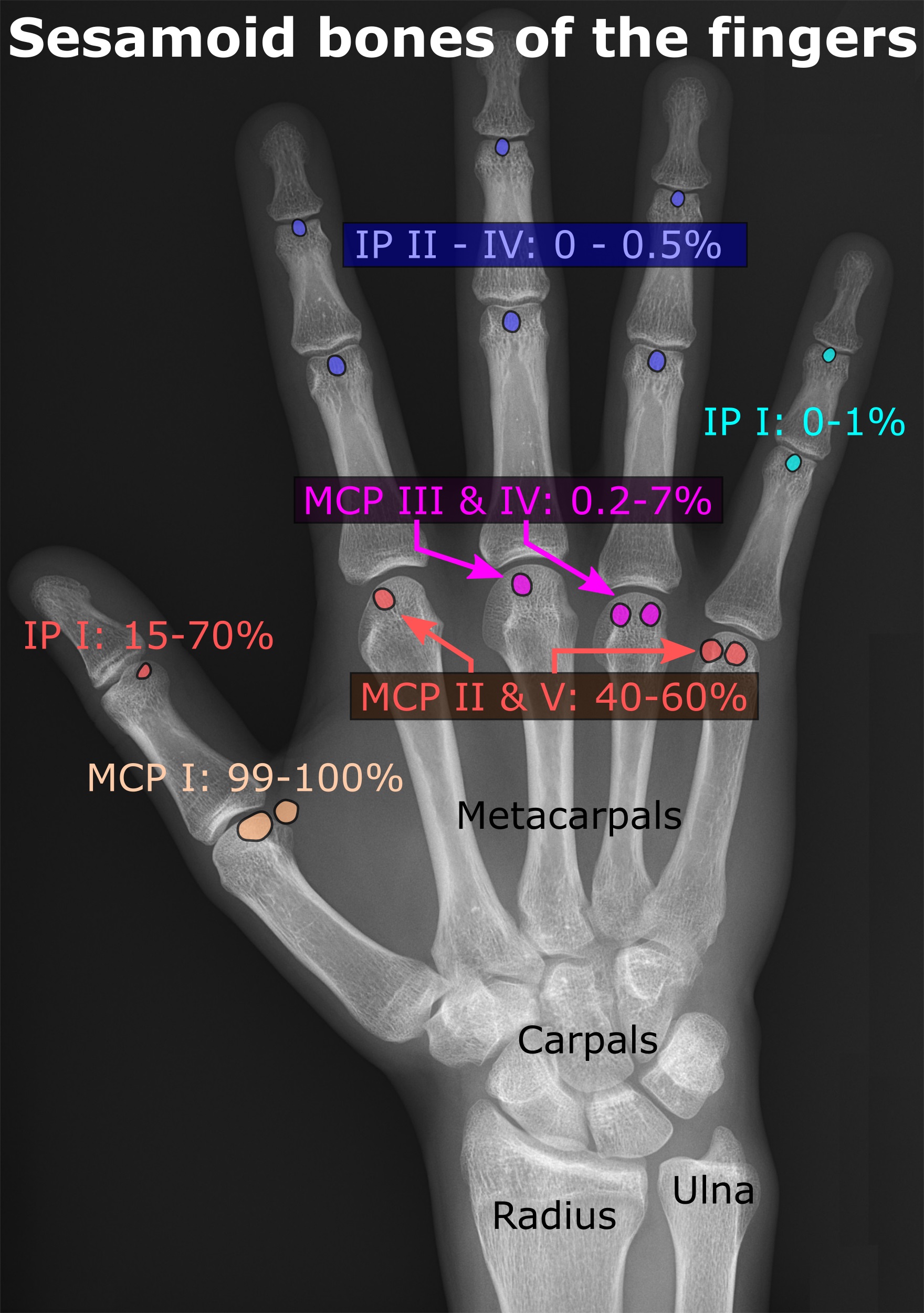
In anatomy, a sesamoid bone () is a bone embedded within a tendon or a muscle. Its name is derived from the Arabic word for ' sesame seed', indicating the small size of most sesamoids. Often, these bones form in response to strain, or can be present as a normal variant. The patella is the largest sesamoid bone in the body. Sesamoids act like pulleys, providing a smooth surface for tendons to slide over, increasing the tendon's ability to transmit muscular forces. Structure Sesamoid bones can be found on joints throughout the body, including: * In the knee—the patella (within the quadriceps tendon). This is the largest sesamoid bone. * In the hand—two sesamoid bones are commonly found in the distal portions of the first metacarpal bone (within the tendons of adductor pollicis and flexor pollicis brevis). There is also commonly a sesamoid bone in distal portions of the second metacarpal bone. * In the wrist—The pisiform of the wrist is a sesamoid bone (within the tend ... [...More Info...] [...Related Items...] OR: [Wikipedia] [Google] [Baidu] |
Sesamoiditis
Sesamoiditis is inflammation of the sesamoid bones. Humans Sesamoiditis occurs on the bottom of the foot, just behind the big toe. There are normally two sesamoid bones on each foot; sometimes sesamoids can be bipartite, which means they each comprise two separate pieces. The sesamoids are roughly the size of jelly beans. The sesamoid bones act as a fulcrum for the flexor tendons, the tendons which bend the big toe downward. Symptoms include inflammation and pain. Sometimes a sesamoid bone is fractured. This can be difficult to pick up on X-ray, so a bone scan or MRI is a better alternative. Among those who are susceptible to the malady are dancers, catchers and pitchers in baseball, soccer players, and football players. Horses In the horse it occurs at the horse's fetlock. The sesamoid bones lie behind the bones of the fetlock, at the back of the joint, and help to keep the tendons and ligaments that run between them correctly functioning. Usually periostitis (new bone gr ... [...More Info...] [...Related Items...] OR: [Wikipedia] [Google] [Baidu] |
Windpuff
Good conformation in the limbs leads to improved movement and decreased likelihood of injuries. Large differences in bone structure and size can be found in horses used for different activities, but correct conformation remains relatively similar across the spectrum. Structural defects, as well as other problems such as injuries and infections, can cause lameness, or movement at an abnormal gait. Injuries to and problems with horse legs can be relatively minor, such as stocking up, which causes swelling without lameness, or quite serious. Even leg injuries that are not immediately fatal may still be life-threatening to horses, as their bodies are adapted to bear weight on all four legs and serious problems can result if this is not possible. Limb anatomy Horses are odd-toed ungulates, or members of the order Perissodactyla. This order also includes the extant species of rhinos and tapirs, and many extinct families and species. Members of this order walk on either one toe (like ... [...More Info...] [...Related Items...] OR: [Wikipedia] [Google] [Baidu] |
Ball (anatomy)
The ball of the foot is the padded portion of the sole between the toes and the arch, underneath the heads of the metatarsal bones. In comparative foot morphology, the ball is most analogous to the metacarpal (forepaw) or metatarsal (hindpaw) pad in many mammals with paws, and serves mostly the same functions. The ball of the foot is of utmost importance when playing sports. Many sports, such as tennis, requires the player to stand on the balls of their feet for increased agility. The ball is a common area in which people develop pain, known as metatarsalgia. People who frequently wear high heels often develop pain in the balls of their feet from the immense amount of pressure that is placed on them for long periods of time, due to the inclination of the shoes. To remedy this, there is a market for ball-of-foot or general foot cushions that are placed into shoes to relieve some of the pressure. Alternately, people can have a procedure done in which a dermal filler is injected ... [...More Info...] [...Related Items...] OR: [Wikipedia] [Google] [Baidu] |
Knuckle
The knuckles are the joints of the fingers. The word is cognate to similar words in other Germanic languages, such as the Dutch "knokkel" (knuckle) or German "Knöchel" (ankle), i.e., ''Knöchlein'', the diminutive of the German word for bone (''Knochen''). Anatomically, it is said that the knuckles consist of the metacarpophalangealUtah Mountain BikingThumb Sprain First as metacarpo. (MCP) and interphalangeal (IP) joints of the finger. The knuckles at the base of the fingers may be referred to as the 1st or major knuckles while the knuckles at the midfinger are known as the 2nd and 3rd, or minor, knuckles. However, the ordinal terms are used inconsistently and may refer to any of the knuckles. Cracking The physical mechanism behind the popping or cracking sound heard when cracking joints such as knuckles has recently been elucidated by cine MRI to be caused by tribonucleation as a gas bubble forms in the synovial fluid that bathes the joint. Despite this evidence, many stil ... [...More Info...] [...Related Items...] OR: [Wikipedia] [Google] [Baidu] |
Metacarpophalangeal Joint
The metacarpophalangeal joints (MCP) are situated between the metacarpal bones and the proximal phalanges of the fingers. These joints are of the condyloid kind, formed by the reception of the rounded heads of the metacarpal bones into shallow cavities on the proximal ends of the proximal phalanges. Being condyloid, they allow the movements of flexion, extension, abduction, adduction and circumduction at the joint. Structure Ligaments Each joint has: * palmar ligaments of metacarpophalangeal articulations * collateral ligaments of metacarpophalangeal articulations Dorsal surfaces The dorsal surfaces of these joints are covered by the expansions of the Extensor tendons, together with some loose areolar tissue which connects the deep surfaces of the tendons to the bones. Function The movements which occur in these joints are flexion, extension, adduction, abduction, and circumduction; the movements of abduction and adduction are very limited, and cannot be performed while th ... [...More Info...] [...Related Items...] OR: [Wikipedia] [Google] [Baidu] |
Abduction (kinesiology)
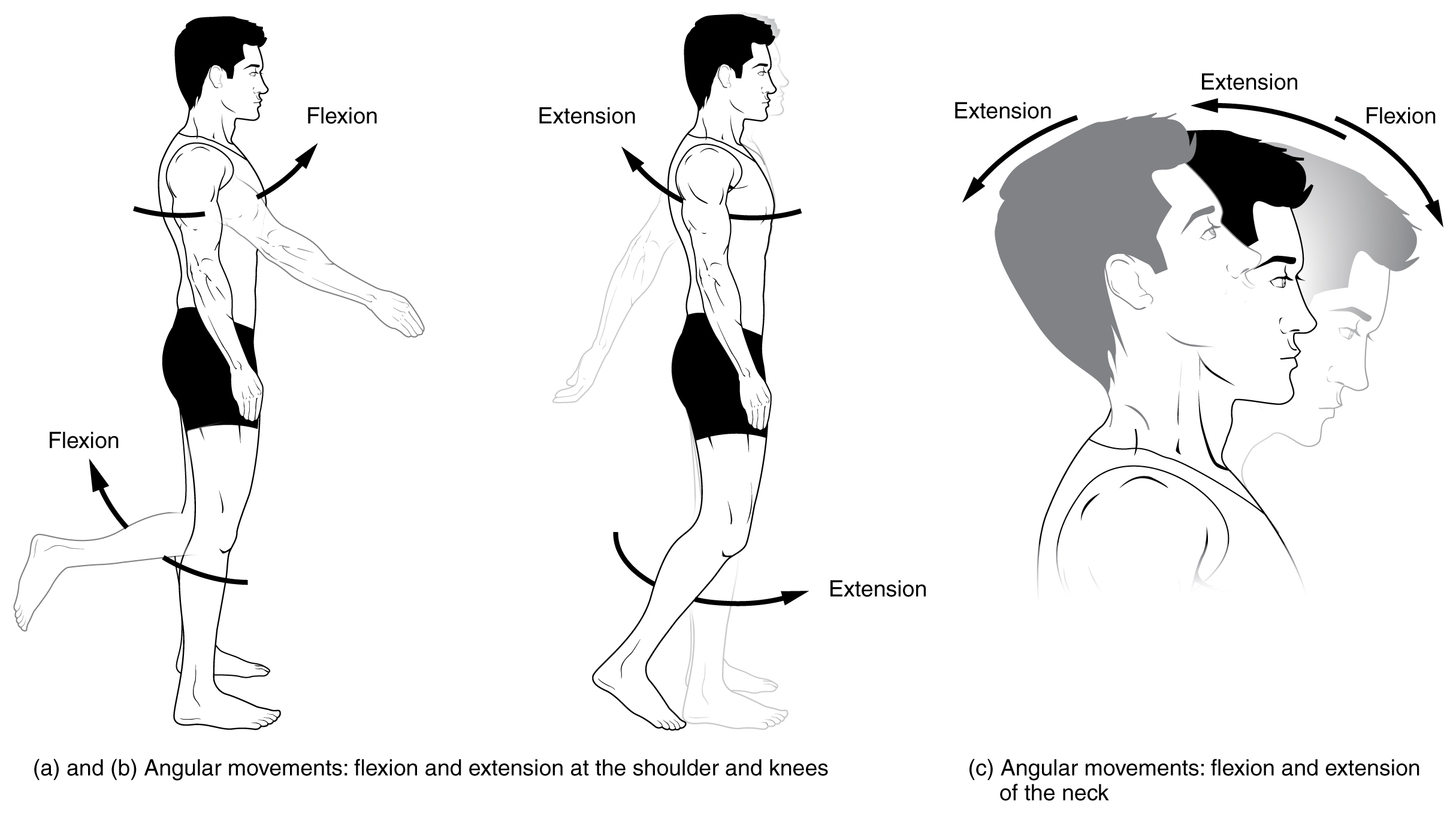
Motion, the process of movement, is described using specific anatomical terms. Motion includes movement of organs, joints, limbs, and specific sections of the body. The terminology used describes this motion according to its direction relative to the anatomical position of the body parts involved. Anatomists and others use a unified set of terms to describe most of the movements, although other, more specialized terms are necessary for describing unique movements such as those of the hands, feet, and eyes. In general, motion is classified according to the anatomical plane it occurs in. ''Flexion'' and ''extension'' are examples of ''angular'' motions, in which two axes of a joint are brought closer together or moved further apart. ''Rotational'' motion may occur at other joints, for example the shoulder, and are described as ''internal'' or ''external''. Other terms, such as ''elevation'' and ''depression'', describe movement above or below the horizontal plane. Many anatomic ... [...More Info...] [...Related Items...] OR: [Wikipedia] [Google] [Baidu] |
Adduction
Motion, the process of movement, is described using specific anatomical terms. Motion includes movement of organs, joints, limbs, and specific sections of the body. The terminology used describes this motion according to its direction relative to the anatomical position of the body parts involved. Anatomists and others use a unified set of terms to describe most of the movements, although other, more specialized terms are necessary for describing unique movements such as those of the hands, feet, and eyes. In general, motion is classified according to the anatomical plane it occurs in. ''Flexion'' and ''extension'' are examples of ''angular'' motions, in which two axes of a joint are brought closer together or moved further apart. ''Rotational'' motion may occur at other joints, for example the shoulder, and are described as ''internal'' or ''external''. Other terms, such as ''elevation'' and ''depression'', describe movement above or below the horizontal plane. Many anatomic ... [...More Info...] [...Related Items...] OR: [Wikipedia] [Google] [Baidu] |
Rotation
Rotation, or spin, is the circular movement of an object around a '' central axis''. A two-dimensional rotating object has only one possible central axis and can rotate in either a clockwise or counterclockwise direction. A three-dimensional object has an infinite number of possible central axes and rotational directions. If the rotation axis passes internally through the body's own center of mass, then the body is said to be ''autorotating'' or '' spinning'', and the surface intersection of the axis can be called a ''pole''. A rotation around a completely external axis, e.g. the planet Earth around the Sun, is called ''revolving'' or ''orbiting'', typically when it is produced by gravity, and the ends of the rotation axis can be called the ''orbital poles''. Mathematics Mathematically, a rotation is a rigid body movement which, unlike a translation, keeps a point fixed. This definition applies to rotations within both two and three dimensions (in a plane and in space, ... [...More Info...] [...Related Items...] OR: [Wikipedia] [Google] [Baidu] |
Extension (kinesiology)
Motion, the process of movement, is described using specific anatomical terms. Motion includes movement of organs, joints, limbs, and specific sections of the body. The terminology used describes this motion according to its direction relative to the anatomical position of the body parts involved. Anatomists and others use a unified set of terms to describe most of the movements, although other, more specialized terms are necessary for describing unique movements such as those of the hands, feet, and eyes. In general, motion is classified according to the anatomical plane it occurs in. ''Flexion'' and ''extension'' are examples of ''angular'' motions, in which two axes of a joint are brought closer together or moved further apart. ''Rotational'' motion may occur at other joints, for example the shoulder, and are described as ''internal'' or ''external''. Other terms, such as ''elevation'' and ''depression'', describe movement above or below the horizontal plane. Many anatomica ... [...More Info...] [...Related Items...] OR: [Wikipedia] [Google] [Baidu] |


