|
Metacarpophalangeal Joint
The metacarpophalangeal joints (MCP) are situated between the metacarpal bones and the proximal phalanges of the fingers. These joints are of the condyloid kind, formed by the reception of the rounded heads of the metacarpal bones into shallow cavities on the proximal ends of the proximal phalanges. Being condyloid, they allow the movements of flexion, extension, abduction, adduction and circumduction at the joint. Structure Ligaments Each joint has: * palmar ligaments of metacarpophalangeal articulations * collateral ligaments of metacarpophalangeal articulations Dorsal surfaces The dorsal surfaces of these joints are covered by the expansions of the Extensor tendons, together with some loose areolar tissue which connects the deep surfaces of the tendons to the bones. Function The movements which occur in these joints are flexion, extension, adduction, abduction, and circumduction; the movements of abduction and adduction are very limited, and cannot be performed wh ... [...More Info...] [...Related Items...] OR: [Wikipedia] [Google] [Baidu] |
Epiphyses
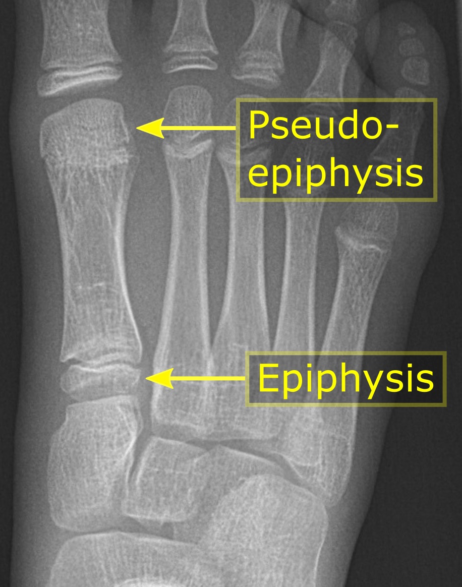
The epiphysis () is the rounded end of a long bone, at its joint with adjacent bone(s). Between the epiphysis and diaphysis (the long midsection of the long bone) lies the metaphysis, including the epiphyseal plate (growth plate). At the joint, the epiphysis is covered with articular cartilage; below that covering is a zone similar to the epiphyseal plate, known as subchondral bone. The epiphysis is filled with red bone marrow, which produces erythrocytes (red blood cells). Structure There are four types of epiphysis: # Pressure epiphysis: The region of the long bone that forms the joint is a pressure epiphysis (e.g. the head of the femur, part of the hip joint complex). Pressure epiphyses assist in transmitting the weight of the human body and are the regions of the bone that are under pressure during movement or locomotion. Another example of a pressure epiphysis is the head of the humerus which is part of the shoulder complex. condyles of femur and tibia also comes u ... [...More Info...] [...Related Items...] OR: [Wikipedia] [Google] [Baidu] |
Interossei
{{short description, Muscles between certain bones Interossei refer to muscles between certain bones. There are many interossei in a human body. Specific interossei include: On the hands * Dorsal interossei muscles of the hand * Palmar interossei muscles File:Gray428.png, Dorsal interossei muscles of the hand File:Gray429.png, Palmar interossei muscles On the feet * Dorsal interossei muscles of the foot * Plantar interossei muscles File:Gray446.png, Dorsal interossei muscles of the foot File:Gray447.png, Plantar interossei muscles Muscular system ... [...More Info...] [...Related Items...] OR: [Wikipedia] [Google] [Baidu] |
Quadrupedalism
Quadrupedalism is a form of locomotion where four limbs are used to bear weight and move around. An animal or machine that usually maintains a four-legged posture and moves using all four limbs is said to be a quadruped (from Latin ''quattuor'' for "four", and ''pes'', ''pedis'' for "foot"). Quadruped animals are found among both vertebrates and invertebrates. Quadrupeds vs. tetrapods Although the words ‘quadruped’ and ‘tetrapod’ are both derived from terms meaning ‘four-footed’, they have distinct meanings. A tetrapod is any member of the taxonomic unit Tetrapoda (which is defined by descent from a specific four-limbed ancestor), whereas a quadruped actually uses four limbs for locomotion. Not all tetrapods are quadrupeds and not all entities that could be described as ‘quadrupedal’ are tetrapods. This last meaning includes certain artificial objects; almost all quadruped ''organisms'' are tetrapods (with the exception of some raptorial arthropods adapte ... [...More Info...] [...Related Items...] OR: [Wikipedia] [Google] [Baidu] |
Osteoarthritis
Osteoarthritis (OA) is a type of degenerative joint disease that results from breakdown of joint cartilage and underlying bone which affects 1 in 7 adults in the United States. It is believed to be the fourth leading cause of disability in the world. The most common symptoms are joint pain and stiffness. Usually the symptoms progress slowly over years. Initially they may occur only after exercise but can become constant over time. Other symptoms may include joint swelling, decreased range of motion, and, when the back is affected, weakness or numbness of the arms and legs. The most commonly involved joints are the two near the ends of the fingers and the joint at the base of the thumbs; the knee and hip joints; and the joints of the neck and lower back. Joints on one side of the body are often more affected than those on the other. The symptoms can interfere with work and normal daily activities. Unlike some other types of arthritis, only the joints, not internal organs, are ... [...More Info...] [...Related Items...] OR: [Wikipedia] [Google] [Baidu] |
Interphalangeal Articulations Of Hand
The interphalangeal joints of the hand are the hinge joints between the phalanges of the fingers that provide flexion towards the palm of the hand. There are two sets in each finger (except in the thumb, which has only one joint): * "proximal interphalangeal joints" (PIJ or PIP), those between the first (also called proximal) and second (intermediate) phalanges * "distal interphalangeal joints" (DIJ or DIP), those between the second (intermediate) and third (distal) phalanges Anatomically, the proximal and distal interphalangeal joints are very similar. There are some minor differences in how the palmar plates are attached proximally and in the segmentation of the flexor tendon sheath, but the major differences are the smaller dimension and reduced mobility of the distal joint. Joint structure The PIP joint exhibits great lateral stability. Its transverse diameter is greater than its antero-posterior diameter and its thick collateral ligaments are tight in all positions duri ... [...More Info...] [...Related Items...] OR: [Wikipedia] [Google] [Baidu] |
Rheumatoid Arthritis
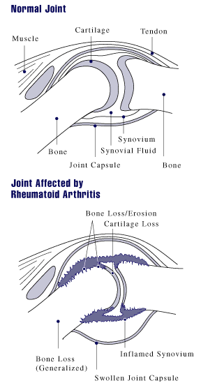
Rheumatoid arthritis (RA) is a long-term autoimmune disorder that primarily affects synovial joint, joints. It typically results in warm, swollen, and painful joints. Pain and stiffness often worsen following rest. Most commonly, the wrist and hands are involved, with the same joints typically involved on both sides of the body. The disease may also affect other parts of the body, including skin, eyes, lungs, heart, nerves and blood. This may result in a anemia, low red blood cell count, pleurisy, inflammation around the lungs, and pericarditis, inflammation around the heart. Fever and low energy may also be present. Often, symptoms come on gradually over weeks to months. While the cause of rheumatoid arthritis is not clear, it is believed to involve a combination of genetics, genetic and environmental factors. The underlying mechanism involves the body's immune system attacking the joints. This results in inflammation and thickening of the synovium, joint capsule. It also affec ... [...More Info...] [...Related Items...] OR: [Wikipedia] [Google] [Baidu] |
Extensor Pollicis Brevis Muscle
In human anatomy, the extensor pollicis brevis is a skeletal muscle on the dorsal side of the forearm. It lies on the medial side of, and is closely connected with, the abductor pollicis longus. The extensor pollicis brevis (EPB) belongs to the deep group of the posterior fascial compartment of the forearm. It is a part of the lateral border of the anatomical snuffbox. Structure The extensor pollicis brevis arises from the ulna distal to the abductor pollicis longus, from the interosseous membrane, and from the dorsal surface of the radius. Its direction is similar to that of the abductor pollicis longus, its tendon passing the same groove on the lateral side of the lower end of the radius, to be inserted into the base of the first phalanx of the thumb. Variation Absence; fusion of tendon with that of the extensor pollicis longus or abductor pollicis longus muscle. Function In a close relationship to the abductor pollicis longus, the extensor pollicis brevis both exten ... [...More Info...] [...Related Items...] OR: [Wikipedia] [Google] [Baidu] |
Extensor Pollicis Longus Muscle
In human anatomy, the extensor pollicis longus muscle (EPL) is a skeletal muscle located dorsally on the forearm. It is much larger than the extensor pollicis brevis, the origin of which it partly covers and acts to stretch the thumb together with this muscle. Structure The extensor pollicis longus arises from the dorsal surface of the ulna and from the interosseous membrane, next to the origins of abductor pollicis longus and extensor pollicis brevis. Passing through the third tendon compartment, lying in a narrow, oblique groove on the back of the lower end of the radius,'' Gray's Anatomy'' 1918, see infobox it crosses the wrist close to the dorsal midline before turning towards the thumb using Lister's tubercle on the distal end of the radius as a pulley. It obliquely crosses the tendons of the extensores carpi radialis longus and brevis, and is separated from the extensor pollicis brevis by a triangular interval, the anatomical snuff box in which the radial artery i ... [...More Info...] [...Related Items...] OR: [Wikipedia] [Google] [Baidu] |
Flexor Pollicis Brevis Muscle
The flexor pollicis brevis is a muscle in the hand that flexes the thumb. It is one of three thenar muscles. It has both a superficial part and a deep part. Origin and insertion The muscle's superficial head arises from the distal edge of the flexor retinaculum and the tubercle of the trapezium, the most lateral bone in the distal row of carpal bones. It passes along the radial side of the tendon of the flexor pollicis longus. The deeper (and medial) head "varies in size and may be absent." It arises from the trapezoid and capitate bones on the floor of the carpal tunnel, as well as the ligaments of the distal carpal row. Both heads become tendinous and insert together into the radial side of the base of the proximal phalanx of the thumb; at the junction between the tendinous heads there is a sesamoid bone.'' Gray's Anatomy'' 1918, see infobox Innervation The superficial head is usually innervated by the lateral terminal branch of the median nerve. The deep part is often in ... [...More Info...] [...Related Items...] OR: [Wikipedia] [Google] [Baidu] |
Flexor Pollicis Longus Muscle
The flexor pollicis longus (; FPL, Latin ''flexor'', bender; ''pollicis'', of the thumb; ''longus'', long) is a muscle in the forearm and hand that flexes the thumb. It lies in the same plane as the flexor digitorum profundus. This muscle is unique to humans, being either rudimentary or absent in other primates. A meta-analysis indicated accessory flexor pollicis longus is present in around 48% of the population. Human anatomy Origin and insertion It arises from the grooved anterior (side of palm) surface of the body of the radius, extending from immediately below the radial tuberosity and oblique line to within a short distance of the pronator quadratus muscle.Gray 1918, ''Flexor Pollicis Longus'', paras 20, 25 An occasionally present accessory long head of the flexor pollicis longus muscle is called 'Gantzer's muscle'. It may cause compression of the anterior interosseous nerve. It arises also from the adjacent part of the interosseous membrane of the forearm, and generall ... [...More Info...] [...Related Items...] OR: [Wikipedia] [Google] [Baidu] |
Extensor Digiti Minimi Muscle
The extensor digiti minimi (extensor digiti quinti proprius) is a slender muscle of the forearm, placed on the ulnar side of the extensor digitorum communis, with which it is generally connected. It arises from the common extensor tendon by a thin tendinous slip and frequently from the intermuscular septa between it and the adjacent muscles. Its tendon passes through a compartment of the extensor retinaculum, posterior to distal radio-ulnar joint, then divides into two as it crosses the dorsum of the hand, and finally joins the extensor digitorum tendon. All three tendons attach to the dorsal digital expansion of the fifth digit (little finger). There may be a slip of tendon to the fourth digit. Variations An additional fibrous slip from the lateral epicondyle; the tendon of insertion may not divide or may send a slip to the ring finger The ring finger, third finger, fourth finger, leech finger, or annulary is the fourth digit of the human hand, located between the midd ... [...More Info...] [...Related Items...] OR: [Wikipedia] [Google] [Baidu] |
Extensor Indicis Proprius
In human anatomy, the extensor indicis roprius'' is a narrow, elongated skeletal muscle in the deep layer of the dorsal forearm, placed medial to, and parallel with, the extensor pollicis longus. Its tendon goes to the index finger, which it extends. Structure It arises from the distal third of the dorsal part of the body of ulna and from the interosseous membrane. It runs through the fourth tendon compartment together with the extensor digitorum, from where it projects into the dorsal aponeurosis of the index finger. Opposite the head of the second metacarpal bone, it joins the ulnar side of the tendon of the extensor digitorum which belongs to the index finger. Like the extensor digiti minimi (i.e. the extensor of the little finger), the tendon of the extensor indicis runs and inserts on the ulnar side of the tendon of the common extensor digitorum. The extensor indicis lacks the juncturae tendinum interlinking the tendons of the extensor digitorum on the dorsal side of the ... [...More Info...] [...Related Items...] OR: [Wikipedia] [Google] [Baidu] |


