|
Esophageal Branches Of The Aorta
The esophageal arteries four or five in number, arise from the front of the aorta, and pass obliquely downward to the esophagus, forming a chain of anastomoses along that tube, anastomosing with the esophageal branches of the inferior thyroid arteries above, and with ascending branches from the left inferior phrenic and left gastric arteries below. These arteries supply the middle third of the esophagus. References External links * - "Branches of the ascending aorta, arch of the aorta, and the descending aorta In human anatomy, the descending aorta is part of the aorta, the largest artery in the body. The descending aorta begins at the aortic arch and runs down through the chest and abdomen. The descending aorta anatomically consists of two portions o ...." Arteries of the thorax {{circulatory-stub ... [...More Info...] [...Related Items...] OR: [Wikipedia] [Google] [Baidu] |
Descending Aorta
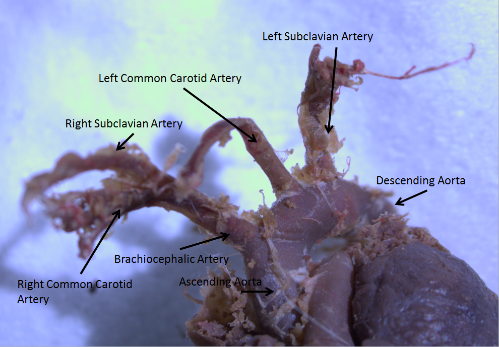
In human anatomy, the descending aorta is part of the aorta, the largest artery in the body. The descending aorta begins at the aortic arch and runs down through the chest and abdomen. The descending aorta anatomically consists of two portions or segments, the thoracic and the abdominal aorta, in correspondence with the two great cavities of the trunk in which it is situated. Within the abdomen, the descending aorta branches into the two common iliac arteries which serve the pelvis and eventually legs. The ductus arteriosus connects to the junction between the pulmonary artery and the descending aorta in foetal life. This artery later regresses as the ligamentum arteriosum. See also *Abbott artery Abbott's artery describes an anomalous artery that arises from the posteromedial aspect of the proximal part of the descending aorta. Normally a minor congenital abnormality, its presence is important during surgical repair of coarctation of the ... References External links ... [...More Info...] [...Related Items...] OR: [Wikipedia] [Google] [Baidu] |
Esophageal Veins
The esophageal veins drain blood from the esophagus to the azygos vein, in the thorax, and to the inferior thyroid vein in the neck. It also drains, although with less significance, to the hemiazygos vein, posterior intercostal vein and bronchial veins. In the abdomen, some drain to the left gastric vein which drains into the portal vein. See also * Esophageal varices Esophageal varices are extremely dilated sub-mucosal veins in the lower third of the esophagus. They are most often a consequence of portal hypertension, commonly due to cirrhosis. People with esophageal varices have a strong tendency to develop ... References External links * * () * https://web.archive.org/web/20080506103555/http://www.med.mun.ca/anatomyts/digest/avein6.htm Veins of the torso {{circulatory-stub ... [...More Info...] [...Related Items...] OR: [Wikipedia] [Google] [Baidu] |
Esophagus
The esophagus (American English) or oesophagus (British English; both ), non-technically known also as the food pipe or gullet, is an organ in vertebrates through which food passes, aided by peristaltic contractions, from the pharynx to the stomach. The esophagus is a fibromuscular tube, about long in adults, that travels behind the trachea and heart, passes through the diaphragm, and empties into the uppermost region of the stomach. During swallowing, the epiglottis tilts backwards to prevent food from going down the larynx and lungs. The word ''oesophagus'' is from Ancient Greek οἰσοφάγος (oisophágos), from οἴσω (oísō), future form of φέρω (phérō, “I carry”) + ἔφαγον (éphagon, “I ate”). The wall of the esophagus from the lumen outwards consists of mucosa, submucosa (connective tissue), layers of muscle fibers between layers of fibrous tissue, and an outer layer of connective tissue. The mucosa is a stratified squamous epithel ... [...More Info...] [...Related Items...] OR: [Wikipedia] [Google] [Baidu] |
Aorta
The aorta ( ) is the main and largest artery in the human body, originating from the left ventricle of the heart and extending down to the abdomen, where it splits into two smaller arteries (the common iliac arteries). The aorta distributes oxygenated blood to all parts of the body through the systemic circulation. Structure Sections In anatomical sources, the aorta is usually divided into sections. One way of classifying a part of the aorta is by anatomical compartment, where the thoracic aorta (or thoracic portion of the aorta) runs from the heart to the diaphragm. The aorta then continues downward as the abdominal aorta (or abdominal portion of the aorta) from the diaphragm to the aortic bifurcation. Another system divides the aorta with respect to its course and the direction of blood flow. In this system, the aorta starts as the ascending aorta, travels superiorly from the heart, and then makes a hairpin turn known as the aortic arch. Following the aortic arch ... [...More Info...] [...Related Items...] OR: [Wikipedia] [Google] [Baidu] |
Esophageal Branches Of The Inferior Thyroid Arteries
The inferior thyroid artery is an artery in the neck. It arises from the thyrocervical trunk and passes upward, in front of the vertebral artery and longus colli muscle. It then turns medially behind the carotid sheath and its contents, and also behind the sympathetic trunk, the middle cervical ganglion resting upon the vessel. Reaching the lower border of the thyroid gland it divides into two branches, which supply the postero-inferior parts of the gland, and anastomose with the superior thyroid artery, and with the corresponding artery of the opposite side. Structure The branches of the inferior thyroid artery are the inferior laryngeal, the oesophageal, the tracheal, the ascending cervical and the pharyngeal arteries. The inferior laryngeal artery climbs the trachea to the back part of the larynx under cover of the inferior pharyngeal constrictor muscle. It is accompanied by the recurrent nerve, and supplies the muscles and mucous membrane of this part, anastomosing wit ... [...More Info...] [...Related Items...] OR: [Wikipedia] [Google] [Baidu] |
Inferior Phrenic Arteries
The inferior phrenic arteries are two small vessels which supply the diaphragm. They present much variety in their origin. Structure Origin The inferior phrenic arteries usually arise between T12 and L2 vertebrae. They may arise separately from the front of the aorta, immediately above the celiac artery, or by a common trunk, which may spring either from the aorta or from the celiac artery. Sometimes one is derived from the aorta, and the other from one of the renal arteries; they rarely arise as separate vessels from the aorta. Branches They diverge from one another across the crura of the diaphragm, and then run obliquely upward and lateralward upon its under surface. * The ''left phrenic'' passes behind the esophagus, and runs forward on the left side of the esophageal hiatus. * The ''right phrenic'' passes behind the inferior vena cava, and along the right side of the foramen which transmits that vein. Near the back part of the central tendon each vessel divides int ... [...More Info...] [...Related Items...] OR: [Wikipedia] [Google] [Baidu] |
Left Gastric
In human anatomy, the left gastric artery arises from the celiac artery and runs along the superior portion of the lesser curvature of the stomach. Branches also supply the lower esophagus. The left gastric artery anastomoses with the right gastric artery, which runs right to left. Important to note is that the esophageal branch of the left gastric artery ascends and passes through the esophageal hiatus. Clinical significance In terms of disease, the left gastric artery may be involved in peptic ulcer disease: if an ulcer erodes through the stomach mucosa into a branch of the artery, this can cause massive blood loss into the stomach, which may result in such symptoms as hematemesis or melaena. Additional images File:Stomach blood supply.svg, Blood supply to the stomach: left and right gastric artery, left and right gastro-omental artery and short gastric artery The short gastric arteries consist of from five to seven small branches, which arise from the end of the splen ... [...More Info...] [...Related Items...] OR: [Wikipedia] [Google] [Baidu] |
Ascending Aorta
The ascending aorta (AAo) is a portion of the aorta commencing at the upper part of the base of the left ventricle, on a level with the lower border of the third costal cartilage behind the left half of the sternum. Structure It passes obliquely upward, forward, and to the right, in the direction of the heart's axis, as high as the upper border of the second right costal cartilage, describing a slight curve in its course, and being situated, about behind the posterior surface of the sternum. The total length is about . Components The aortic root is the portion of the aorta beginning at the aortic annulus and extending to the sinotubular junction. It is sometimes regarded as a part of the ascending aorta, and sometimes regarded as a separate entity from the rest of the ascending aorta. Between each commissure of the aortic valve and opposite the cusps of the aortic valve, three small dilatations called the aortic sinuses. The sinotubular junction is the point in the ascendi ... [...More Info...] [...Related Items...] OR: [Wikipedia] [Google] [Baidu] |
Aortic Arch
The aortic arch, arch of the aorta, or transverse aortic arch () is the part of the aorta between the ascending and descending aorta. The arch travels backward, so that it ultimately runs to the left of the trachea. Structure The aorta begins at the level of the upper border of the second/third sternocostal articulation of the right side, behind the ventricular outflow tract and pulmonary trunk. The right atrial appendage overlaps it. The first few centimeters of the ascending aorta and pulmonary trunk lies in the same pericardial sheath. and runs at first upward, arches over the pulmonary trunk, right pulmonary artery, and right main bronchus to lie behind the right second coastal cartilage. The right lung and sternum lies anterior to the aorta at this point. The aorta then passes posteriorly and to the left, anterior to the trachea, and arches over left main bronchus and left pulmonary artery, and reaches to the left side of the T4 vertebral body. Apart from T4 vertebral body ... [...More Info...] [...Related Items...] OR: [Wikipedia] [Google] [Baidu] |


