|
Extensor Digitorum Brevis Manus
Extensor digitorum brevis manus is an extra or accessory muscle on the backside (dorsum) of the hand. It was first described by Albinus in 1758. The muscles lies in the fourth extensor compartment of the wrist, and is relatively rare. It has a prevalence of 4% in the general population according to a meta-analysis. This muscle is commonly misdiagnosed as a ganglion cysta, synovial nodule or cyst. Structure The extensor digitorum brevis manus usually originates from the dorsal aspect (backside) of the wrist, either from the joint capsule, the distal end (the most distant end) of the radius, the metacarpal, or from the radiocarpal ligament in the area of the fourth extensor compartment. Many variations of the muscle have been described in the literature. It could have up to four tendons with a single tendon inserting to the index or the middle finger being the two most common variations. At the insertion the tendon of the extensor digitorum brevis manus often joins the extensor i ... [...More Info...] [...Related Items...] OR: [Wikipedia] [Google] [Baidu] |
Muscular System
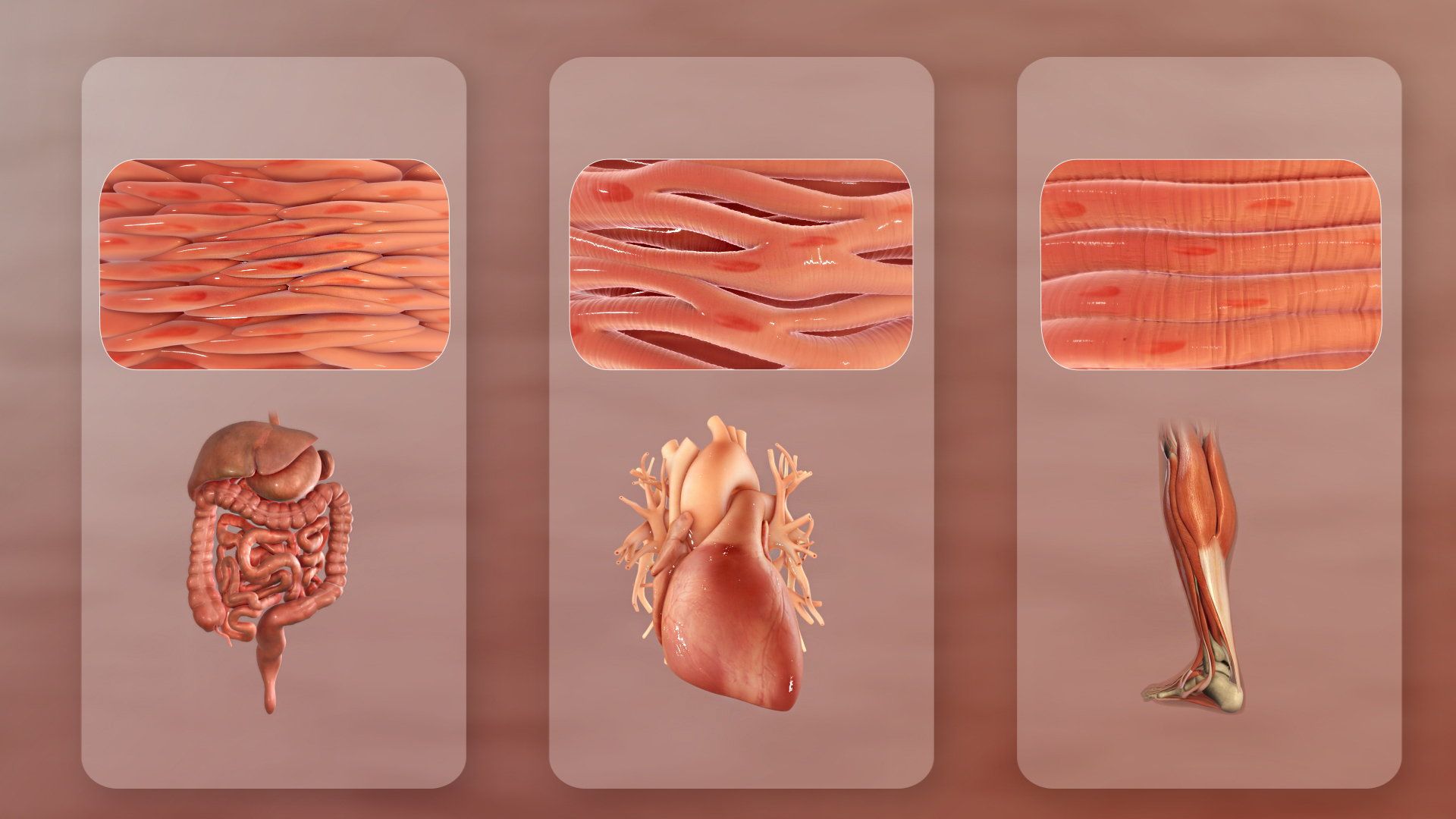
The muscular system is an organ (anatomy), organ system consisting of skeletal muscle, skeletal, smooth muscle, smooth, and cardiac muscle, cardiac muscle. It permits movement of the body, maintains posture, and circulates blood throughout the body. The muscular systems in vertebrates are controlled through the nervous system although some muscles (such as the cardiac muscle) can be completely autonomous. Together with the skeletal system in the human, it forms the musculoskeletal system, which is responsible for the movement of the human body, body. Types There are three distinct types of muscle: skeletal muscle, cardiac muscle, cardiac or heart muscle, and smooth muscle, smooth (non-striated) muscle. Muscles provide strength, balance, posture, movement, and heat for the body to keep warm. There are over 650 muscles in the human body. A kind of elastic tissue makes up each muscle, which consists of thousands, or tens of thousands, of small muscle fibers. Each fiber comprises ... [...More Info...] [...Related Items...] OR: [Wikipedia] [Google] [Baidu] |
Embryonic Development
An embryo is an initial stage of development of a multicellular organism. In organisms that reproduce sexually, embryonic development is the part of the life cycle that begins just after fertilization of the female egg cell by the male sperm cell. The resulting fusion of these two cells produces a single-celled zygote that undergoes many cell divisions that produce cells known as blastomeres. The blastomeres are arranged as a solid ball that when reaching a certain size, called a morula, takes in fluid to create a cavity called a blastocoel. The structure is then termed a blastula, or a blastocyst in mammals. The mammalian blastocyst hatches before implantating into the endometrial lining of the womb. Once implanted the embryo will continue its development through the next stages of gastrulation, neurulation, and organogenesis. Gastrulation is the formation of the three germ layers that will form all of the different parts of the body. Neurulation forms the nervous syst ... [...More Info...] [...Related Items...] OR: [Wikipedia] [Google] [Baidu] |
Accessory Muscle
An accessory muscle is a relatively rare anatomical variation where duplication of a muscle may appear anywhere in the muscular system. Treatment is not indicated unless the accessory muscle interferes with normal function. Examples are the sternalis muscle, accessory soleus muscle, extensor digitorum brevis manus and epitrochleoanconeus muscle. An accessory muscle can also refer to a muscle that is not primarily responsible for movement but does provide assistance. Examples of such muscles are the accessory muscles of respiration where the sternocleidomastoid and the scalene muscles (anterior, middle and posterior scalene) are typically considered accessory muscles of respiration.Netter FH. Atlas of Human Anatomy 3rd ed. Icon Learning Systems. Teterboro, New Jersey 2003 - plate 191 See also * Accessory bone * List of muscles of the human body This is a table of skeletal muscles of the human anatomy. There are around 650 skeletal muscles within the typical human body. Almo ... [...More Info...] [...Related Items...] OR: [Wikipedia] [Google] [Baidu] |
Hand
A hand is a prehensile, multi-fingered appendage located at the end of the forearm or forelimb of primates such as humans, chimpanzees, monkeys, and lemurs. A few other vertebrates such as the koala (which has two opposable thumbs on each "hand" and fingerprints extremely similar to human fingerprints) are often described as having "hands" instead of paws on their front limbs. The raccoon is usually described as having "hands" though opposable thumbs are lacking. Some evolutionary anatomists use the term ''hand'' to refer to the appendage of digits on the forelimb more generally—for example, in the context of whether the three digits of the bird hand involved the same homologous loss of two digits as in the dinosaur hand. The human hand usually has five digits: four fingers plus one thumb; these are often referred to collectively as five fingers, however, whereby the thumb is included as one of the fingers. It has 27 bones, not including the sesamoid bone, the number o ... [...More Info...] [...Related Items...] OR: [Wikipedia] [Google] [Baidu] |
List Of Anatomical Variations
This article lists anatomical variations that are not deemed inherently pathological. {{incomplete list, date=December 2013 Accessory features Bones * Cervical rib * Fabella * Foramen tympanicum * Supracondylar process of the humerus * Sternal foramen * Stafne bone cavity * Episternal ossicles * Fossa navicularis magna * Transverse basilar fissure - or ''Saucer's fissure'' * Canalis basilaris medianus * Craniopharyngeal canal * Intermediate condylar canal * Foramen arcuale * Os odontoideum * Os acromiale * Ossiculum terminale (of dens) * Scapular foramina and tunnels Muscles * Accessory soleus muscle * Axillary arch * Epitrochleoanconeus muscle - or ''anconeous epitrochlearis'' * Extensor medii proprius muscle * Extensor digitorum brevis manus muscle * Extensor indicis et medii communis muscle * Extensor pollicis et indicis communis muscle * Extensor carpi radialis tertius muscle - or ''extensor carpi radialis accessorius'' * Linburg-Comstock variation - or conjoin ... [...More Info...] [...Related Items...] OR: [Wikipedia] [Google] [Baidu] |
Extensor Indicis Et Medii Communis Muscle
The extensor indicis et medii communis is a rare anatomical variant in the extensor compartment of forearm. This additional muscle lies in the deep extensor layer next to the extensor indicis proprius and the extensor pollicis longus. The characteristics of this anomalous muscle resemble those of the extensor indicis proprius, with split tendons to the index and the middle finger. This muscle can also be considered as a variation of the aberrant extensor medii proprius. Structure The extensor indicis et medii communis originates from the distal third of ulna next to the extensor indicis proprius. After passing the wrist joint through the fourth extensor compartment, the tendon splits into two to insert to the extensor expansion of the index and the middle finger. Prevalence The extensor indicis et medii communis has an incidence between 0% and 6.5%. Meta-analysis showed that the muscle was present in average of 1.6% of the total 3,760 hands, and was more prevalent in North A ... [...More Info...] [...Related Items...] OR: [Wikipedia] [Google] [Baidu] |
Extensor Indicis Muscle
In human anatomy, the extensor indicis roprius'' is a narrow, elongated skeletal muscle in the deep layer of the dorsal forearm, placed medial to, and parallel with, the extensor pollicis longus. Its tendon goes to the index finger, which it extends. Structure It arises from the distal third of the dorsal part of the body of ulna and from the interosseous membrane. It runs through the fourth tendon compartment together with the extensor digitorum, from where it projects into the dorsal aponeurosis of the index finger. Opposite the head of the second metacarpal bone, it joins the ulnar side of the tendon of the extensor digitorum which belongs to the index finger. Like the extensor digiti minimi (i.e. the extensor of the little finger), the tendon of the extensor indicis runs and inserts on the ulnar side of the tendon of the common extensor digitorum. The extensor indicis lacks the juncturae tendinum interlinking the tendons of the extensor digitorum on the dorsal side of the h ... [...More Info...] [...Related Items...] OR: [Wikipedia] [Google] [Baidu] |
Lipoma
A lipoma is a benign tumor made of fat tissue. They are generally soft to the touch, movable, and painless. They usually occur just under the skin, but occasionally may be deeper. Most are less than in size. Common locations include upper back, shoulders, and abdomen. It is possible to have a number of lipomas. The cause is generally unclear. Risk factors include family history, obesity, and lack of exercise. Diagnosis is typically based on a physical exam. Occasionally medical imaging or tissue biopsy is used to confirm the diagnosis. Treatment is typically by observation or surgical removal. Rarely, the condition may recur following removal, but this can generally be managed with repeat surgery. They are not generally associated with a future risk of cancer. Lipomas have a prevalence of roughly 2 out of every 100 people. Lipomas typically occur in adults between 40 and 60 years of age. Males are more often affected than females. They are the most common noncancerous soft-t ... [...More Info...] [...Related Items...] OR: [Wikipedia] [Google] [Baidu] |
Pathology
Pathology is the study of the causes and effects of disease or injury. The word ''pathology'' also refers to the study of disease in general, incorporating a wide range of biology research fields and medical practices. However, when used in the context of modern medical treatment, the term is often used in a narrower fashion to refer to processes and tests that fall within the contemporary medical field of "general pathology", an area which includes a number of distinct but inter-related medical specialties that diagnose disease, mostly through analysis of tissue, cell, and body fluid samples. Idiomatically, "a pathology" may also refer to the predicted or actual progression of particular diseases (as in the statement "the many different forms of cancer have diverse pathologies", in which case a more proper choice of word would be " pathophysiologies"), and the affix ''pathy'' is sometimes used to indicate a state of disease in cases of both physical ailment (as in cardiomy ... [...More Info...] [...Related Items...] OR: [Wikipedia] [Google] [Baidu] |
Swelling (medical)
Edema, also spelled oedema, and also known as fluid retention, dropsy, hydropsy and swelling, is the build-up of fluid in the body's tissue. Most commonly, the legs or arms are affected. Symptoms may include skin which feels tight, the area may feel heavy, and joint stiffness. Other symptoms depend on the underlying cause. Causes may include venous insufficiency, heart failure, kidney problems, low protein levels, liver problems, deep vein thrombosis, infections, angioedema, certain medications, and lymphedema. It may also occur after prolonged sitting or standing and during menstruation or pregnancy. The condition is more concerning if it starts suddenly, or pain or shortness of breath is present. Treatment depends on the underlying cause. If the underlying mechanism involves sodium retention, decreased salt intake and a diuretic may be used. Elevating the legs and support stockings may be useful for edema of the legs. Older people are more commonly affected. The word i ... [...More Info...] [...Related Items...] OR: [Wikipedia] [Google] [Baidu] |
Anterior Interosseous Artery
The anterior interosseous artery (volar interosseous artery) is an artery in the forearm. It is a branch of the common interosseous artery. Course It passes down the forearm on the palmar surface of the interosseous membrane. It is accompanied by the palmar interosseous branch of the median nerve, and overlapped by the contiguous margins of the flexor digitorum profundus and flexor pollicis longus muscles, giving off in this situation muscular branches, and the nutrient arteries of the radius and ulna. At the upper border of the pronator quadratus muscle it pierces the interosseous membrane and reaches the back of the forearm, where it anastomoses with the dorsal interosseous artery. It then descends, in company with the terminal portion of the dorsal interosseous nerve, to the back of the wrist to join the dorsal carpal network. The anterior interosseous artery may give off a slender branch, the median artery, which accompanies the median nerve, and gives offsets to its sub ... [...More Info...] [...Related Items...] OR: [Wikipedia] [Google] [Baidu] |


.jpg)