|
Extensor Indicis Et Medii Communis Muscle
The extensor indicis et medii communis is a rare anatomical variant in the extensor compartment of forearm. This additional muscle lies in the deep extensor layer next to the extensor indicis proprius and the extensor pollicis longus. The characteristics of this anomalous muscle resemble those of the extensor indicis proprius, with split tendons to the index and the middle finger. This muscle can also be considered as a variation of the aberrant extensor medii proprius. Structure The extensor indicis et medii communis originates from the distal third of ulna next to the extensor indicis proprius. After passing the wrist joint through the fourth extensor compartment, the tendon splits into two to insert to the extensor expansion of the index and the middle finger. Prevalence The extensor indicis et medii communis has an incidence between 0% and 6.5%. Meta-analysis showed that the muscle was present in average of 1.6% of the total 3,760 hands, and was more prevalent in North A ... [...More Info...] [...Related Items...] OR: [Wikipedia] [Google] [Baidu] |
Ulna
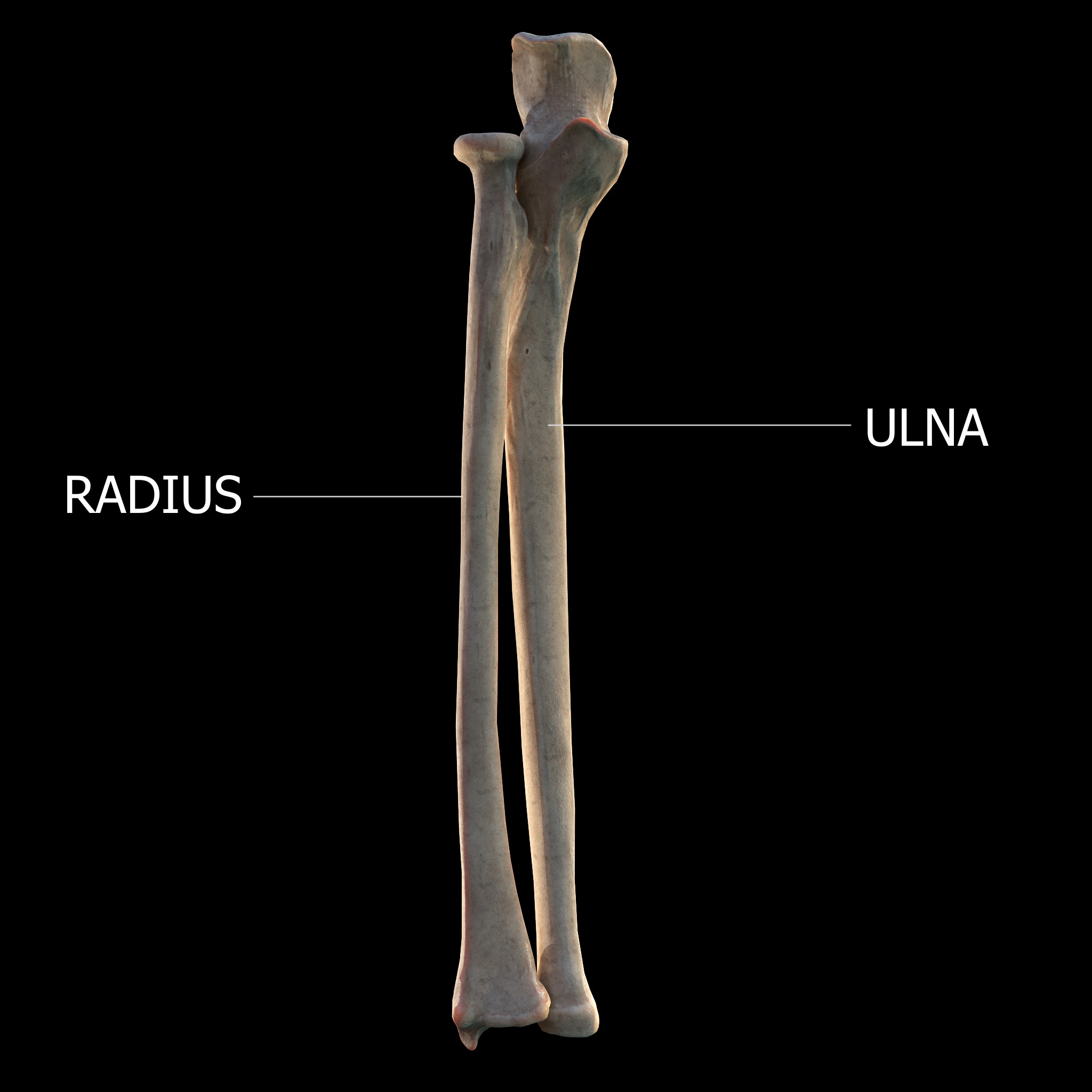
The ulna (''pl''. ulnae or ulnas) is a long bone found in the forearm that stretches from the elbow to the smallest finger, and when in anatomical position, is found on the medial side of the forearm. That is, the ulna is on the same side of the forearm as the little finger. It runs parallel to the radius, the other long bone in the forearm. The ulna is usually slightly longer than the radius, but the radius is thicker. Therefore, the radius is considered to be the larger of the two. Structure The ulna is a long bone found in the forearm that stretches from the elbow to the smallest finger, and when in anatomical position, is found on the medial side of the forearm. It is broader close to the elbow, and narrows as it approaches the wrist. Close to the elbow, the ulna has a bony process, the olecranon process, a hook-like structure that fits into the olecranon fossa of the humerus. This prevents hyperextension and forms a hinge joint with the trochlea of the humerus. There is ... [...More Info...] [...Related Items...] OR: [Wikipedia] [Google] [Baidu] |
Extensor Pollicis Longus Muscle
In human anatomy, the extensor pollicis longus muscle (EPL) is a skeletal muscle located dorsally on the forearm. It is much larger than the extensor pollicis brevis, the origin of which it partly covers and acts to stretch the thumb together with this muscle. Structure The extensor pollicis longus arises from the dorsal surface of the ulna and from the interosseous membrane, next to the origins of abductor pollicis longus and extensor pollicis brevis. Passing through the third tendon compartment, lying in a narrow, oblique groove on the back of the lower end of the radius,''Gray's Anatomy'' 1918, see infobox it crosses the wrist close to the dorsal midline before turning towards the thumb using Lister's tubercle on the distal end of the radius as a pulley. It obliquely crosses the tendons of the extensores carpi radialis longus and brevis, and is separated from the extensor pollicis brevis by a triangular interval, the anatomical snuff box in which the radial artery is found. ... [...More Info...] [...Related Items...] OR: [Wikipedia] [Google] [Baidu] |
List Of Anatomical Variations
This article lists anatomical variations that are not deemed inherently pathological. {{incomplete list, date=December 2013 Accessory features Bones * Cervical rib * Fabella * Foramen tympanicum * Supracondylar process of the humerus * Sternal foramen * Stafne bone cavity * Episternal ossicles * Fossa navicularis magna * Transverse basilar fissure - or ''Saucer's fissure'' * Canalis basilaris medianus * Craniopharyngeal canal * Intermediate condylar canal * Foramen arcuale * Os odontoideum * Os acromiale * Ossiculum terminale (of dens) * Scapular foramina and tunnels Muscles * Accessory soleus muscle * Axillary arch * Epitrochleoanconeus muscle - or ''anconeous epitrochlearis'' * Extensor medii proprius muscle * Extensor digitorum brevis manus muscle * Extensor indicis et medii communis muscle * Extensor pollicis et indicis communis muscle * Extensor carpi radialis tertius muscle - or ''extensor carpi radialis accessorius'' * Linburg-Comstock variation - or conjoin ... [...More Info...] [...Related Items...] OR: [Wikipedia] [Google] [Baidu] |
Extensor Digitorum Brevis Manus
Extensor digitorum brevis manus is an extra or accessory muscle on the backside (dorsum) of the hand. It was first described by Albinus in 1758. The muscles lies in the fourth extensor compartment of the wrist, and is relatively rare. It has a prevalence of 4% in the general population according to a meta-analysis. This muscle is commonly misdiagnosed as a ganglion cysta, synovial nodule or cyst. Structure The extensor digitorum brevis manus usually originates from the dorsal aspect (backside) of the wrist, either from the joint capsule, the distal end (the most distant end) of the radius, the metacarpal, or from the radiocarpal ligament in the area of the fourth extensor compartment. Many variations of the muscle have been described in the literature. It could have up to four tendons with a single tendon inserting to the index or the middle finger being the two most common variations. At the insertion the tendon of the extensor digitorum brevis manus often joins the extensor i ... [...More Info...] [...Related Items...] OR: [Wikipedia] [Google] [Baidu] |
Meta-analysis
A meta-analysis is a statistical analysis that combines the results of multiple scientific studies. Meta-analyses can be performed when there are multiple scientific studies addressing the same question, with each individual study reporting measurements that are expected to have some degree of error. The aim then is to use approaches from statistics to derive a pooled estimate closest to the unknown common truth based on how this error is perceived. Meta-analytic results are considered the most trustworthy source of evidence by the evidence-based medicine literature.Herrera Ortiz AF., Cadavid Camacho E, Cubillos Rojas J, Cadavid Camacho T, Zoe Guevara S, Tatiana Rincón Cuenca N, Vásquez Perdomo A, Del Castillo Herazo V, & Giraldo Malo R. A Practical Guide to Perform a Systematic Literature Review and Meta-analysis. Principles and Practice of Clinical Research. 2022;7(4):47–57. https://doi.org/10.21801/ppcrj.2021.74.6 Not only can meta-analyses provide an estimate of the un ... [...More Info...] [...Related Items...] OR: [Wikipedia] [Google] [Baidu] |
Incidence (epidemiology)
In epidemiology, incidence is a measure of the probability of occurrence of a given medical condition in a population within a specified period of time. Although sometimes loosely expressed simply as the number of new cases during some time period, it is better expressed as a proportion or a rate with a denominator. Incidence proportion Incidence proportion (IP), also known as cumulative incidence, is defined as the probability that a particular event, such as occurrence of a particular disease, has occurred before a given time. It is calculated dividing the number of new cases during a given period by the number of subjects at risk in the population initially at risk at the beginning of the study. Where the period of time considered is an entire lifetime, the incidence proportion is called lifetime risk. For example, if a population initially contains 1,000 persons and 28 develop a condition since the disease first occurred until two years later, the cumulative incidence prop ... [...More Info...] [...Related Items...] OR: [Wikipedia] [Google] [Baidu] |
Extensor Expansion
An extensor expansion (extensor hood, dorsal expansion, dorsal hood, dorsal aponeurosis) is the special connective attachments by which the extensor tendons insert into the phalanges. These flattened tendons (aponeurosis) of extensor muscles span the proximal and middle phalanges. At the distal end of the metacarpal, the extensor tendon will expand to form a hood, which covers the back and sides of the head of the metacarpal and the proximal phalanx. Bands The expansion soon divides into three bands: * lateral bands pass on either side of the proximal phalanx and stretch all the way to the distal phalanx. The lumbricals of the hand, extensor indicis muscle, dorsal interossei of the hand, and palmar interossei In human anatomy, the palmar or volar interossei (interossei volares in older literature) are three small, unipennate muscles in the hand that lie between the metacarpal bones and are attached to the index, ring, and little fingers. They are small ... insert on these ba ... [...More Info...] [...Related Items...] OR: [Wikipedia] [Google] [Baidu] |
Posterior Compartment Of The Forearm
The posterior compartment of the forearm (or extensor compartment) contains twelve muscles which primarily extend the wrist and digits. It is separated from the anterior compartment by the interosseous membrane between the radius and ulna. Structure Muscles There are generally twelve muscles in the posterior compartment of the forearm, which can be further divided into superficial, intermediate, and deep. Most of the muscles in the superficial and the intermediate layers share a common origin which is the outer part of the elbow, the lateral epicondyle of humerus. The deep muscles arise from the distal part of the ulna and the surrounding interosseous membrane. The brachioradialis, flexor of the elbow, is unusual in that it is located in the posterior compartment, but it is actually a muscle of flexor / anterior compartment of the forearm. The anconeus, assisting in extension of the elbow joint, is by some considered part of the posterior compartment of the arm. The majority o ... [...More Info...] [...Related Items...] OR: [Wikipedia] [Google] [Baidu] |
Extensor Medii Proprius Muscle
The extensor medii proprius (so called the ''extensor digiti medii'') is a rare anatomical variant in the extensor compartment of the forearm. The aberrant muscle is analogous to the extensor indicis with the insertion being the middle finger instead of the index finger. Structure The extensor medii proprius originates from the distal third of ulna near the extensor indicis and the adjacent interosseous membrane. It passes through the fourth extensor compartment along with the extensor indicis and the extensor digitorum. It inserts to the extensor expansion of the middle finger usually on the ulnar side of the tendon of the extensor digitorum of the middle finger, though, insertion deep to the extensor digirorum tendon was seen. Insertion to the fibrous tissue proximal to the metacarpophalangeal joint of the middle finger was also reported. Prevalence The reported incidence of the extensor medii proprius in cadaveric dissections ranges from 0% to 12%. Meta-analysis showed th ... [...More Info...] [...Related Items...] OR: [Wikipedia] [Google] [Baidu] |
Extensor Indicis Muscle
In human anatomy, the extensor indicis roprius'' is a narrow, elongated skeletal muscle in the deep layer of the dorsal forearm, placed medial to, and parallel with, the extensor pollicis longus. Its tendon goes to the index finger, which it extends. Structure It arises from the distal third of the dorsal part of the body of ulna and from the interosseous membrane. It runs through the fourth tendon compartment together with the extensor digitorum, from where it projects into the dorsal aponeurosis of the index finger. Opposite the head of the second metacarpal bone, it joins the ulnar side of the tendon of the extensor digitorum which belongs to the index finger. Like the extensor digiti minimi (i.e. the extensor of the little finger), the tendon of the extensor indicis runs and inserts on the ulnar side of the tendon of the common extensor digitorum. The extensor indicis lacks the juncturae tendinum interlinking the tendons of the extensor digitorum on the dorsal side of the h ... [...More Info...] [...Related Items...] OR: [Wikipedia] [Google] [Baidu] |
Interosseous Membrane
An interosseous membrane is a thick dense fibrous sheet of connective tissue that spans the space between two bones, forming a type of syndesmosis joint. Interosseous membranes in the human body: * Interosseous membrane of forearm * Interosseous membrane of leg The interosseous membrane of the leg (middle tibiofibular ligament) extends between the interosseous crests of the tibia and fibula, helps stabilize the Tib-Fib relationship and separates the muscles on the front from those on the back of the leg. ... Gallery File:5 ligaments of interosseous membrane of forearm.png, Five ligaments of interosseous membrane of forearm:* Central band (key portion to be reconstructed in case of injury)* Accessory band * Distal oblique bundle * Proximal oblique cord* Dorsal oblique accessory cord Notes External links * * {{Authority control Skeletal system ... [...More Info...] [...Related Items...] OR: [Wikipedia] [Google] [Baidu] |
Forearm
The forearm is the region of the upper limb between the elbow and the wrist. The term forearm is used in anatomy to distinguish it from the arm, a word which is most often used to describe the entire appendage of the upper limb, but which in anatomy, technically, means only the region of the upper arm, whereas the lower "arm" is called the forearm. It is homologous to the region of the leg that lies between the knee and the ankle joints, the crus. The forearm contains two long bones, the radius and the ulna, forming the two radioulnar joints. The interosseous membrane connects these bones. Ultimately, the forearm is covered by skin, the anterior surface usually being less hairy than the posterior surface. The forearm contains many muscles, including the flexors and extensors of the wrist, flexors and extensors of the digits, a flexor of the elbow (brachioradialis), and pronators and supinators that turn the hand to face down or upwards, respectively. In cross-section, the for ... [...More Info...] [...Related Items...] OR: [Wikipedia] [Google] [Baidu] |


