|
Ephaptic Coupling
Ephaptic coupling is a form of communication within the nervous system and is distinct from direct communication systems like electrical synapses and chemical synapses. The phrase may refer to the coupling of adjacent (touching) nerve fibers caused by the exchange of ions between the cells, or it may refer to coupling of nerve fibers as a result of local electric fields.Aur D., Jog, MS. (2010) ''Neuroelectrodynamics: Understanding the brain language'', IOS Press, In either case ephaptic coupling can influence the synchronization and timing of action potential firing in neurons. Research suggests that myelination may inhibit ephaptic interactions. History and etymology The idea that the electrical activity generated by nervous tissue may influence the activity of surrounding nervous tissue is one that dates back to the late 19th century. Early experiments, like those by Emil du Bois-Reymond, demonstrated that the firing of a primary nerve may induce the firing of an adjacent ... [...More Info...] [...Related Items...] OR: [Wikipedia] [Google] [Baidu] [Amazon] |
Nervous System
In biology, the nervous system is the complex system, highly complex part of an animal that coordinates its behavior, actions and sense, sensory information by transmitting action potential, signals to and from different parts of its body. The nervous system detects environmental changes that impact the body, then works in tandem with the endocrine system to respond to such events. Nervous tissue first arose in Ediacara biota, wormlike organisms about 550 to 600 million years ago. In Vertebrate, vertebrates, it consists of two main parts, the central nervous system (CNS) and the peripheral nervous system (PNS). The CNS consists of the brain and spinal cord. The PNS consists mainly of nerves, which are enclosed bundles of the long fibers, or axons, that connect the CNS to every other part of the body. Nerves that transmit signals from the brain are called motor nerves (efferent), while those nerves that transmit information from the body to the CNS are called sensory nerves (aff ... [...More Info...] [...Related Items...] OR: [Wikipedia] [Google] [Baidu] [Amazon] |
Electrical Conduction System Of The Heart
The cardiac conduction system (CCS, also called the electrical conduction system of the heart) transmits the Cardiac action potential, signals generated by the sinoatrial node – the heart's Cardiac pacemaker, pacemaker, to cause the heart muscle to Muscle contraction, contract, and pump blood through the body's circulatory system. The Cardiac pacemaker, pacemaking signal travels through the right atrium to the atrioventricular node, along the bundle of His, and through the bundle branches to Purkinje fibers in the Ventricle (heart), walls of the ventricles. The Purkinje fibers transmit the signals more rapidly to stimulate contraction of the ventricles. The conduction system consists of specialized Cardiomyocyte, heart muscle cells, situated within the myocardium. There is a cardiac skeleton, skeleton of fibrous tissue that surrounds the conduction system which can be seen on an ECG. Dysfunction of the conduction system can cause Heart arrhythmia, irregular heart rhythms includ ... [...More Info...] [...Related Items...] OR: [Wikipedia] [Google] [Baidu] [Amazon] |
NeuroElectroDynamics
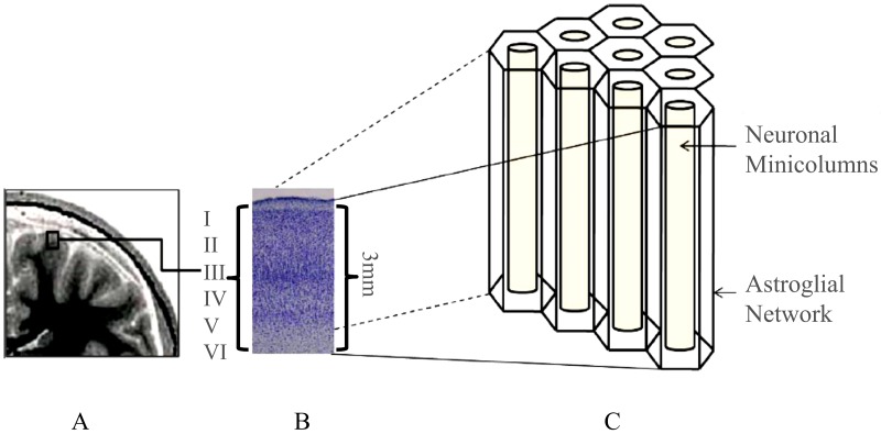
Neural coding (or neural representation) is a neuroscience field concerned with characterising the hypothetical relationship between the stimulus and the neuronal responses, and the relationship among the electrical activities of the neurons in the ensemble. Based on the theory that sensory and other information is represented in the brain by networks of neurons, it is believed that neurons can encode both digital and analog information. Overview Neurons have an ability uncommon among the cells of the body to propagate signals rapidly over large distances by generating characteristic electrical pulses called action potentials: voltage spikes that can travel down axons. Sensory neurons change their activities by firing sequences of action potentials in various temporal patterns, with the presence of external sensory stimuli, such as light, sound, taste, smell and touch. Information about the stimulus is encoded in this pattern of action potentials and transmitted into and arou ... [...More Info...] [...Related Items...] OR: [Wikipedia] [Google] [Baidu] [Amazon] |
Electroencephalography
Electroencephalography (EEG) is a method to record an electrogram of the spontaneous electrical activity of the brain. The biosignal, bio signals detected by EEG have been shown to represent the postsynaptic potentials of pyramidal neurons in the neocortex and allocortex. It is typically non-invasive, with the EEG electrodes placed along the scalp (commonly called "scalp EEG") using the 10–20 system (EEG), International 10–20 system, or variations of it. Electrocorticography, involving surgical placement of electrodes, is sometimes called Electrocorticography, "intracranial EEG". Clinical interpretation of EEG recordings is most often performed by visual inspection of the tracing or quantitative EEG, quantitative EEG analysis. Voltage fluctuations measured by the EEG bioamplifier, bio amplifier and electrodes allow the evaluation of normal Brain activity and meditation, brain activity. As the electrical activity monitored by EEG originates in neurons in the underlying Huma ... [...More Info...] [...Related Items...] OR: [Wikipedia] [Google] [Baidu] [Amazon] |
Saltatory Conduction
In neuroscience, saltatory conduction () is the propagation of action potentials along myelinated axons from one node of Ranvier to the next, increasing the conduction velocity of action potentials. The uninsulated nodes of Ranvier are the only places along the axon where ions are exchanged across the axon membrane, regenerating the action potential between regions of the axon that are insulated by myelin, unlike electrical conduction in a simple circuit. Mechanism Myelinated axons only allow action potentials to occur at the unmyelinated nodes of Ranvier that occur between the myelinated internodes. It is by this restriction that saltatory conduction propagates an action potential along the axon of a neuron at rates significantly higher than would be possible in unmyelinated axons (150 m/s compared from 0.5 to 10 m/s). As sodium rushes into the node it creates an electrical force which pushes on the ions already inside the axon. This rapid conduction of electrical s ... [...More Info...] [...Related Items...] OR: [Wikipedia] [Google] [Baidu] [Amazon] |
Teleostei
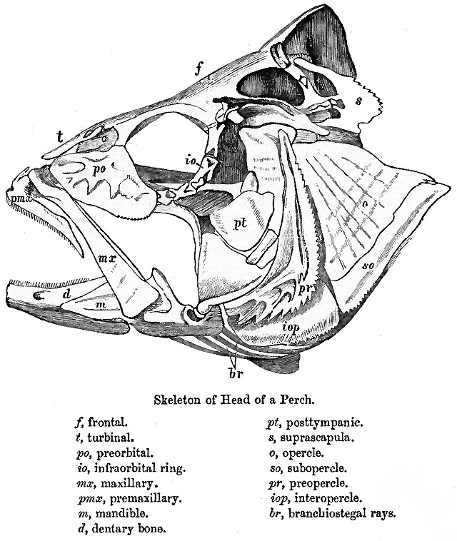
Teleostei (; Ancient Greek, Greek ''teleios'' "complete" + ''osteon'' "bone"), members of which are known as teleosts (), is, by far, the largest group of ray-finned fishes (class Actinopterygii), with 96% of all neontology, extant species of fish. The Teleostei, which is variously considered a Division (zoology), division or an infraclass in different taxonomic systems, include over 26,000 species that are arranged in about 40 order (biology), orders and 448 family (biology), families. Teleosts range from giant oarfish measuring or more, and ocean sunfish weighing over , to the minute male anglerfish ''Photocorynus spiniceps'', just long. Including not only torpedo-shaped fish built for speed, teleosts can be flattened vertically or horizontally, be elongated cylinders or take specialised shapes as in anglerfish and seahorses. The difference between teleosts and other bony fish lies mainly in their jaw bones; teleosts have a movable premaxilla and corresponding modifications ... [...More Info...] [...Related Items...] OR: [Wikipedia] [Google] [Baidu] [Amazon] |
Basket Cell
Basket cells are inhibitory GABAergic interneurons of the brain, found throughout different regions of the cortex and cerebellum. Anatomy and physiology Basket cells are multipolar GABAergic interneurons that function to make inhibitory synapses and control the overall potentials of target cells. In general, dendrites of basket cells are free branching, contain smooth spines, and extend from 3 to 9 mm. Axons are highly branched, ranging in total from 20 to 50mm in total length. The branched axonal arborizations give rise to the name as they appear as baskets surrounding the soma of the target cell. Basket cells form axo-somatic synapses, meaning their synapses target somas of other cells. By controlling the somas of other neurons, basket cells can directly control the action potential discharge rate of target cells. Basket cells can be found throughout the brain, in among other the cortex, hippocampus, amygdala, basal ganglia, and the cerebellum. Cortex In the cortex, basket cel ... [...More Info...] [...Related Items...] OR: [Wikipedia] [Google] [Baidu] [Amazon] |
Purkinje Cell
Purkinje cells or Purkinje neurons, named for Czech physiologist Jan Evangelista Purkyně who identified them in 1837, are a unique type of prominent, large neuron located in the Cerebellum, cerebellar Cortex (anatomy), cortex of the brain. With their flask-shaped cell bodies, many branching Dendrite, dendrites, and a single long axon, these cells are essential for controlling motor activity. Purkinje cells mainly release GABA (gamma-aminobutyric acid) neurotransmitter, which inhibits some neurons to reduce nerve impulse transmission. Purkinje cells efficiently control and coordinate the body's motor motions through these inhibitory actions. Structure These Cell (biology), cells are some of the largest neurons in the human brain (Betz cells being the largest), with an intricately elaborate dendrite, dendritic arbor, characterized by a large number of dendritic spines. Purkinje cells are found within the Purkinje layer in the cerebellum. Purkinje cells are aligned like domi ... [...More Info...] [...Related Items...] OR: [Wikipedia] [Google] [Baidu] [Amazon] |
Cable Theory
In neuroscience, classical cable theory uses mathematical models to calculate the electric current (and accompanying voltage) along passive neurites, particularly the dendrites that receive synaptic inputs at different sites and times. Estimates are made by modeling dendrites and axons as cylinders composed of segments with capacitances c_m and resistances r_m combined in parallel (see Fig. 1). The capacitance of a neuronal fiber comes about because electrostatic forces are acting through the very thin lipid bilayer (see Figure 2). The resistance in series along the fiber r_l is due to the axoplasm's significant resistance to movement of electric charge. History Cable theory in computational neuroscience has roots leading back to the 1850s, when Professor William Thomson (later known as Lord Kelvin) began developing mathematical models of signal decay in submarine (underwater) telegraphic cables. The models resembled the partial differential equations used by Fourier to d ... [...More Info...] [...Related Items...] OR: [Wikipedia] [Google] [Baidu] [Amazon] |
Calyx Of Held
The calyx of Held is a particularly large excitatory synapse in the mammalian auditory nervous system, so named after Hans Held who first described it in his 1893 article ''Die centrale Gehörleitung''Held, H. "Die centrale Gehörleitung" Arch. Anat. Physiol. Anat. Abt, 1893 because of its resemblance to the calyx of a flower. Globular bushy cells in the anteroventral cochlear nucleus (AVCN) send axons to the contralateral medial nucleus of the trapezoid body (MNTB), where they synapse via these calyces on MNTB principal cells. These principal cells then project to the ipsilateral lateral superior olive (LSO), where they inhibit postsynaptic neurons and provide a basis for interaural level detection (ILD), required for high frequency sound localization. This synapse has been described as the largest in the brain. The related endbulb of Held is also a large axon terminal synapse (15–30 μm in diameter) found in another auditory brainstem structure, namely the anterov ... [...More Info...] [...Related Items...] OR: [Wikipedia] [Google] [Baidu] [Amazon] |
Ciliary Ganglion
The ciliary ganglion is a parasympathetic ganglion located just behind the eye in the posterior orbit. It is 1–2 mm in diameter and in humans contains approximately 2,500 neurons. The ganglion contains postganglionic parasympathetic neurons. These neurons supply the pupillary sphincter muscle, which constricts the pupil, and the ciliary muscle which contracts to make the lens more convex. Both of these muscles are involuntary since they are controlled by the parasympathetic division of the autonomic nervous system. The ciliary ganglion is one of four parasympathetic ganglia of the head. The others are the submandibular ganglion, pterygopalatine ganglion, and otic ganglion. Structure The ciliary ganglion contains postganglionic parasympathetic neurons that supply the ciliary muscle and the pupillary sphincter muscle. Because of the much larger size of the ciliary muscle, 95% of the neurons in the ciliary ganglion innervate it compared to the pupillary sphincter. Roots T ... [...More Info...] [...Related Items...] OR: [Wikipedia] [Google] [Baidu] [Amazon] |


