|
Emissary Veins
The emissary veins connect the extracranial venous system with the intracranial venous sinuses. They connect the veins outside the cranium to the venous sinuses inside the cranium. They drain from the scalp, through the skull, into the larger meningeal veins and dural venous sinuses. Emissary veins have an important role in selective cooling of the head. They also serve as routes where infections are carried into the cranial cavity from the extracranial veins to the intracranial veins. There are several types of emissary veins including posterior condyloid, mastoid, occipital and parietal emissary vein. Structure There are also emissary veins passing through the foramen ovale, jugular foramen, foramen lacerum, and hypoglossal canal. Function Because the emissary veins are valveless, they are an important part in selective brain cooling through bidirectional flow of cooler blood from the evaporating surface of the head. In general, blood flow is from external to internal but t ... [...More Info...] [...Related Items...] OR: [Wikipedia] [Google] [Baidu] |
Human Skull
The skull is a bone protective cavity for the brain. The skull is composed of four types of bone i.e., cranial bones, facial bones, ear ossicles and hyoid bone. However two parts are more prominent: the cranium and the mandible. In humans, these two parts are the neurocranium and the viscerocranium ( facial skeleton) that includes the mandible as its largest bone. The skull forms the anterior-most portion of the skeleton and is a product of cephalisation—housing the brain, and several sensory structures such as the eyes, ears, nose, and mouth. In humans these sensory structures are part of the facial skeleton. Functions of the skull include protection of the brain, fixing the distance between the eyes to allow stereoscopic vision, and fixing the position of the ears to enable sound localisation of the direction and distance of sounds. In some animals, such as horned ungulates (mammals with hooves), the skull also has a defensive function by providing the mount (on the front ... [...More Info...] [...Related Items...] OR: [Wikipedia] [Google] [Baidu] |
Cavernous Sinus
The cavernous sinus within the human head is one of the dural venous sinuses creating a cavity called the lateral sellar compartment bordered by the temporal bone of the skull and the sphenoid bone, lateral to the sella turcica. Structure The cavernous sinus is one of the dural venous sinuses of the head. It is a network of veins that sit in a cavity. It sits on both sides of the sphenoidal bone and pituitary gland, approximately 1 × 2 cm in size in an adult. The carotid siphon of the internal carotid artery, and cranial nerves III, IV, V (branches V1 and V2) and VI all pass through this blood filled space. Both sides of cavernous sinus is connected to each other via intercavernous sinuses. The cavernous sinus lies in between the inner and outer layers of dura mater. Nearby structures * Above: optic tract, optic chiasma, internal carotid artery. * Inferiorly: foramen lacerum, and the junction of the body and greater wing of sphenoid bone. * Medially: pituitary gla ... [...More Info...] [...Related Items...] OR: [Wikipedia] [Google] [Baidu] |
Vacuum Extraction
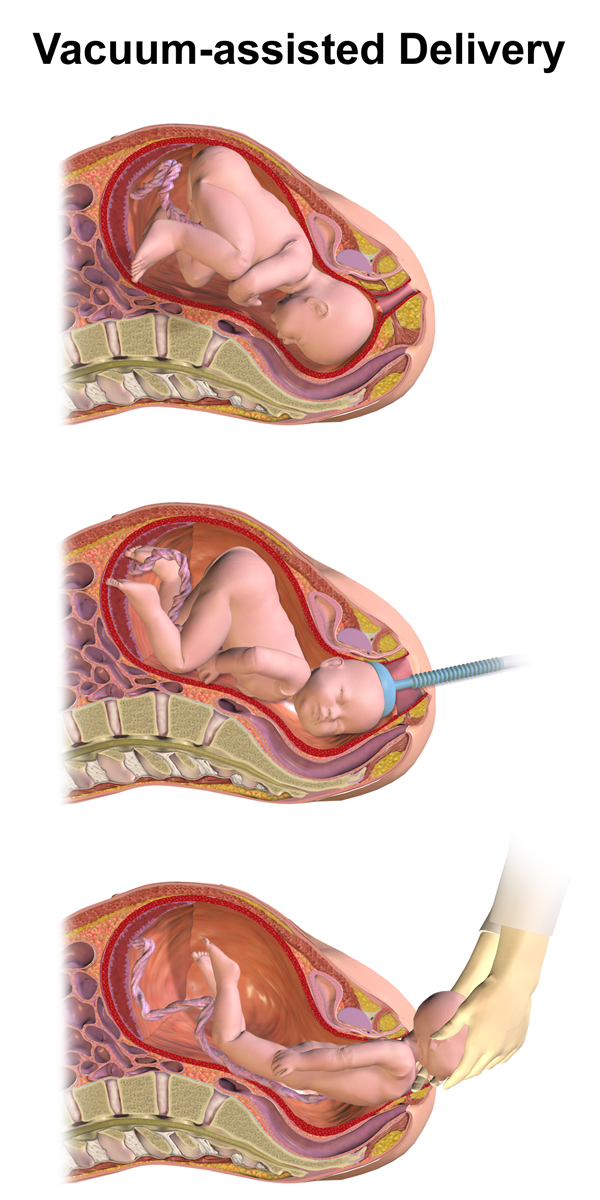
Vacuum extraction (VE), also known as ventouse, is a method to assist delivery of a baby using a vacuum device. It is used in the second stage of labor if it has not progressed adequately. It may be an alternative to a forceps delivery and caesarean section. It cannot be used when the baby is in the breech position or for premature births. The use of VE is generally safe, but it can occasionally have negative effects on either the mother or the child. The term comes from the French word for "suction cup". Medical uses There are several indications to use a vacuum extraction to aid delivery: * Maternal exhaustion * Prolonged second stage of labor * Foetal distress in the second stage of labor, generally indicated by changes in the foetal heart-rate (usually measured on a CTG) * Maternal illness where prolonged "bearing down" or pushing efforts would be risky (e.g. cardiac conditions, blood pressure, aneurysm, glaucoma). If these conditions are known about before the birth, or ... [...More Info...] [...Related Items...] OR: [Wikipedia] [Google] [Baidu] |
Subgaleal Hemorrhage
Subgaleal hemorrhage, also known as subgaleal hematoma, is bleeding in the potential space between the skull periosteum and the scalp galea aponeurosis. Symptoms The diagnosis is generally clinical, with a fluctuant boggy mass developing over the scalp (especially over the occiput) with superficial skin bruising. The swelling develops gradually 12–72 hours after delivery, although it may be noted immediately after delivery in severe cases. Subgaleal hematoma growth is insidious, as it spreads across the whole calvaria and may not be recognized for hours to days. If enough blood accumulates, a visible fluid wave may be seen. Patients may develop periorbital ecchymosis ("raccoon eyes"). Patients with subgaleal hematoma may present with hemorrhagic shock given the volume of blood that can be lost into the potential space between the skull periosteum and the scalp galea aponeurosis, which has been found to be as high as 20-40% of the neonatal blood volume in some studies. The swel ... [...More Info...] [...Related Items...] OR: [Wikipedia] [Google] [Baidu] |
Meningitis
Meningitis is acute or chronic inflammation of the protective membranes covering the brain and spinal cord, collectively called the meninges. The most common symptoms are fever, headache, and neck stiffness. Other symptoms include confusion or altered consciousness, nausea, vomiting, and an inability to tolerate light or loud noises. Young children often exhibit only nonspecific symptoms, such as irritability, drowsiness, or poor feeding. A non-blanching rash (a rash that does not fade when a glass is rolled over it) may also be present. The inflammation may be caused by infection with viruses, bacteria or other microorganisms. Non-infectious causes include malignancy (cancer), subarachnoid haemorrhage, chronic inflammatory disease (sarcoidosis) and certain drugs. Meningitis can be life-threatening because of the inflammation's proximity to the brain and spinal cord; therefore, the condition is classified as a medical emergency. A lumbar puncture, in which a needle is inserte ... [...More Info...] [...Related Items...] OR: [Wikipedia] [Google] [Baidu] |
Cavernous Sinus Thrombosis
The cavernous sinus within the human head is one of the dural venous sinuses creating a cavity called the lateral sellar compartment bordered by the temporal bone of the skull and the sphenoid bone, lateral to the sella turcica. Structure The cavernous sinus is one of the dural venous sinuses of the head. It is a network of veins that sit in a cavity. It sits on both sides of the sphenoidal bone and pituitary gland, approximately 1 × 2 cm in size in an adult. The carotid siphon of the internal carotid artery, and cranial nerves III, IV, V (branches V1 and V2) and VI all pass through this blood filled space. Both sides of cavernous sinus is connected to each other via intercavernous sinuses. The cavernous sinus lies in between the inner and outer layers of dura mater. Nearby structures * Above: optic tract, optic chiasma, internal carotid artery. * Inferiorly: foramen lacerum, and the junction of the body and greater wing of sphenoid bone. * Medially: pituitary gla ... [...More Info...] [...Related Items...] OR: [Wikipedia] [Google] [Baidu] |
Internal Carotid
The internal carotid artery (Latin: arteria carotis interna) is an artery in the neck which supplies the anterior circulation of the brain. In human anatomy, the internal and external carotids arise from the common carotid arteries, where these bifurcate at cervical vertebrae C3 or C4. The internal carotid artery supplies the brain, including the eyes, while the external carotid nourishes other portions of the head, such as the face, scalp, skull, and meninges. Classification Terminologia Anatomica in 1998 subdivided the artery into four parts: "cervical", "petrous", "cavernous", and "cerebral". However, in clinical settings, the classification system of the internal carotid artery usually follows the 1996 recommendations by Bouthillier, describing seven anatomical segments of the internal carotid artery, each with a corresponding alphanumeric identifier—C1 cervical, C2 petrous, C3 lacerum, C4 cavernous, C5 clinoid, C6 ophthalmic, and C7 communicating. The Bouthillier nomenclatu ... [...More Info...] [...Related Items...] OR: [Wikipedia] [Google] [Baidu] |
Cranial Nerve
Cranial nerves are the nerves that emerge directly from the brain (including the brainstem), of which there are conventionally considered twelve pairs. Cranial nerves relay information between the brain and parts of the body, primarily to and from regions of the head and neck, including the special senses of vision, taste, smell, and hearing. The cranial nerves emerge from the central nervous system above the level of the first vertebra of the vertebral column. Each cranial nerve is paired and is present on both sides. There are conventionally twelve pairs of cranial nerves, which are described with Roman numerals I–XII. Some considered there to be thirteen pairs of cranial nerves, including cranial nerve zero. The numbering of the cranial nerves is based on the order in which they emerge from the brain and brainstem, from front to back. The terminal nerves (0), olfactory nerves (I) and optic nerves (II) emerge from the cerebrum, and the remaining ten pairs arise from t ... [...More Info...] [...Related Items...] OR: [Wikipedia] [Google] [Baidu] |
Pterygoid Plexus
The pterygoid plexus (; in Merriam-Webster Online Dictionary '. from ''pteryx'', "wing" and ''eidos'', "shape") is a of considerable size, and is situated between the and |
Meningeal Vein
In neuroanatomy, dura mater is a thick membrane made of dense irregular connective tissue that surrounds the brain and spinal cord. It is the outermost of the three layers of membrane called the meninges that protect the central nervous system. The other two meningeal layers are the arachnoid mater and the pia mater. It envelops the arachnoid mater, which is responsible for keeping in the cerebrospinal fluid. It is derived primarily from the neural crest cell population, with postnatal contributions of the paraxial mesoderm. Structure The dura mater has several functions and layers. The dura mater is a membrane that envelops the arachnoid mater. It surrounds and supports the dural sinuses (also called dural venous sinuses, cerebral sinuses, or cranial sinuses) and carries blood from the brain toward the heart. Cranial dura mater has two layers called ''lamellae'', a superficial layer (also called the periosteal layer), which serves as the skull's inner periosteum, called the end ... [...More Info...] [...Related Items...] OR: [Wikipedia] [Google] [Baidu] |
Zygomatic Arch
In anatomy, the zygomatic arch, or cheek bone, is a part of the skull formed by the zygomatic process of the temporal bone (a bone extending forward from the side of the skull, over the opening of the ear) and the temporal process of the zygomatic bone (the side of the cheekbone), the two being united by an oblique suture (the zygomaticotemporal suture); the tendon of the temporal muscle passes medial to (i.e. through the middle of) the arch, to gain insertion into the coronoid process of the mandible (jawbone). The jugal point is the point at the anterior (towards face) end of the upper border of the zygomatic arch where the masseteric and maxillary edges meet at an angle, and where it meets the process of the zygomatic bone. The arch is typical of '' Synapsida'' (“fused arch”), a clade of amniotes that includes mammals and their extinct relatives, such as ''Moschops'' and '' Dimetrodon''. Structure The zygomatic process of the temporal arises by two roots: * an ''anter ... [...More Info...] [...Related Items...] OR: [Wikipedia] [Google] [Baidu] |
Sphenoidal Emissary Foramen
In the base of the skull, in the great wings of the sphenoid bone, medial to the foramen ovale, a small aperture, the sphenoidal emissary foramen, may occasionally be seen (it is often absent) opposite the root of the pterygoid process. When present, it opens below near the scaphoid fossa. Vesalius was the first to describe and illustrate this foramen, and it thus sometimes bears the name of foramen Vesalii (meaning foramen of Vesalius). Other names include foramen venosum and canaliculus sphenoidalis. Importance If at all present, the sphenoidal emissary foramen gives passage to a small vein (vein of Vesalius) that connects the pterygoid plexus with the cavernous sinus. The importance of this passage lies in the fact that an infected thrombus from an extracranial source may reach the cavernous sinus. The mean area of the foramen is small, which may suggest that it plays a minor role in the dynamics of blood circulation in the venous system of the head. Structure The sphenoidal ... [...More Info...] [...Related Items...] OR: [Wikipedia] [Google] [Baidu] |


