|
Embryonal Carcinoma
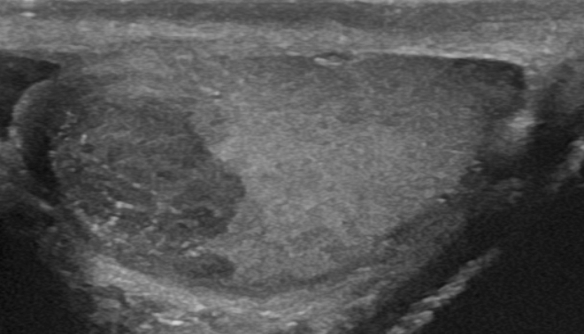
Embryonal carcinoma is a relatively uncommon type of germ cell tumour that occurs in the ovaries and testes. Signs and symptoms The presenting features may be a palpable testicular mass or asymmetric testicular enlargement in some cases. The tumour may present as signs and symptoms relating to the presence of widespread metastases, without any palpable lump in the testis. The clinical features associated with metastasising embryonal carcinoma may include low back pain, dyspnoea, cough, haemoptysis, haematemesis and neurologic abnormalities. Males with pure embryonal carcinoma tend to have a normal amount of the protein alpha-fetoprotein in the fluid component of their blood. The finding of elevated amounts of alpha-fetoprotein is more suggestive of a mixed germ cell tumour, with the alpha-fetoprotein being released by the yolk sac tumour component. Diagnosis The gross examination usually shows a two to three centimetre pale grey, poorly defined tumour with associated haemor ... [...More Info...] [...Related Items...] OR: [Wikipedia] [Google] [Baidu] |
Micrograph
A micrograph or photomicrograph is a photograph or digital image taken through a microscope or similar device to show a magnified image of an object. This is opposed to a macrograph or photomacrograph, an image which is also taken on a microscope but is only slightly magnified, usually less than 10 times. Micrography is the practice or art of using microscopes to make photographs. A micrograph contains extensive details of microstructure. A wealth of information can be obtained from a simple micrograph like behavior of the material under different conditions, the phases found in the system, failure analysis, grain size estimation, elemental analysis and so on. Micrographs are widely used in all fields of microscopy. Types Photomicrograph A light micrograph or photomicrograph is a micrograph prepared using an optical microscope, a process referred to as ''photomicroscopy''. At a basic level, photomicroscopy may be performed simply by connecting a camera to a microscope, th ... [...More Info...] [...Related Items...] OR: [Wikipedia] [Google] [Baidu] |
Embryonal Carcinoma - High Mag
An embryo is an initial stage of development of a multicellular organism. In organisms that reproduce sexually, embryonic development is the part of the life cycle that begins just after fertilization of the female egg cell by the male sperm cell. The resulting fusion of these two cells produces a single-celled zygote that undergoes many cell divisions that produce cells known as blastomeres. The blastomeres are arranged as a solid ball that when reaching a certain size, called a morula, takes in fluid to create a cavity called a blastocoel. The structure is then termed a blastula, or a blastocyst in mammals. The mammalian blastocyst hatches before implantating into the endometrial lining of the womb. Once implanted the embryo will continue its development through the next stages of gastrulation, neurulation, and organogenesis. Gastrulation is the formation of the three germ layers that will form all of the different parts of the body. Neurulation forms the nervous system, an ... [...More Info...] [...Related Items...] OR: [Wikipedia] [Google] [Baidu] |
Yolk Sac Tumor
Endodermal sinus tumor (EST) is a member of the germ cell tumor group of cancers. It is the most common testicular tumor in children under three, and is also known as infantile embryonal carcinoma. This age group has a very good prognosis. In contrast to the pure form typical of infants, adult endodermal sinus tumors are often found in combination with other kinds of germ cell tumor, particularly teratoma and embryonal carcinoma. While pure teratoma is usually benign, endodermal sinus tumor is malignant. Cause Causes for this cancer are poorly understood. Diagnosis The histology of EST is variable, but usually includes malignant endodermal cells. These cells secrete alpha-fetoprotein (AFP), which can be detected in tumor tissue, serum, cerebrospinal fluid, urine and, in the rare case of fetal EST, in amniotic fluid. When there is incongruence between biopsy and AFP test results for EST, the result indicating presence of EST dictates treatment. This is because EST often occurs as s ... [...More Info...] [...Related Items...] OR: [Wikipedia] [Google] [Baidu] |
Teratocarcinoma
Germ cell tumor (GCT) is a neoplasm derived from germ cells. Germ-cell tumors can be cancerous or benign. Germ cells normally occur inside the gonads (ovary and testis). GCTs that originate outside the gonads may be birth defects resulting from errors during development of the embryo. Classification GCTs are classified by their histology, regardless of location in the body. However, as more information about the genetics of these tumors become available, they may be classified based on specific gene mutations that characterize specific tumors. They are broadly divided in two classes: * The germinomatous or seminomatous germ-cell tumors (GGCT, SGCT) include only germinoma and its synonyms dysgerminoma and seminoma. * The nongerminomatous or nonseminomatous germ-cell tumors (NGGCT, NSGCT) include all other germ-cell tumors, pure and mixed. The two classes reflect an important clinical difference. Compared with germinomatous tumors, nongerminomatous tumors tend to grow faster, have ... [...More Info...] [...Related Items...] OR: [Wikipedia] [Google] [Baidu] |
Seminoma
A seminoma is a germ cell tumor of the testicle or, more rarely, the mediastinum or other extra-gonadal locations. It is a Malignancy, malignant neoplasm and is one of the most treatable and curable cancers, with a survival rate above 95% if discovered in early stages. Testicular seminoma originates in the Germinal epithelium (male), germinal epithelium of the seminiferous tubules. About half of germ cell tumors of the testicles are seminomas. Treatment usually requires removal of one testicle. However, fertility usually isn't affected. All other sexual functions will remain intact. Signs and symptoms The average age of diagnosis is between 35 and 50 years. This is about 5 to 10 years older than men with other germ cell tumors of the testes. In most cases, they produce masses that are readily felt on testicular self-examination; however, in up to 11 percent of cases, there may be no mass able to be felt, or there may be testicular atrophy. Testicular pain is reported in up to ... [...More Info...] [...Related Items...] OR: [Wikipedia] [Google] [Baidu] |
Metastases
Metastasis is a pathogenic agent's spread from an initial or primary site to a different or secondary site within the host's body; the term is typically used when referring to metastasis by a cancerous tumor. The newly pathological sites, then, are metastases (mets). It is generally distinguished from cancer invasion, which is the direct extension and penetration by cancer cells into neighboring tissues. Cancer occurs after cells are genetically altered to proliferate rapidly and indefinitely. This uncontrolled proliferation by mitosis produces a primary heterogeneic tumour. The cells which constitute the tumor eventually undergo metaplasia, followed by dysplasia then anaplasia, resulting in a malignant phenotype. This malignancy allows for invasion into the circulation, followed by invasion to a second site for tumorigenesis. Some cancer cells known as circulating tumor cells acquire the ability to penetrate the walls of lymphatic or blood vessels, after which they are abl ... [...More Info...] [...Related Items...] OR: [Wikipedia] [Google] [Baidu] |
Histology
Histology, also known as microscopic anatomy or microanatomy, is the branch of biology which studies the microscopic anatomy of biological tissues. Histology is the microscopic counterpart to gross anatomy, which looks at larger structures visible without a microscope. Although one may divide microscopic anatomy into ''organology'', the study of organs, ''histology'', the study of tissues, and ''cytology'', the study of cells, modern usage places all of these topics under the field of histology. In medicine, histopathology is the branch of histology that includes the microscopic identification and study of diseased tissue. In the field of paleontology, the term paleohistology refers to the histology of fossil organisms. Biological tissues Animal tissue classification There are four basic types of animal tissues: muscle tissue, nervous tissue, connective tissue, and epithelial tissue. All animal tissues are considered to be subtypes of these four principal tissue types ... [...More Info...] [...Related Items...] OR: [Wikipedia] [Google] [Baidu] |
Alpha Fetoprotein
Alpha-fetoprotein (AFP, α-fetoprotein; also sometimes called alpha-1-fetoprotein, alpha-fetoglobulin, or alpha fetal protein) is a protein that in humans is encoded by the ''AFP'' gene. The ''AFP'' gene is located on the ''q'' arm of chromosome 4 (4q25). Maternal AFP serum level is used to screen for Down syndrome, neural tube defects, and other chromosomal abnormalities. AFP is a major plasma protein produced by the yolk sac and the fetal liver during fetal development. It is thought to be the fetal analog of serum albumin. AFP binds to copper, nickel, fatty acids and bilirubin and is found in monomeric, dimeric and trimeric forms. Structure AFP is a glycoprotein of 591 amino acids and a carbohydrate moiety. Function The function of AFP in adult humans is unknown. AFP is the most abundant plasma protein found in the human fetus. Maternal plasma levels peak near the end of the first trimester, and begin decreasing prenatally at that time, then decrease rapidly after birt ... [...More Info...] [...Related Items...] OR: [Wikipedia] [Google] [Baidu] |
Human Chorionic Gonadotropin
Human chorionic gonadotropin (hCG) is a hormone for the maternal recognition of pregnancy produced by trophoblast cells that are surrounding a growing embryo (syncytiotrophoblast initially), which eventually forms the placenta after implantation. The presence of hCG is detected in some pregnancy tests (HCG pregnancy strip tests). Some cancerous tumors produce this hormone; therefore, elevated levels measured when the patient is not pregnant may lead to a cancer diagnosis and, if high enough, paraneoplastic syndromes, however, it is not known whether this production is a contributing cause, or an effect of carcinogenesis. The pituitary analog of hCG, known as luteinizing hormone (LH), is produced in the pituitary gland of males and females of all ages. Various endogenous forms of hCG exist. The measurement of these diverse forms is used in the diagnosis of pregnancy and a variety of disease states. Preparations of hCG from various sources have also been used therapeutically, by ... [...More Info...] [...Related Items...] OR: [Wikipedia] [Google] [Baidu] |
Median
In statistics and probability theory, the median is the value separating the higher half from the lower half of a data sample, a population, or a probability distribution. For a data set, it may be thought of as "the middle" value. The basic feature of the median in describing data compared to the mean (often simply described as the "average") is that it is not skewed by a small proportion of extremely large or small values, and therefore provides a better representation of a "typical" value. Median income, for example, may be a better way to suggest what a "typical" income is, because income distribution can be very skewed. The median is of central importance in robust statistics, as it is the most resistant statistic, having a breakdown point of 50%: so long as no more than half the data are contaminated, the median is not an arbitrarily large or small result. Finite data set of numbers The median of a finite list of numbers is the "middle" number, when those numbers are list ... [...More Info...] [...Related Items...] OR: [Wikipedia] [Google] [Baidu] |
Embryoma
Embryoma is a mass of rapidly growing cells believed to originate in embryonic (fetal) tissue. Embryonal tumors may be benign or malignant, and include neuroblastomas and Wilms tumor Wilms' tumor or Wilms tumor, also known as nephroblastoma, is a cancer of the kidneys that typically occurs in children, rarely in adults.; and occurs most commonly as a renal tumor in child patients. It is named after Max Wilms, the German sur ...s. Also called embryoma. Embryomas have been defined as: "Adult neoplasms expressing one or more embryo-exclusive genes." Embryomas can appear in the lungs. It is not a precise term, and it is not commonly used in modern medical literature. Embryomas have been defined as: "Adult neoplasms expressing one or more embryo-exclusive genes". References External links Embryonal tumorentry in the public domain NCI Dictionary of Cancer Terms Male genital neoplasia {{oncology-stub ... [...More Info...] [...Related Items...] OR: [Wikipedia] [Google] [Baidu] |
Polyembryoma
Polyembryoma is a rare, very aggressive form of germ cell tumor usually found in the ovaries. Polyembryoma has features of both yolk sac tumour and undifferentiated teratoma/embryonal carcinoma, with a characteristic finding of embryoid bodies lying in a loose mesenchymal stroma. It has been found in association with Klinefelter syndrome Klinefelter syndrome (KS), also known as 47,XXY, is an aneuploid genetic condition where a male has an additional copy of the X chromosome. The primary features are infertility and small, poorly functioning testicles. Usually, symptoms are su .... References External links Germ cell neoplasia Gynaecological neoplasia {{oncology-stub ... [...More Info...] [...Related Items...] OR: [Wikipedia] [Google] [Baidu] |


