|
Dystrophic Calcification
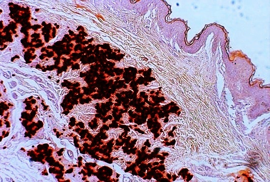
Dystrophic calcification (DC) is the calcification occurring in degenerated or necrotic tissue, as in hyalinized scars, degenerated foci in leiomyomas, and caseous nodules. This occurs as a reaction to tissue damage, including as a consequence of medical device implantation. Dystrophic calcification can occur even if the amount of calcium in the blood is not elevated (a systemic mineral imbalance would elevate calcium levels in the blood and all tissues) and cause metastatic calcification. Basophilic calcium salt deposits aggregate, first in the mitochondria, then progressively throughout the cell. These calcifications are an indication of previous microscopic cell injury, occurring in areas of cell necrosis when activated phosphatases bind calcium ions to phospholipids in the membrane. Calcification in dead tissue #Caseous necrosis in T.B. is most common site of dystrophic calcification. #Liquefactive necrosis in chronic abscesses may get calcified. #Fat necrosis following acu ... [...More Info...] [...Related Items...] OR: [Wikipedia] [Google] [Baidu] |
Schistosoma
''Schistosoma'' is a genus of trematodes, commonly known as blood flukes. They are parasitic flatworms responsible for a highly significant group of infections in humans termed '' schistosomiasis'', which is considered by the World Health Organization as the second-most socioeconomically devastating parasitic disease (after malaria), with hundreds of millions infected worldwide. Adult flatworms parasitize blood capillaries of either the mesenteries or plexus of the bladder, depending on the infecting species. They are unique among trematodes and any other flatworms in that they are dioecious with distinct sexual dimorphism between male and female. Thousands of eggs are released and reach either the bladder or the intestine (according to the infecting species), and these are then excreted in urine or feces to fresh water. Larvae must then pass through an intermediate snail host, before the next larval stage of the parasite emerges that can infect a new mammalian host by directl ... [...More Info...] [...Related Items...] OR: [Wikipedia] [Google] [Baidu] |
Calcinosis
Calcinosis is the formation of calcium deposits in any soft tissue. It is a rare condition that has many different causes. These range from infection and injury to systemic diseases like kidney failure. Types Dystrophic calcification The most common type of calcinosis is dystrophic calcification. This type of calcification can occur as a response to any soft tissue damage, including that involved in implantation of medical devices. Metastatic calcification Metastatic calcification involves a systemic calcium excess imbalance, which can be caused by hypercalcemia, kidney failure, milk-alkali syndrome, lack or excess of other minerals, or other causes. Tumoral calcinosis The cause of the rare condition of tumoral calcinosis is not entirely understood. It is generally characterized by large, globular calcifications near joints. See also * Calcification * Calcinosis cutis * Dermatomyositis * Fahr's syndrome * Hyperphosphatemia * Primrose syndrome * Scleroderma Scleroderma is ... [...More Info...] [...Related Items...] OR: [Wikipedia] [Google] [Baidu] |
Bruch's Membrane
Bruch's membrane is the innermost layer of the choroid of the eye. It is also called the ''vitreous lamina'' or ''Membrane vitriae'', because of its glassy microscopic appearance. It is 2–4 μm thick. Layers Bruch's membrane consists of five layers (from inside to outside): #the basement membrane of the retinal pigment epithelium #the inner collagenous zone #a central band of elastic fibers #the outer collagenous zone #the basement membrane of the choriocapillaris The retinal pigment epithelium transports metabolic waste from the photoreceptors across Bruch's membrane to the choroid. Embryology Bruch's membrane is present by midterm in fetal development as an elastic sheet. Pathology Bruch's membrane thickens with age, slowing the transport of metabolites. This may lead to the formation of drusen in age-related macular degeneration. There is also a buildup of deposits (Basal Linear Deposits or BLinD and Basal Lamellar Deposits BLamD) on and within the membrane, primarily co ... [...More Info...] [...Related Items...] OR: [Wikipedia] [Google] [Baidu] |
Angioid Streaks
Angioid streaks, also called Knapp streaks or Knapp striae, are small breaks in Bruch's membrane, an elastic tissue containing membrane of the retina that may become calcified and crack. Up to 50% of angioid streak cases are idiopathic. It may occur secondary to blunt trauma, or it may be associated with many systemic diseases. The condition is usually asymptomatic, but decrease in vision may occur due to choroidal neovascularization. Clinical features Angioid streaks are often associated with pseudoxanthoma elasticum, but have been found to occur in conjunction with other disorders, including Paget's disease, sickle cell disease and Ehlers–Danlos syndrome. These streaks can have a negative impact on vision due to choroidal neovascularization or choroidal rupture. Also, vision can be impaired if the streaks progress to the fovea and damage the retinal pigment epithelium. Signs Retinal fundus examination may reveal grey or dark red spoke like lesions around optic disk and radi ... [...More Info...] [...Related Items...] OR: [Wikipedia] [Google] [Baidu] |
Pseudoxanthoma Elasticum
Pseudoxanthoma elasticum (PXE) is a genetic disease that causes mineralization of elastic fibers in some tissues. The most common problems arise in the skin and eyes, and later in blood vessels in the form of premature atherosclerosis. PXE is caused by autosomal recessive mutations in the '' ABCC6'' gene on the short arm of chromosome 16 (16p13.1). Signs and symptoms Usually, pseudoxanthoma elasticum affects the skin first, often in childhood or early adolescence. Small, yellowish papular lesions form and cutaneous laxity mainly affect the neck, axillae (armpits), groin, and flexural creases (the inside parts of the elbows and knees). Skin may become lax and redundant. Many individuals have "oblique mental creases" (horizontal grooves of the chin) PXE first affects the retina through a dimpling of the Bruch membrane (a thin membrane separating the blood vessel-rich layer from the pigmented layer of the retina), that is only visible during ophthalmologic examinations. This is ca ... [...More Info...] [...Related Items...] OR: [Wikipedia] [Google] [Baidu] |
Calcinosis Cutis
Calcinosis cutis is a type of calcinosis wherein calcium deposits form in the skin. A variety of factors can result in this condition. The most common source is dystrophic calcification, which occurs in soft tissue as a response to injury. In addition, calcinosis is seen in Limited Cutaneous Systemic Sclerosis, also known as CREST syndrome (the "C" in CREST). In dogs, calcinosis cutis is found in young, large breed dogs and is thought to occur after a traumatic injury. Causes Calcinosis may result from a variety of causes such as: *Trauma to the region *Inflammation (bug bites, acne) *Varicose veins *Infections *Tumors (malignant or benign) *Diseases of connective tissue *Hypercalcemia *Hyperphosphatemia Calcinosis cutis is associated with systemic sclerosis. Diagnosis Types Calcinosis cutis may be divided into the following types: * Dystrophic calcinosis cutis * Metastatic calcinosis cutis * Iatrogenic calcinosis cutis * Traumatic calcinosis cutis * Idiopathic calcino ... [...More Info...] [...Related Items...] OR: [Wikipedia] [Google] [Baidu] |
Cyst
A cyst is a closed sac, having a distinct envelope and cell division, division compared with the nearby Biological tissue, tissue. Hence, it is a cluster of Cell (biology), cells that have grouped together to form a sac (like the manner in which water molecules group together to form a bubble); however, the distinguishing aspect of a cyst is that the cells forming the "shell" of such a sac are distinctly abnormal (in both appearance and behaviour) when compared with all surrounding cells for that given location. A cyst may contain air, fluids, or semi-solid material. A collection of pus is called an abscess, not a cyst. Once formed, a cyst may resolve on its own. When a cyst fails to resolve, it may need to be removed surgically, but that would depend upon its type and location. Cancer-related cysts are formed as a defense mechanism for the body following the development of mutations that lead to an uncontrolled cellular division. Once that mutation has occurred, the affected cell ... [...More Info...] [...Related Items...] OR: [Wikipedia] [Google] [Baidu] |
Aorta
The aorta ( ) is the main and largest artery in the human body, originating from the left ventricle of the heart and extending down to the abdomen, where it splits into two smaller arteries (the common iliac arteries). The aorta distributes oxygenated blood to all parts of the body through the systemic circulation. Structure Sections In anatomical sources, the aorta is usually divided into sections. One way of classifying a part of the aorta is by anatomical compartment, where the thoracic aorta (or thoracic portion of the aorta) runs from the heart to the diaphragm. The aorta then continues downward as the abdominal aorta (or abdominal portion of the aorta) from the diaphragm to the aortic bifurcation. Another system divides the aorta with respect to its course and the direction of blood flow. In this system, the aorta starts as the ascending aorta, travels superiorly from the heart, and then makes a hairpin turn known as the aortic arch. Following the aortic arch ... [...More Info...] [...Related Items...] OR: [Wikipedia] [Google] [Baidu] |
Atheroma
An atheroma, or atheromatous plaque, is an abnormal and reversible accumulation of material in the inner layer of an arterial wall. The material consists of mostly macrophage cells, or debris, containing lipids, calcium and a variable amount of fibrous connective tissue. The accumulated material forms a swelling in the artery wall, which may intrude into the lumen of the artery, narrowing it and restricting blood flow. Atheroma is the pathological basis for the disease entity atherosclerosis, a subtype of arteriosclerosis. Signs and symptoms For most people, the first symptoms result from atheroma progression within the heart arteries, most commonly resulting in a heart attack and ensuing debility. The heart arteries are difficult to track because they are small (from about 5 mm down to microscopic), they are hidden deep within the chest and they never stop moving. Additionally, all mass-applied clinical strategies focus on both minimal cost and the overall safety of ... [...More Info...] [...Related Items...] OR: [Wikipedia] [Google] [Baidu] |
Hyaline
A hyaline substance is one with a glassy appearance. The word is derived from el, ὑάλινος, translit=hyálinos, lit=transparent, and el, ὕαλος, translit=hýalos, lit=crystal, glass, label=none. Histopathology Hyaline cartilage is named after its glassy appearance on fresh gross pathology. On light microscopy of H&E stained slides, the extracellular matrix of hyaline cartilage looks homogeneously pink, and the term "hyaline" is used to describe similarly homogeneously pink material besides the cartilage. Hyaline material is usually acellular and proteinaceous. For example, arterial hyaline is seen in aging, high blood pressure, diabetes mellitus and in association with some drugs (e.g. calcineurin inhibitors). It is bright pink with PAS staining. Ichthyology and entomology In ichthyology and entomology, ''hyaline'' denotes a colorless, transparent substance, such as unpigmented fins of fishes or clear insect wings. Resh, Vincent H. and R. T. Cardé, Eds. Encyclo ... [...More Info...] [...Related Items...] OR: [Wikipedia] [Google] [Baidu] |
Cardiovascular Calcification - Sergio Bertazzo
The blood circulatory system is a system of organs that includes the heart, blood vessels, and blood which is circulated throughout the entire body of a human or other vertebrate. It includes the cardiovascular system, or vascular system, that consists of the heart and blood vessels (from Greek ''kardia'' meaning ''heart'', and from Latin ''vascula'' meaning ''vessels''). The circulatory system has two divisions, a systemic circulation or circuit, and a pulmonary circulation or circuit. Some sources use the terms ''cardiovascular system'' and ''vascular system'' interchangeably with the ''circulatory system''. The network of blood vessels are the great vessels of the heart including large elastic arteries, and large veins; other arteries, smaller arterioles, capillaries that join with venules (small veins), and other veins. The circulatory system is closed in vertebrates, which means that the blood never leaves the network of blood vessels. Some invertebrates such as arthr ... [...More Info...] [...Related Items...] OR: [Wikipedia] [Google] [Baidu] |


