|
Dissecting Aneurysm
A dissection is a tear within the wall of a blood vessel, which allows blood to separate the wall layers. Usually, a dissection is an arterial wall dissection, but vein wall dissections (VWD) have been documented. By separating a portion of the wall of the artery (a layer of the tunica media or in some cases tunica intima), a dissection creates two lumens or passages within the vessel, the native or true lumen, and the "false lumen" created by the new space within the wall of the artery. It is not yet clear if the tear in the innermost layer, the tunica intima, is secondary to the tear in the tunica media. Dissections originating in the tunica media are caused by disruption of the vasa vasorum. It is thought that dysfunction in the vasa vasorum is an underlying cause of dissections. Description Dissections become threatening to the health of the organism when growth of the false lumen prevents perfusion of the true lumen and the end organs perfused by the true lumen. For exampl ... [...More Info...] [...Related Items...] OR: [Wikipedia] [Google] [Baidu] |
Vascular Surgery
Vascular surgery is a surgical subspecialty in which diseases of the vascular system, or arteries, veins and lymphatic circulation, are managed by medical therapy, minimally-invasive catheter procedures and surgical reconstruction. The specialty evolved from general and cardiac surgery and includes treatment of the body's other major and essential veins and arteries. Open surgery techniques, as well as endovascular techniques are used to treat vascular diseases. The vascular surgeon is trained in the diagnosis and management of diseases affecting all parts of the vascular system excluding the coronaries and intracranial vasculature. Vascular surgeons often assist other physicians to address traumatic vascular injury, hemorrhage control, and safe exposure of vascular structures. History Early leaders of the field included Russian surgeon Nikolai Korotkov, noted for developing early surgical techniques, American interventional radiologist Charles Theodore Dotter who is credited ... [...More Info...] [...Related Items...] OR: [Wikipedia] [Google] [Baidu] |
Coronary Artery Dissection
Spontaneous coronary artery dissection (SCAD) is an uncommon but potentially lethal condition in which one of the arteries that supply the heart spontaneously develops a blood collection, or hematoma, within the artery wall. This leads to a separation. SCAD is member of the vascular dissection (medical) disease family. SCAD is a major cause of heart attacks in young, otherwise healthy women who usually lacking typical cardiovascular risk factors. While the exact cause is not yet known, SCAD is likely related to changes that occur during and after pregnancy, as well as other diseases. These changes lead to the dissection of the wall which restricts blood flow to the heart and causes symptoms. SCAD is often diagnosed in the cath lab with angiography, though more advanced confirmatory tests exist. While the risk of death due to SCAD is low, it has a relatively high rate of recurrence leading to further heart attack-like symptoms in the future. It was first described in 1931. Signs ... [...More Info...] [...Related Items...] OR: [Wikipedia] [Google] [Baidu] |
Collagen
Collagen () is the main structural protein in the extracellular matrix found in the body's various connective tissues. As the main component of connective tissue, it is the most abundant protein in mammals, making up from 25% to 35% of the whole-body protein content. Collagen consists of amino acids bound together to form a triple helix of elongated fibril known as a collagen helix. It is mostly found in connective tissue such as cartilage, bones, tendons, ligaments, and skin. Depending upon the degree of mineralization, collagen tissues may be rigid (bone) or compliant (tendon) or have a gradient from rigid to compliant (cartilage). Collagen is also abundant in corneas, blood vessels, the gut, intervertebral discs, and the dentin in teeth. In muscle tissue, it serves as a major component of the endomysium. Collagen constitutes one to two percent of muscle tissue and accounts for 6% of the weight of the skeletal muscle tissue. The fibroblast is the most common cell tha ... [...More Info...] [...Related Items...] OR: [Wikipedia] [Google] [Baidu] |
Vascular Smooth Muscle Cell
Vascular smooth muscle is the type of smooth muscle that makes up most of the walls of blood vessels. Structure Vascular smooth muscle refers to the particular type of smooth muscle found within, and composing the majority of the wall of blood vessels. Nerve supply Vascular smooth muscle is innervated primarily by the sympathetic nervous system through adrenergic receptors (adrenoceptors). The three types present are: Alpha-1 adrenergic receptor, alpha-1, Alpha-2 adrenergic receptor, alpha-2 and beta-2 adrenergic receptors, . The main endogeny, endogenous agonist of these cell receptors is norepinephrine (NE). The adrenergic receptors exert opposite physiologic effects in the vascular smooth muscle under activation: * alpha-1 receptors. Under NE binding alpha-1 receptors cause vasoconstriction ( contraction of the vascular smooth muscle cells decreasing the diameter of the vessels). Thesea receptors are activated in response to shock or low blood pressure as a defensive reac ... [...More Info...] [...Related Items...] OR: [Wikipedia] [Google] [Baidu] |
Extracellular Matrix
In biology, the extracellular matrix (ECM), also called intercellular matrix, is a three-dimensional network consisting of extracellular macromolecules and minerals, such as collagen, enzymes, glycoproteins and hydroxyapatite that provide structural and biochemical support to surrounding cells. Because multicellularity evolved independently in different multicellular lineages, the composition of ECM varies between multicellular structures; however, cell adhesion, cell-to-cell communication and differentiation are common functions of the ECM. The animal extracellular matrix includes the interstitial matrix and the basement membrane. Interstitial matrix is present between various animal cells (i.e., in the intercellular spaces). Gels of polysaccharides and fibrous proteins fill the interstitial space and act as a compression buffer against the stress placed on the ECM. Basement membranes are sheet-like depositions of ECM on which various epithelial cells rest. Each type of conn ... [...More Info...] [...Related Items...] OR: [Wikipedia] [Google] [Baidu] |
TGF-β
Transforming growth factor beta (TGF-β) is a multifunctional cytokine belonging to the transforming growth factor superfamily that includes three different mammalian isoforms (TGF-β 1 to 3, HGNC symbols TGFB1, TGFB2, TGFB3) and many other signaling proteins. TGFB proteins are produced by all white blood cell lineages. Activated TGF-β complexes with other factors to form a serine/threonine kinase complex that binds to TGF-β receptors. TGF-β receptors are composed of both type 1 and type 2 receptor subunits. After the binding of TGF-β, the type 2 receptor kinase phosphorylates and activates the type 1 receptor kinase that activates a signaling cascade. This leads to the activation of different downstream substrates and regulatory proteins, inducing transcription of different target genes that function in differentiation, chemotaxis, proliferation, and activation of many immune cells. TGF-β is secreted by many cell types, including macrophages, in a latent form in whi ... [...More Info...] [...Related Items...] OR: [Wikipedia] [Google] [Baidu] |
Cervical Artery Dissection (other)
Cervical artery dissection is dissection of one of the layers that compose the carotid and vertebral artery in the neck (cervix). They include: * Carotid artery dissection, a separation of the layers of the artery wall supplying oxygen-bearing blood to the head and brain. * Vertebral artery dissection, a flap-like tear of the inner lining of the vertebral artery that supply blood to the brain and spinal cord. Cervical dissections can be broadly classified as either "spontaneous" or traumatic. Cervical artery dissections are a significant cause of strokes in young adults. A dissection typically results in a tear in one of the layers of the arterial wall. The result of this tear is often an intramural hematoma and/or aneurysmal dilation in the arteries leading to the intracranial area. Signs and symptoms of a cervical artery dissection are often non-specific and can be localized or generalized. There is no specific treatment, although most patients are either given an anti-platelet ... [...More Info...] [...Related Items...] OR: [Wikipedia] [Google] [Baidu] |
Vertebral Artery
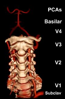
The vertebral arteries are major arteries of the neck. Typically, the vertebral arteries originate from the subclavian arteries. Each vessel courses superiorly along each side of the neck, merging within the skull to form the single, midline basilar artery. As the supplying component of the ''vertebrobasilar vascular system'', the vertebral arteries supply blood to the upper spinal cord, brainstem, cerebellum, and posterior part of brain. Structure The vertebral arteries usually arise from the posterosuperior aspect of the central subclavian arteries on each side of the body, then enter deep to the transverse process at the level of the 6th cervical vertebrae (C6), or occasionally (in 7.5% of cases) at the level of C7. They then proceed superiorly, in the transverse foramen of each cervical vertebra. Once they have passed through the transverse foramen of C1 (also known as the atlas), the vertebral arteries travel across the posterior arch of C1 and through the suboccip ... [...More Info...] [...Related Items...] OR: [Wikipedia] [Google] [Baidu] |
Vertebral Artery Dissection
Vertebral artery dissection (VAD) is a flap-like tear of the inner lining of the vertebral artery, which is located in the neck and supplies blood to the brain. After the tear, blood enters the arterial wall and forms a blood clot, thickening the artery wall and often impeding blood flow. The symptoms of vertebral artery dissection include head and neck pain and intermittent or permanent stroke symptoms such as difficulty speaking, impaired coordination and visual loss. It is usually diagnosed with a contrast-enhanced CT or MRI scan. Vertebral dissection may occur after physical trauma to the neck, such as a blunt injury (e.g. traffic collision), or strangulation, or after sudden neck movements, i.e. coughing, but may also happen spontaneously. 1–4% of spontaneous cases have a clear underlying connective tissue disorder affecting the blood vessels. Treatment is usually with either antiplatelet drugs such as aspirin or with anticoagulants such as heparin or warfarin ... [...More Info...] [...Related Items...] OR: [Wikipedia] [Google] [Baidu] |
Carotid Artery
Carotid artery may refer to: * Common carotid artery, often "carotids" or "carotid", an artery on each side of the neck which divides into the external carotid artery and internal carotid artery * External carotid artery The external carotid artery is a major artery of the head and neck. It arises from the common carotid artery when it splits into the external and internal carotid artery. External carotid artery supplies blood to the face and neck. Structure ..., an artery on each side of the head and neck supplying blood to the face, scalp, skull, neck and meninges * Internal carotid artery, an artery on each side of the head and neck supplying blood to the brain {{SIA ... [...More Info...] [...Related Items...] OR: [Wikipedia] [Google] [Baidu] |
Carotid Artery Dissection
Carotid artery dissection is a separation of the layers of the artery wall supplying oxygen-bearing blood to the head and brain and is the most common cause of stroke in young adults. (Dissection is a blister-like de-lamination between the outer and inner walls of a blood vessel, generally originating with a partial leak in the inner lining.) Dissection may occur after physical trauma to the neck, such as a blunt injury (e.g. traffic collision), strangulation, but may also happen spontaneously. Signs and symptoms The signs and symptoms of carotid artery dissection may be divided into ischemic and non-ischemic categories: Non-ischemic signs and symptoms: * Localised headache, particularly around one of the eyes * Neck pain * Swollen tongue * Decreased pupil size with drooping of the upper eyelid (Horner syndrome) * Pulsatile tinnitus Ischemic signs and symptoms: * Temporary vision loss * Ischemic stroke Causes Dissection in ultrasound The causes of internal caroti ... [...More Info...] [...Related Items...] OR: [Wikipedia] [Google] [Baidu] |
Pulmonary Artery
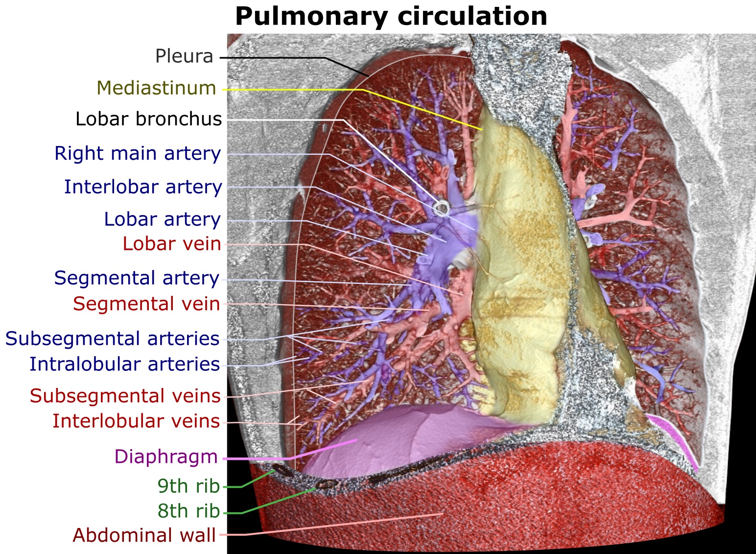
A pulmonary artery is an artery in the pulmonary circulation that carries deoxygenated blood from the right side of the heart to the lungs. The largest pulmonary artery is the ''main pulmonary artery'' or ''pulmonary trunk'' from the heart, and the smallest ones are the arterioles, which lead to the capillaries that surround the pulmonary alveoli. Structure The pulmonary arteries are blood vessels that carry systemic venous blood from the right ventricle of the heart to the microcirculation of the lungs. Unlike in other organs where arteries supply oxygenated blood, the blood carried by the pulmonary arteries is deoxygenated, as it is venous blood returning to the heart. The main pulmonary arteries emerge from the right side of the heart, and then split into smaller arteries that progressively divide and become arterioles, eventually narrowing into the capillary microcirculation of the lungs where gas exchange occurs. Pulmonary trunk In order of blood flow, the pulmonar ... [...More Info...] [...Related Items...] OR: [Wikipedia] [Google] [Baidu] |


