|
Diffuse Alveolar Damage
Diffuse alveolar damage (DAD) is a histologic term used to describe specific changes that occur to the structure of the lungs during injury or disease. Most often DAD is described in association with the early stages of acute respiratory distress syndrome (ARDS). It is important to note that DAD can be seen in situations other than ARDS (such as acute interstitial pneumonia) and that ARDS can occur without DAD. Definitions * Diffuse alveolar damage (DAD): an acute lung condition with the presence of hyaline membranes. These hyaline membranes are made up of dead cells, surfactant, and proteins. The hyaline membranes deposit along the walls of the alveoli, where gas exchange typically occurs, thereby making gas exchange difficult. * Acute respiratory distress syndrome (ARDS): a potentially life-threatening condition where the alveoli are damaged thereby letting fluid leak into the lungs which makes it difficult to exchange gases and oxygenate the blood. It is the general practice of ... [...More Info...] [...Related Items...] OR: [Wikipedia] [Google] [Baidu] |
Micrograph
A micrograph or photomicrograph is a photograph or digital image taken through a microscope or similar device to show a magnified image of an object. This is opposed to a macrograph or photomacrograph, an image which is also taken on a microscope but is only slightly magnified, usually less than 10 times. Micrography is the practice or art of using microscopes to make photographs. A micrograph contains extensive details of microstructure. A wealth of information can be obtained from a simple micrograph like behavior of the material under different conditions, the phases found in the system, failure analysis, grain size estimation, elemental analysis and so on. Micrographs are widely used in all fields of microscopy. Types Photomicrograph A light micrograph or photomicrograph is a micrograph prepared using an optical microscope, a process referred to as ''photomicroscopy''. At a basic level, photomicroscopy may be performed simply by connecting a camera to a microscope, th ... [...More Info...] [...Related Items...] OR: [Wikipedia] [Google] [Baidu] |
Alveolar Macrophage
An alveolar macrophage, pulmonary macrophage, (or dust cell) is a type of macrophage, a professional phagocyte, found in the airways and at the level of the alveoli in the lungs, but separated from their walls. Activity of the alveolar macrophage is relatively high, because they are located at one of the major boundaries between the body and the outside world. They are responsible for removing particles such as dust or microorganisms from the respiratory surfaces. Alveolar macrophages are frequently seen to contain granules of exogenous material such as particulate carbon that they have picked up from respiratory surfaces. Such black granules may be especially common in smoker's lungs or long-term city dwellers. The alveolar macrophage is the third cell type in the alveolus, the others are the type I and type II pneumocytes. Comparison of pigmented pulmonary macrophages Function Alveolar macrophages are phagocytes that play a critical role in homeostasis, host defen ... [...More Info...] [...Related Items...] OR: [Wikipedia] [Google] [Baidu] |
Tracheal Tube
A tracheal tube is a catheter that is inserted into the trachea for the primary purpose of establishing and maintaining a patent airway and to ensure the adequate exchange of oxygen and carbon dioxide. Many different types of tracheal tubes are available, suited for different specific applications: * An endotracheal tube is a specific type of tracheal tube that is nearly always inserted through the mouth (orotracheal) or nose (nasotracheal). * A tracheostomy tube is another type of tracheal tube; this curved metal or plastic tube may be inserted into a tracheostomy stoma (following a tracheotomy) to maintain a patent lumen. * A tracheal button is a rigid plastic cannula about 1 inch in length that can be placed into the tracheostomy after removal of a tracheostomy tube to maintain patency of the lumen. History Portex Medical (England and France) produced the first cuff-less plastic 'Ivory' endotracheal tubes. Ivan Magill later added a cuff (these were glued on by hand to mak ... [...More Info...] [...Related Items...] OR: [Wikipedia] [Google] [Baidu] |
False Positives And False Negatives
A false positive is an error in binary classification in which a test result incorrectly indicates the presence of a condition (such as a disease when the disease is not present), while a false negative is the opposite error, where the test result incorrectly indicates the absence of a condition when it is actually present. These are the two kinds of errors in a binary test, in contrast to the two kinds of correct result (a and a ). They are also known in medicine as a false positive (or false negative) diagnosis, and in statistical classification as a false positive (or false negative) error. In statistical hypothesis testing the analogous concepts are known as type I and type II errors, where a positive result corresponds to rejecting the null hypothesis, and a negative result corresponds to not rejecting the null hypothesis. The terms are often used interchangeably, but there are differences in detail and interpretation due to the differences between medical testing and statist ... [...More Info...] [...Related Items...] OR: [Wikipedia] [Google] [Baidu] |
Biopsy
A biopsy is a medical test commonly performed by a surgeon, interventional radiologist, or an interventional cardiologist. The process involves extraction of sample cells or tissues for examination to determine the presence or extent of a disease. The tissue is then fixed, dehydrated, embedded, sectioned, stained and mounted before it is generally examined under a microscope by a pathologist; it may also be analyzed chemically. When an entire lump or suspicious area is removed, the procedure is called an excisional biopsy. An incisional biopsy or core biopsy samples a portion of the abnormal tissue without attempting to remove the entire lesion or tumor. When a sample of tissue or fluid is removed with a needle in such a way that cells are removed without preserving the histological architecture of the tissue cells, the procedure is called a needle aspiration biopsy. Biopsies are most commonly performed for insight into possible cancerous or inflammatory conditions. History T ... [...More Info...] [...Related Items...] OR: [Wikipedia] [Google] [Baidu] |
Preterm Birth
Preterm birth, also known as premature birth, is the Childbirth, birth of a baby at fewer than 37 weeks Gestational age (obstetrics), gestational age, as opposed to full-term delivery at approximately 40 weeks. Extreme preterm is less than 28 weeks, very early preterm birth is between 28 and 32 weeks, early preterm birth occurs between 32 and 36 weeks, late preterm birth is between 34 and 36 weeks' gestation. These babies are also known as premature babies or colloquially preemies (American English) or premmies (Australian English). Symptoms of preterm labor include uterine contractions which occur more often than every ten minutes and/or the leaking of fluid from the vagina before 37 weeks. Premature infants are at greater risk for cerebral palsy, delays in development, hearing problems and problems with their Visual impairment, vision. The earlier a baby is born, the greater these risks will be. The cause of spontaneous preterm birth is often not known. Risk factors include dia ... [...More Info...] [...Related Items...] OR: [Wikipedia] [Google] [Baidu] |
Infant Respiratory Distress Syndrome
Infantile respiratory distress syndrome (IRDS), also called respiratory distress syndrome of newborn, or increasingly surfactant deficiency disorder (SDD), and previously called hyaline membrane disease (HMD), is a syndrome in premature infants caused by developmental insufficiency of pulmonary surfactant production and structural immaturity in the lungs. It can also be a consequence of neonatal infection and can result from a genetic problem with the production of surfactant-associated proteins. IRDS affects about 1% of newborns and is the leading cause of death in preterm infants. Data has shown the choice of elective caesarean sections to strikingly increase the incidence of respiratory distress in term infants; dating back to 1995, the UK first documented 2,000 annual caesarean section births requiring neonatal admission for respiratory distress. The incidence decreases with advancing gestational age, from about 50% in babies born at 26–28 weeks to about 25% at 30–31 weeks ... [...More Info...] [...Related Items...] OR: [Wikipedia] [Google] [Baidu] |
Sepsis
Sepsis, formerly known as septicemia (septicaemia in British English) or blood poisoning, is a life-threatening condition that arises when the body's response to infection causes injury to its own tissues and organs. This initial stage is followed by suppression of the immune system. Common signs and symptoms include fever, tachycardia, increased heart rate, hyperventilation, increased breathing rate, and mental confusion, confusion. There may also be symptoms related to a specific infection, such as a cough with pneumonia, or dysuria, painful urination with a pyelonephritis, kidney infection. The very young, old, and people with a immunodeficiency, weakened immune system may have no symptoms of a specific infection, and the hypothermia, body temperature may be low or normal instead of having a fever. Severe sepsis causes organ dysfunction, poor organ function or blood flow. The presence of Hypotension, low blood pressure, high blood Lactic acid, lactate, or Oliguria, low urine o ... [...More Info...] [...Related Items...] OR: [Wikipedia] [Google] [Baidu] |
Pneumonia
Pneumonia is an inflammatory condition of the lung primarily affecting the small air sacs known as alveoli. Symptoms typically include some combination of productive or dry cough, chest pain, fever, and difficulty breathing. The severity of the condition is variable. Pneumonia is usually caused by infection with viruses or bacteria, and less commonly by other microorganisms. Identifying the responsible pathogen can be difficult. Diagnosis is often based on symptoms and physical examination. Chest X-rays, blood tests, and culture of the sputum may help confirm the diagnosis. The disease may be classified by where it was acquired, such as community- or hospital-acquired or healthcare-associated pneumonia. Risk factors for pneumonia include cystic fibrosis, chronic obstructive pulmonary disease (COPD), sickle cell disease, asthma, diabetes, heart failure, a history of smoking, a poor ability to cough (such as following a stroke), and a weak immune system. Vaccines to ... [...More Info...] [...Related Items...] OR: [Wikipedia] [Google] [Baidu] |
Transplant Rejection
Transplant rejection occurs when Organ transplant, transplanted tissue is rejected by the recipient's immune system, which destroys the transplanted tissue. Transplant rejection can be lessened by determining the molecular similitude between donor and recipient and by use of immunosuppressant drugs after transplant. Types of transplant rejection Transplant rejection can be classified into three types: hyperacute, acute, and chronic. These types are differentiated by how quickly the recipient's immune system is activated and the specific aspect or aspects of immunity involved. Hyperacute rejection Hyperacute rejection is a form of rejection that manifests itself in the minutes to hours following transplantation. It is caused by the presence of pre-existing Antibody, antibodies in the recipient that recognize antigens in the donor organ. These antigens are located on the endothelial lining of blood vessels within the transplanted organ and, once antibodies bind, will lead to the ... [...More Info...] [...Related Items...] OR: [Wikipedia] [Google] [Baidu] |
Idiopathic Pulmonary Fibrosis
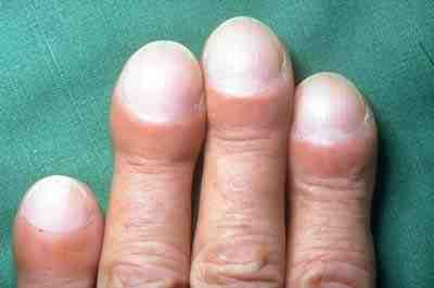
Idiopathic pulmonary fibrosis (IPF), or (formerly) fibrosing alveolitis, is a rare, progressive illness of the respiratory system, characterized by the thickening and stiffening of lung tissue, associated with the formation of scar tissue. It is a type of chronic scarring lung disease characterized by a progressive and irreversible decline in lung function. The tissue in the lungs becomes thick and stiff, which affects the tissue that surrounds the air sacs in the lungs. Symptoms typically include gradual onset of shortness of breath and a dry cough. Other changes may include feeling tired, and abnormally large and dome shaped finger and toenails (nail clubbing). Complications may include pulmonary hypertension, heart failure, pneumonia or pulmonary embolism. The cause is unknown, hence the term idiopathic. Risk factors include cigarette smoking, acid reflux disease (GERD), certain viral infections, and genetic predisposition. The underlying mechanism involves scarring of th ... [...More Info...] [...Related Items...] OR: [Wikipedia] [Google] [Baidu] |
Acute Interstitial Pneumonitis
Acute interstitial pneumonitis is a rare, severe lung disease that usually affects otherwise healthy individuals. There is no known cause or cure. Acute interstitial pneumonitis is often categorized as both an interstitial lung disease and a form of acute respiratory distress syndrome (ARDS) but it is distinguished from the ''chronic'' forms of interstitial pneumonia such as idiopathic pulmonary fibrosis. Symptoms and signs The most common symptoms of acute interstitial pneumonitis are highly productive cough with expectoration of thick mucus, fever, and difficulties breathing. These often occur over a period of one to two weeks before medical attention is sought. The presence of fluid means the person experiences a feeling similar to 'drowning'. Difficulties breathing can quickly progress to an inability to breathe without support (respiratory failure). Acute interstitial pneumonitis typically progresses rapidly, with hospitalization and mechanical ventilation often required o ... [...More Info...] [...Related Items...] OR: [Wikipedia] [Google] [Baidu] |


