|
Development Of The Urinary And Reproductive Organs
The development of the urinary system begins during prenatal development, and relates to the development of the urogenital system – both the organs of the urinary system and the sex organs of the reproductive system. The development continues as a part of sexual differentiation. The urinary and reproductive organs are developed from the intermediate mesoderm. The permanent organs of the adult are preceded by a set of structures which are purely embryonic, and which with the exception of the ducts disappear almost entirely before birth. These embryonic structures are on either side; the pronephros, the mesonephros and the metanephros of the kidney, and the Wolffian and Müllerian ducts of the sex organ. The pronephros disappears very early; the structural elements of the mesonephros mostly degenerate, but the gonad is developed in their place, with which the Wolffian duct remains as the duct in males, and the Müllerian as that of the female. Some of the tubules of the mesonephro ... [...More Info...] [...Related Items...] OR: [Wikipedia] [Google] [Baidu] |
Prenatal Development
Prenatal development () includes the development of the embryo and of the fetus during a viviparous animal's gestation. Prenatal development starts with fertilization, in the germinal stage of embryonic development, and continues in fetal development until birth. In human pregnancy, prenatal development is also called antenatal development. The development of the human embryo follows fertilization, and continues as fetal development. By the end of the tenth week of gestational age the embryo has acquired its basic form and is referred to as a fetus. The next period is that of fetal development where many organs become fully developed. This fetal period is described both topically (by organ) and chronologically (by time) with major occurrences being listed by gestational age. The very early stages of embryonic development are the same in all mammals, but later stages of development, and the length of gestation varies. Terminology In the human: Different terms are used to ... [...More Info...] [...Related Items...] OR: [Wikipedia] [Google] [Baidu] |
Dorsum (anatomy)
Standard anatomical terms of location are used to unambiguously describe the anatomy of animals, including humans. The terms, typically derived from Latin or Greek roots, describe something in its standard anatomical position. This position provides a definition of what is at the front ("anterior"), behind ("posterior") and so on. As part of defining and describing terms, the body is described through the use of anatomical planes and anatomical axes. The meaning of terms that are used can change depending on whether an organism is bipedal or quadrupedal. Additionally, for some animals such as invertebrates, some terms may not have any meaning at all; for example, an animal that is radially symmetrical will have no anterior surface, but can still have a description that a part is close to the middle ("proximal") or further from the middle ("distal"). International organisations have determined vocabularies that are often used as standard vocabularies for subdisciplines of anatom ... [...More Info...] [...Related Items...] OR: [Wikipedia] [Google] [Baidu] |
Ostium Of The Fallopian Tube
The fallopian tubes, also known as uterine tubes, oviducts or salpinges (singular salpinx), are paired tubes in the human female that stretch from the uterus to the ovaries. The fallopian tubes are part of the female reproductive system. In other mammals they are only called oviducts. Each tube is a muscular hollow organ that is on average between 10 and 14 cm in length, with an external diameter of 1 cm. It has four described parts: the intramural part, isthmus, ampulla, and infundibulum with associated fimbriae. Each tube has two openings a proximal opening nearest and opening to the uterus, and a distal opening furthest and opening to the abdomen. The fallopian tubes are held in place by the mesosalpinx, a part of the broad ligament mesentery that wraps around the tubes. Another part of the broad ligament, the mesovarium suspends the ovaries in place. An egg cell is transported from an ovary to a fallopian tube where it may be fertilized in the ampulla of the tub ... [...More Info...] [...Related Items...] OR: [Wikipedia] [Google] [Baidu] |
Müllerian Ducts
Paramesonephric ducts (or Müllerian ducts) are paired ducts of the embryo that run down the lateral sides of the genital ridge and terminate at the sinus tubercle in the primitive urogenital sinus. In the female, they will develop to form the fallopian tubes, uterus, cervix, and the upper one-third of the vagina. Development The female reproductive system is composed of two embryological segments: the urogenital sinus and the paramesonephric ducts. The two are conjoined at the sinus tubercle. Paramesonephric ducts are present on the embryo of both sexes. Only in females do they develop into reproductive organs. They degenerate in males of certain species, but the adjoining mesonephric ducts develop into male reproductive organs. The sex based differences in the contributions of the paramesonephric ducts to reproductive organs is based on the presence, and degree of presence, of Anti-Müllerian hormone. During the formation of the reproductive system, the paramesonephric ducts ar ... [...More Info...] [...Related Items...] OR: [Wikipedia] [Google] [Baidu] |
Development Of The Suspensory Ligament Of The Ovary
The suspensory ligament of the ovary, also infundibulopelvic ligament (commonly abbreviated IP ligament or simply IP), is a fold of peritoneum that extends out from the ovary to the wall of the pelvis. Some sources consider it a part of the broad ligament of uterus while other sources just consider it a "termination" of the ligament. It is not considered a true ligament in that it does not physically support any anatomical structures; however it is an important landmark and it houses the ovarian vessels. The suspensory ligament is directed upward over the iliac vessels. Structure It contains the ovarian artery, ovarian vein, ovarian nerve plexus, at |
Seminal Vesicle
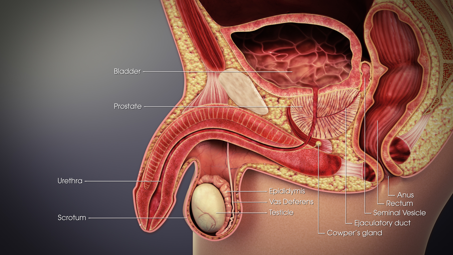
The seminal vesicles (also called vesicular glands, or seminal glands) are a pair of two convoluted tubular glands that lie behind the urinary bladder of some male mammals. They secrete fluid that partly composes the semen. The vesicles are 5–10 cm in size, 3–5 cm in diameter, and are located between the bladder and the rectum. They have multiple outpouchings which contain secretory glands, which join together with the vas deferens at the ejaculatory duct. They receive blood from the vesiculodeferential artery, and drain into the vesiculodeferential veins. The glands are lined with column-shaped and cuboidal cells. The vesicles are present in many groups of mammals, but not marsupials, monotremes or carnivores. Inflammation of the seminal vesicles is called seminal vesiculitis, most often is due to bacterial infection as a result of a sexually transmitted disease or following a surgical procedure. Seminal vesiculitis can cause pain in the lower abdomen, scrotum, ... [...More Info...] [...Related Items...] OR: [Wikipedia] [Google] [Baidu] |
Ejaculatory Duct
The ejaculatory ducts (''ductus ejaculatorii'') are paired structures in male anatomy. Each ejaculatory duct is formed by the union of the vas deferens with the duct of the seminal vesicle. They pass through the prostate, and open into the urethra above the seminal colliculus. During ejaculation, semen passes through the prostate gland, enters the urethra and exits the body via the urinary meatus. Function Ejaculation Ejaculation occurs in two stages, the emission stage and the expulsion stage.Rathus, S. A., Nevid, J. S., Fichner-Rathus, L., Herold, E. S. (2010). ''Human Sexuality in a World of Diversity''. Pearsons Education Canada, Pearson Canada Inc. Toronto, ON. The emission stage involves the workings of several structures of the ejaculatory duct; contractions of the prostate gland, the seminal vesicles, the bulbourethral gland and the vas deferens push fluids into the prostatic urethra. The semen is stored here until ejaculation occurs. Muscles at the base of the penis co ... [...More Info...] [...Related Items...] OR: [Wikipedia] [Google] [Baidu] |
Ductus Deferens
The vas deferens or ductus deferens is part of the male reproductive system of many vertebrates. The ducts transport sperm from the epididymis to the ejaculatory ducts in anticipation of ejaculation. The vas deferens is a partially coiled tube which exits the abdominal cavity through the inguinal canal. Etymology ''Vas deferens'' is Latin, meaning "carrying-away vessel"; the plural version is ''vasa deferentia''. ''Ductus deferens'' is also Latin, meaning "carrying-away duct"; the plural version is ''ducti deferentes''. Structure There are two vasa deferentia, connecting the left and right epididymis with the seminal vesicles to form the ejaculatory duct in order to move spermatozoon, sperm. The (human) vas deferens measures 30–35 cm in length, and 2–3 mm in diameter. The vas deferens is continuous proximally with the tail of the epididymis. The vas deferens exhibits a tortuous, convoluted initial/proximal section (which measures 2–3 cm in length). Distall ... [...More Info...] [...Related Items...] OR: [Wikipedia] [Google] [Baidu] |
Epididymis
The epididymis (; plural: epididymides or ) is a tube that connects a testicle to a vas deferens in the male reproductive system. It is a single, narrow, tightly-coiled tube in adult humans, in length. It serves as an interconnection between the multiple efferent ducts at the rear of a testicle (proximally), and the vas deferens (distally). Anatomy The epididymis is situated posterior and somewhat lateral to the testis. The epididymis is invested completely by the tunica vaginalis (which is continuous with the tunica vaginalis covering the testis). The epididymis can be divided into three main regions: * The head ( la, caput). The head of the epididymis receives spermatozoa via the efferent ducts of the mediastinium of the testis at the superior pole of the testis. The head is characterized histologically by a thick epithelium with long stereocilia (described below) and a little smooth muscle. It is involved in absorbing fluid to make the sperm more concentrated. The concentrat ... [...More Info...] [...Related Items...] OR: [Wikipedia] [Google] [Baidu] |
Mesonephros
The mesonephros ( el, middle kidney) is one of three excretory system, excretory organs that develop in vertebrates. It serves as the main excretory organ of aquatic vertebrates and as a temporary kidney in reptiles, birds, and mammals. The mesonephros is included in the Wolffian body after Caspar Friedrich Wolff who described it in 1759. (The Wolffian body is composed of: mesonephros + paramesonephrotic blastema) Structure The mesonephros acts as a structure similar to the human kidney, kidney that, in humans, functions between the sixth and tenth weeks of embryological life. Despite the similarity in structure, function, and terminology, however, the mesonephric nephrons do not form any part of the mature kidney or nephrons. In humans, the mesonephros consists of units which are similar in structure and function to nephrons of the adult kidney. Each of these consists of a glomerulus, a tuft of capillary, capillaries which arises from lateral branches of dorsal aorta and drains i ... [...More Info...] [...Related Items...] OR: [Wikipedia] [Google] [Baidu] |
Abdominal Cavity
The abdominal cavity is a large body cavity in humans and many other animals that contains many organs. It is a part of the abdominopelvic cavity. It is located below the thoracic cavity, and above the pelvic cavity. Its dome-shaped roof is the thoracic diaphragm, a thin sheet of muscle under the lungs, and its floor is the pelvic inlet, opening into the pelvis. Structure Organs Organs of the abdominal cavity include the stomach, liver, gallbladder, spleen, pancreas, small intestine, kidneys, large intestine, and adrenal glands. Peritoneum The abdominal cavity is lined with a protective membrane termed the peritoneum. The inside wall is covered by the parietal peritoneum. The kidneys are located behind the peritoneum, in the retroperitoneum, outside the abdominal cavity. The viscera are also covered by visceral peritoneum. Between the visceral and parietal peritoneum is the peritoneal cavity, which is a potential space. It contains a serous fluid called peritoneal fluid tha ... [...More Info...] [...Related Items...] OR: [Wikipedia] [Google] [Baidu] |


