|
Dentinogenesis
{{Refimprove, date=September 2014 Dentinogenesis is the formation of dentin, a substance that forms the majority of teeth. Dentinogenesis is performed by odontoblasts, which are a special type of biological cell on the outer wall of dental pulps, and it begins at the late bell stage of a tooth development. The different stages of dentin formation after differentiation of the cell result in different types of dentin: mantle dentin, primary dentin, secondary dentin, and tertiary dentin. Odontoblast differentiation Odontoblasts differentiate from cells of the dental papilla. This is an expression of signaling molecules and growth factors of the inner enamel epithelium (IEE). Formation of mantle dentin They begin secreting an organic matrix around the area directly adjacent to the IEE, closest to the area of the future cusp of a tooth. The organic matrix contains collagen fibers with large diameters (0.1-0.2 μm in diameter). The odontoblasts begin to move toward the center of the to ... [...More Info...] [...Related Items...] OR: [Wikipedia] [Google] [Baidu] |
Dentinogenesis Imperfecta
Dentinogenesis imperfecta (DI) is a genetic disorder of tooth development. It is inherited in an autosomal dominant pattern, as a result of mutations on chromosome 4q21, in the dentine sialophosphoprotein gene (DSPP). It is one of the most frequently occurring autosomal dominant features in humans. Dentinogenesis imperfecta affects an estimated 1 in 6,000-8,000 people. This condition can cause teeth to be discolored (most often a blue-gray or yellow-brown color) and translucent, giving teeth an opalescent sheen. Teeth are also weaker than normal, making them prone to rapid wear, breakage, and loss. These problems can affect baby (primary/deciduous) teeth alone, or both baby teeth and adult (permanent) teeth, with the baby teeth usually more severely affected. Although genetic factors are the main contributor for the disease, any environmental or systemic upset that impedes calcification or metabolisation of calcium can also result in anomalous dentine. Classification Shield cl ... [...More Info...] [...Related Items...] OR: [Wikipedia] [Google] [Baidu] |
Dentin
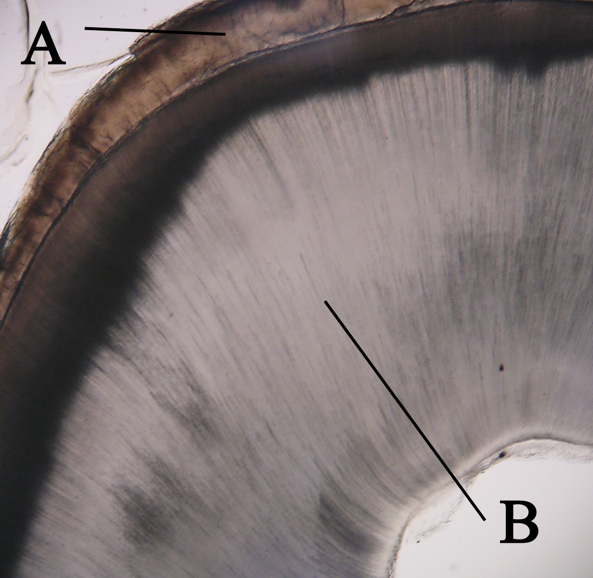
Dentin () (American English) or dentine ( or ) (British English) ( la, substantia eburnea) is a calcified tissue of the body and, along with enamel, cementum, and pulp, is one of the four major components of teeth. It is usually covered by enamel on the crown and cementum on the root and surrounds the entire pulp. By volume, 45% of dentin consists of the mineral hydroxyapatite, 33% is organic material, and 22% is water. Yellow in appearance, it greatly affects the color of a tooth due to the translucency of enamel. Dentin, which is less mineralized and less brittle than enamel, is necessary for the support of enamel. Dentin rates approximately 3 on the Mohs scale of mineral hardness. There are two main characteristics which distinguish dentin from enamel: firstly, dentin forms throughout life; secondly, dentin is sensitive and can become hypersensitive to changes in temperature due to the sensory function of odontoblasts, especially when enamel recedes and dentin channels becom ... [...More Info...] [...Related Items...] OR: [Wikipedia] [Google] [Baidu] |
Tooth Development
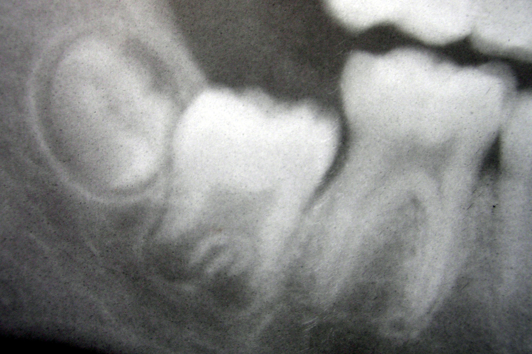
Tooth development or odontogenesis is the complex process by which teeth form from embryonic cells, grow, and erupt into the mouth. For human teeth to have a healthy oral environment, all parts of the tooth must develop during appropriate stages of fetal development. Primary (baby) teeth start to form between the sixth and eighth week of prenatal development, and permanent teeth begin to form in the twentieth week.Ten Cate's Oral Histology, Nanci, Elsevier, 2013, pages 70-94 If teeth do not start to develop at or near these times, they will not develop at all, resulting in hypodontia or anodontia. A significant amount of research has focused on determining the processes that initiate tooth development. It is widely accepted that there is a factor within the tissues of the first pharyngeal arch that is necessary for the development of teeth. Overview The tooth germ is an aggregation of cells that eventually forms a tooth.University of Texas Medical Branch. These cells are de ... [...More Info...] [...Related Items...] OR: [Wikipedia] [Google] [Baidu] |
Cervical Loop
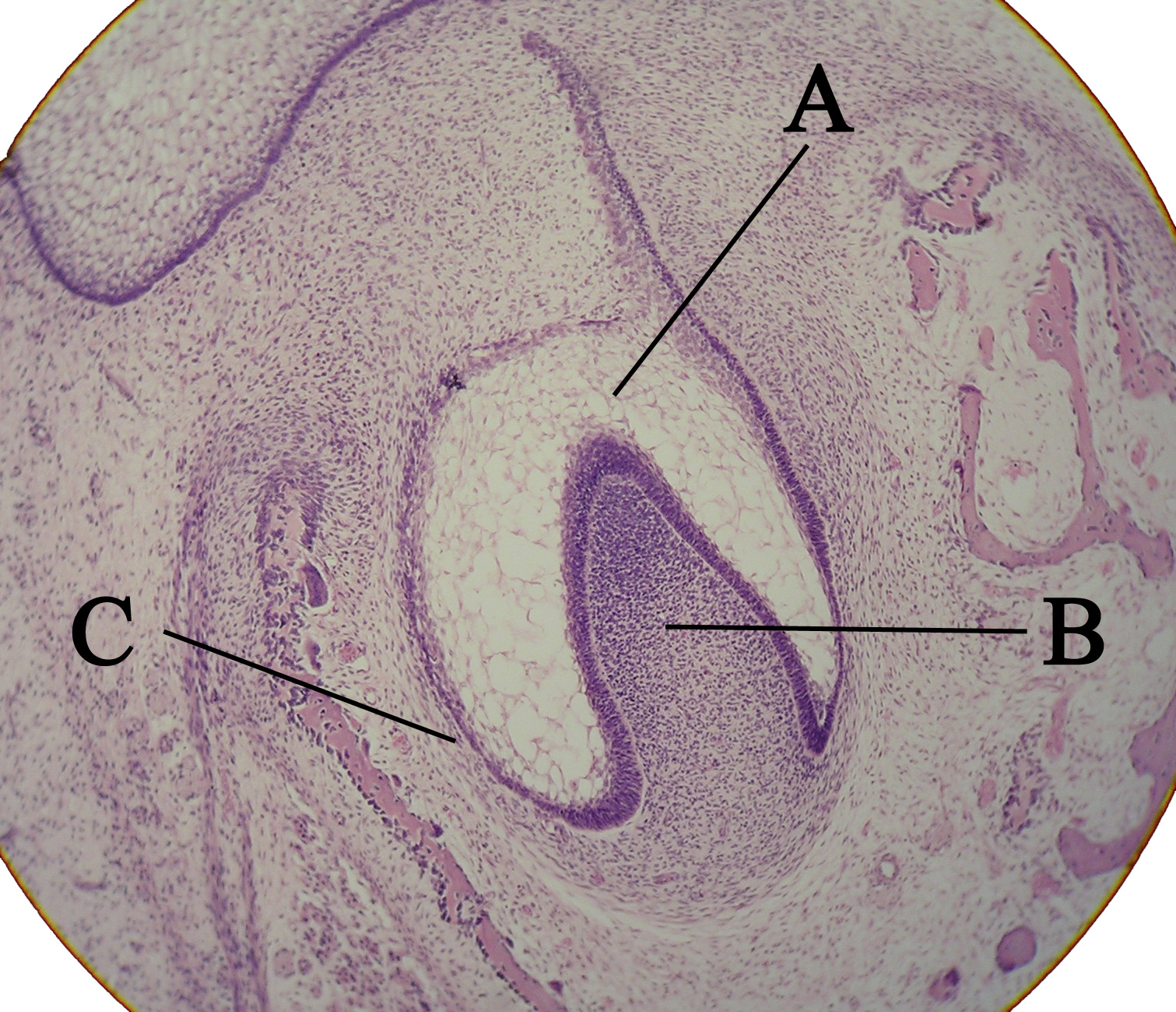
The cervical loop is the location on an enamel organ in a developing tooth where the outer enamel epithelium and the inner enamel epithelium join. The cervical loop is a histologic term indicating a specific epithelial structure at the apical side of the tooth germ, consisting of loosely aggregated stellate reticulum in the center surrounded by stratum intermedium. These tissues are enveloped by a basal layer of epithelium known on the outside of the tooth as outer enamel epithelium and on the inside as inner enamel epithelium. During root formation the inner layers of epithelium disappear and only the basal layers are left creating Hertwig's epithelial root sheath (HERS). At this point it is usually referred to as HERS instead of the cervical loop to indicate the structural difference. Cervical loop as epithelial stem cell niche It is thought that the central epithelial tissue of the cervical loop, the stellate reticulum, acts as a stem cell In multicellular organisms, stem ... [...More Info...] [...Related Items...] OR: [Wikipedia] [Google] [Baidu] |
Odontoblasts
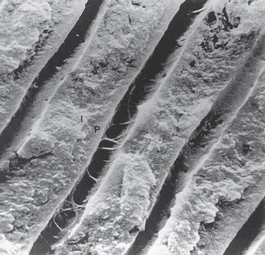
In vertebrates, an odontoblast is a cell of neural crest origin that is part of the outer surface of the dental pulp, and whose biological function is dentinogenesis, which is the formation of dentin, the substance beneath the tooth enamel on the crown and the cementum on the root. Structure Odontoblasts are large columnar cells, whose cell bodies are arranged along the interface between dentin and pulp, from the crown to cervix to the root apex in a mature tooth. The cell is rich in endoplasmic reticulum and Golgi complex, especially during primary dentin formation, which allows it to have a high secretory capacity; it first forms the collagenous matrix to form predentin, then mineral levels to form the mature dentin. Odontoblasts form approximately 4 μm of predentin daily during tooth development.Ten Cate's Oral Histology, Nanci, Elsevier, 2013, page 170 During secretion after differentiation from the outer cells of the dental papilla, it is noted that it is polarized so its nu ... [...More Info...] [...Related Items...] OR: [Wikipedia] [Google] [Baidu] |
Dental Caries
Tooth decay, also known as cavities or caries, is the breakdown of teeth due to acids produced by bacteria. The cavities may be a number of different colors from yellow to black. Symptoms may include pain and difficulty with eating. Complications may include inflammation of the tissue around the tooth, tooth loss and infection or abscess formation. The cause of cavities is acid from bacteria dissolving the hard tissues of the teeth ( enamel, dentin and cementum). The acid is produced by the bacteria when they break down food debris or sugar on the tooth surface. Simple sugars in food are these bacteria's primary energy source and thus a diet high in simple sugar is a risk factor. If mineral breakdown is greater than build up from sources such as saliva, caries results. Risk factors include conditions that result in less saliva such as: diabetes mellitus, Sjögren syndrome and some medications. Medications that decrease saliva production include antihistamines and antide ... [...More Info...] [...Related Items...] OR: [Wikipedia] [Google] [Baidu] |
Attrition (dental)
Dental attrition is a type of tooth wear caused by tooth-to-tooth contact, resulting in loss of tooth tissue, usually starting at the incisal or occlusal surfaces. Tooth wear is a physiological process and is commonly seen as a normal part of aging. Advanced and excessive wear and tooth surface loss can be defined as pathological in nature, requiring intervention by a dental practitioner. The pathological wear of the tooth surface can be caused by bruxism, which is clenching and grinding of the teeth. If the attrition is severe, the enamel can be completely worn away leaving underlying dentin exposed, resulting in an increased risk of dental caries and dentin hypersensitivity. It is best to identify pathological attrition at an early stage to prevent unnecessary loss of tooth structure as enamel does not regenerate. Signs and symptoms Attrition occurs as a result of opposing tooth surfaces contacting. The contact can affect cuspal, incisal and proximal surface areas. Indicatio ... [...More Info...] [...Related Items...] OR: [Wikipedia] [Google] [Baidu] |
Tertiary Dentin
Tertiary dentin (including reparative dentin or sclerotic dentin) forms as a reaction to stimulation, including caries, wear and fractures. Tertiary dentin is therefore a mechanism for a tooth to ‘heal’, with new material formation protecting the pulp chamber and ultimately therefore protects the tooth and individual against abscesses and infection. This form of dentine can be easily distinguished on the surface of a tooth, and is much darker in appearance compared to primary dentine. Tertiary dentine will often not be visible on the surface of a tooth, but because it is more dense it can be viewed on a Micro-CT scan of the tooth. Wear on the surface of a tooth can lead to the exposure of the underlying dentine. When wear is severe tertiary dentine may form to help protect the pulp chamber. Frequency of tertiary dentin in different species of primate suggests teeth 'heal' at different rates in different species. Gorillas have a high rate of tertiary dentin formation, with over ... [...More Info...] [...Related Items...] OR: [Wikipedia] [Google] [Baidu] |
Dentin Phosphoprotein
Dentin phosphoprotein, or phosphophoryn, is one of three Protein, proteins formed from dentin sialophosphoprotein (protein), dentin sialophosphoprotein and is important in the regulation of mineralization (biology), mineralization of dentin. Phosphophoryn is the most Acid, acidic protein ever discovered and has an isoelectric point of 1. This extreme acidity is achieved by its amino acid sequence. Many portions of its chain are repeating -D-S-S- (aspartate, aspartic acid-serine-serine) sequences. In protein chemistry, net acidity equates to negative charge. Being highly negative, dentin phosphoprotein is able to attract large amounts of calcium. ''IIn vitro, n vitro'' studies also indicate phosphophoryn can initiate hydroxyapatite formation.Nanci, Antonio. Ten Cate's Oral Histology: Development, Structure, and Function. 7th ed. St. Louis, MO: Mosby Elsevier, 2008. Print. References Teeth Proteins {{protein-stub ... [...More Info...] [...Related Items...] OR: [Wikipedia] [Google] [Baidu] |
Hertwig's Epithelial Root Sheath
The Hertwig epithelial root sheath (HERS) or epithelial root sheath is a proliferation of epithelial cells located at the cervical loop of the enamel organ in a developing tooth. Hertwig epithelial root sheath initiates the formation of dentin in the root of a tooth by causing the differentiation of odontoblasts from the dental papilla. The root sheath eventually disintegrates with the periodontal ligament, but residual pieces that do not completely disappear are seen as epithelial cell rests of Malassez (ERM). These rests can become cystic, presenting future periodontal infections. Structure Hertwig epithelial root sheath is derived from the inner and outer enamel epithelium of the enamel organ.Illustrated Dental Embryology, Histology, and Anatomy, Bath-Balogh and Fehrenbach, Elsevier, 2011, p. 66 Function The sheath is also responsible for multiple or accessory roots (medial growth) and lateral or accessory canals in the root (break in epithelium).Ten Cate's Oral Histology ... [...More Info...] [...Related Items...] OR: [Wikipedia] [Google] [Baidu] |


