|
Odontoblasts
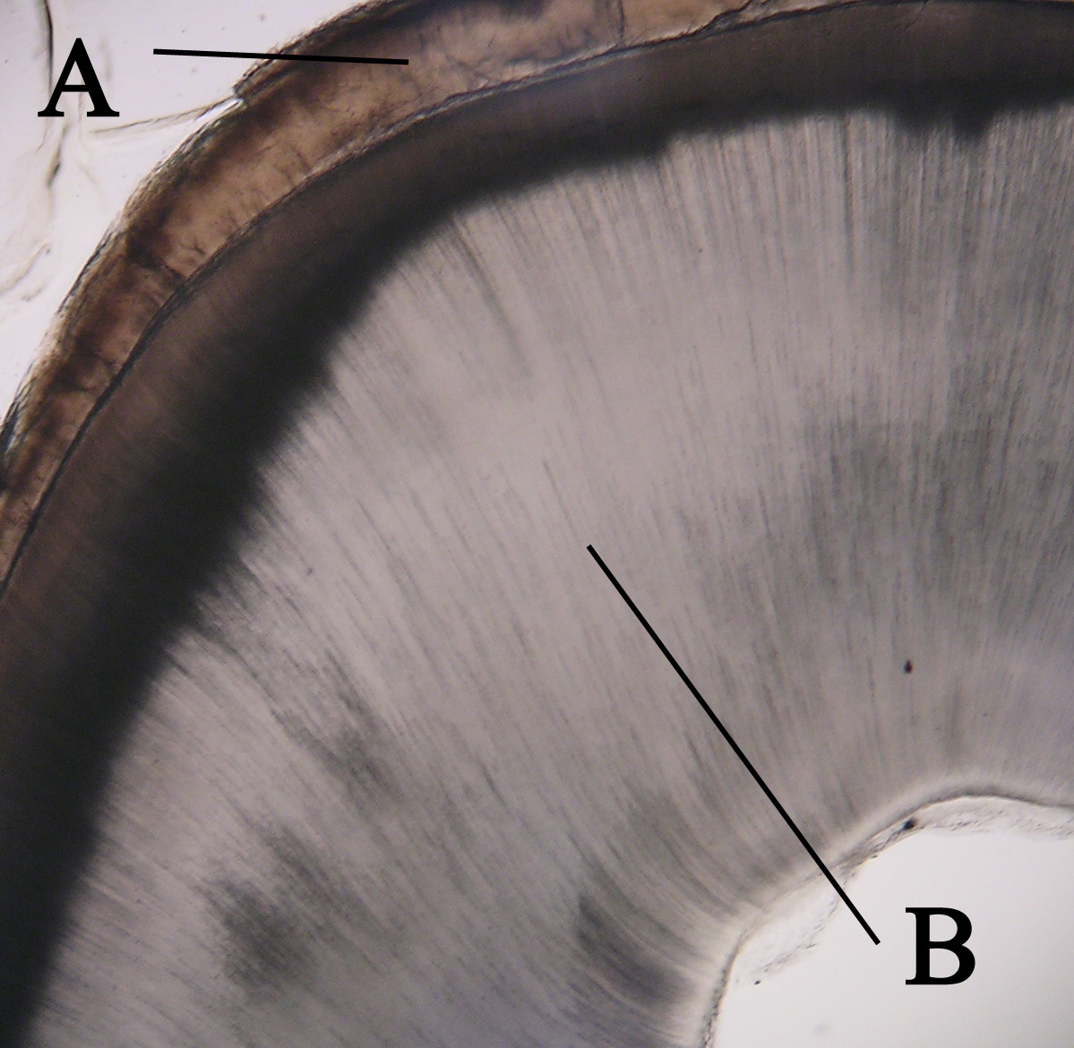
In vertebrates, an odontoblast is a Cell (biology), cell of neural crest origin that is part of the outer surface of the pulp (tooth), dental pulp, and whose biological function is dentinogenesis, which is the formation of dentin, the substance beneath the tooth enamel on the crown and the cementum on the root. Structure Odontoblasts are large columnar cells, whose cell bodies are arranged along the interface between dentin and pulp, from the crown to the cervix to the Root apex (dental), root apex in a mature tooth. The cell is rich in endoplasmic reticulum and Golgi complex, especially during primary dentin formation, which allows it to have a high secretory capacity; it first forms the collagenous matrix to form predentin, then mineral levels to form the mature dentin. Odontoblasts form approximately 4 μm of predentin daily during tooth development.Ten Cate's Oral Histology, Nanci, Elsevier, 2013, page 170 During secretion after differentiation from the outer cells of the dent ... [...More Info...] [...Related Items...] OR: [Wikipedia] [Google] [Baidu] |
Dentin
Dentin ( ) (American English) or dentine ( or ) (British English) () is a calcified tissue (biology), tissue of the body and, along with tooth enamel, enamel, cementum, and pulp (tooth), pulp, is one of the four major components of teeth. It is usually covered by enamel on the crown and cementum on the root and surrounds the entire pulp. By volume, 45% of dentin consists of the mineral hydroxyapatite, 33% is organic material, and 22% is water. Yellow in appearance, it greatly affects the color of a tooth due to the translucency of enamel. Dentin, which is less mineralized and less brittle than enamel, is necessary for the support of enamel. Dentin rates approximately 3 on the Mohs scale of mineral hardness. There are two main characteristics which distinguish dentin from enamel: firstly, dentin forms throughout life; secondly, dentin is sensitive and can become hypersensitive to changes in temperature due to the sensory function of odontoblasts, especially when enamel recedes an ... [...More Info...] [...Related Items...] OR: [Wikipedia] [Google] [Baidu] |
Pulp (tooth)
The pulp is the connective tissue, nerves, blood vessels, and odontoblasts that comprise the innermost layer of a tooth. The pulp's activity and signalling processes regulate its behaviour. Anatomy The pulp is the neurovascular bundle central to each tooth, permanent tooth, permanent or primary tooth, primary. It is composed of a central pulp chamber, pulp horns, and radicular canals. The large mass of the pulp is contained within the pulp chamber, which is contained in and mimics the overall shape of the crown of the tooth.Fehrenbach, MJ. and Popowics, T. (2026), ''Illustrated Dental Embryology, Histology, and Anatomy'', Elsevier, page 185-86. Because of the continuous deposition of the dentine, the pulp chamber becomes smaller with the age. This is not uniform throughout the coronal pulp but progresses faster on the floor than on the roof or sidewalls. Radicular pulp canals extend down from the cervical region of the crown to the Root apex (dental), root apex. They are not ... [...More Info...] [...Related Items...] OR: [Wikipedia] [Google] [Baidu] |
Dentinogenesis
{{Refimprove, date=September 2014 In animal tooth development, dentinogenesis is the formation of dentin, a substance that forms the majority of teeth. Dentinogenesis is performed by odontoblasts, which are a special type of biological cell on the outer wall of dental pulps, and it begins at the late bell stage of a tooth development. The different stages of dentin formation after differentiation of the cell result in different types of dentin: mantle dentin, primary dentin, secondary dentin, and tertiary dentin. Odontoblast differentiation Odontoblasts differentiate from cells of the dental papilla. This is an expression of signaling molecules and growth factors of the inner enamel epithelium (IEE). Formation of mantle dentin They begin secreting an organic matrix around the area directly adjacent to the IEE, closest to the area of the future cusp of a tooth. The organic matrix contains collagen fibers with large diameters (0.1-0.2 μm in diameter). The odontoblasts begin to ... [...More Info...] [...Related Items...] OR: [Wikipedia] [Google] [Baidu] |
Reelin
Reelin, encoded by the ''RELN'' gene, is a large secreted extracellular matrix glycoprotein that helps regulate processes of neuronal migration and positioning in the developing brain by controlling cell–cell interactions. Besides this important role in early Developmental biology, development, reelin continues to work in the adult brain. It modulates synaptic plasticity by enhancing the induction and maintenance of long-term potentiation. It also stimulates dendrite and dendritic spine development in the hippocampus, and regulates the continuing migration of neuroblasts generated in adult neurogenesis sites of the subventricular zone, subventricular and subgranular zones. It is found not only in the brain but also in the liver, Thyroid, thyroid gland, adrenal gland, fallopian tube, breast and in comparatively lower levels across a range of anatomical regions. Reelin has been suggested to be implicated in pathogenesis of several brain diseases. The expression of the protein has ... [...More Info...] [...Related Items...] OR: [Wikipedia] [Google] [Baidu] |
Ameloblast
Ameloblasts are cells present only during tooth development that deposit tooth enamel, which is the hard outermost layer of the tooth forming the surface of the crown. Structure Each ameloblast is a columnar cell approximately 4 micrometers in diameter, 40 micrometers in length and is hexagonal in cross section. The secretory end of the ameloblast ends in a six-sided pyramid-like projection known as the Tomes' process. The angulation of the Tomes' process is significant in the orientation of enamel rods, the basic unit of tooth enamel. Distal terminal bars are junctional complexes that separate the Tomes' processes from ameloblast proper. Development Ameloblasts are derived from oral epithelium tissue of ectodermal origin. Their differentiation from preameloblasts (whose origin is from inner enamel epithelium) is a result of signaling from the ectomesenchymal cells of the dental papilla. Initially the preameloblasts will differentiate into presecretory ameloblasts and then ... [...More Info...] [...Related Items...] OR: [Wikipedia] [Google] [Baidu] |
Neural Crest
The neural crest is a ridge-like structure that is formed transiently between the epidermal ectoderm and neural plate during vertebrate development. Neural crest cells originate from this structure through the epithelial-mesenchymal transition, and in turn give rise to a diverse cell lineage—including melanocytes, craniofacial cartilage and bone, smooth muscle, dentin, peripheral and enteric neurons, adrenal medulla and glia. After gastrulation, the neural crest is specified at the border of the neural plate and the non-neural ectoderm. During neurulation, the borders of the neural plate, also known as the neural folds, converge at the dorsal midline to form the neural tube. Subsequently, neural crest cells from the roof plate of the neural tube undergo an epithelial to mesenchymal transition, delaminating from the neuroepithelium and migrating through the periphery, where they differentiate into varied cell types. The emergence of the neural crest was important in v ... [...More Info...] [...Related Items...] OR: [Wikipedia] [Google] [Baidu] |
Radula
The radula (; : radulae or radulas) is an anatomical structure used by mollusks for feeding, sometimes compared to a tongue. It is a minutely toothed, chitinous ribbon, which is typically used for scraping or cutting food before the food enters the esophagus. The radula is unique to the mollusks, and is found in every class of mollusk except the bivalves, which instead use cilia, waving filaments that bring minute organisms to the mouth. Within the gastropods, the radula is used in feeding by both herbivorous and carnivorous snails and slugs. The arrangement of teeth ( denticles) on the radular ribbon varies considerably from one group to another. In most of the more ancient lineages of gastropods, the radula is used to graze, by scraping diatoms and other microscopic algae off rock surfaces and other substrates. Predatory marine snails such as the Naticidae use the radula plus an acidic secretion to bore through the shell of other mollusks. Other predatory marine snails, ... [...More Info...] [...Related Items...] OR: [Wikipedia] [Google] [Baidu] |
Uterus
The uterus (from Latin ''uterus'', : uteri or uteruses) or womb () is the hollow organ, organ in the reproductive system of most female mammals, including humans, that accommodates the embryonic development, embryonic and prenatal development, fetal development of one or more Fertilized egg, fertilized eggs until birth. The uterus is a hormone-responsive sex organ that contains uterine gland, glands in its endometrium, lining that secrete uterine milk for embryonic nourishment. (The term ''uterus'' is also applied to analogous structures in some non-mammalian animals.) In humans, the lower end of the uterus is a narrow part known as the Uterine isthmus, isthmus that connects to the cervix, the anterior gateway leading to the vagina. The upper end, the body of the uterus, is connected to the fallopian tubes at the uterine horns; the rounded part, the fundus, is above the openings to the fallopian tubes. The connection of the uterine cavity with a fallopian tube is called the utero ... [...More Info...] [...Related Items...] OR: [Wikipedia] [Google] [Baidu] |
List Of Human Cell Types Derived From The Germ Layers
This is a list of Cell (biology), cells in humans derived from the three embryonic germ layers – ectoderm, mesoderm, and endoderm. Cells derived from ectoderm Surface ectoderm Skin * Trichocyte (human), Trichocyte * Keratinocyte Anterior pituitary * Gonadotropic cell, Gonadotrope * Corticotropic cell, Corticotrope * Thyrotropic cell, Thyrotrope * Somatotropic cell, Somatotrope * Prolactin cell, Lactotroph Tooth enamel * Ameloblast Neural crest Peripheral nervous system * Neuron * Neuroglia, Glia ** Schwann cell ** Satellite glial cell Neuroendocrine system * Chromaffin cell * Glomus cell Skin * Melanocyte ** Nevus cell * Merkel cell Teeth * Odontoblast * Cementoblast Eyes * Corneal keratocyte Smooth muscle Neural tube Central nervous system * Neuron * Glia ** Astrocyte ** Ependyma, Ependymocytes ** Müller glia (retina) ** Oligodendrocyte ** Oligodendrocyte progenitor cell ** Pituicyte (posterior pituitary) Pineal gland * Pinealocyte Cells derived from mesoderm ... [...More Info...] [...Related Items...] OR: [Wikipedia] [Google] [Baidu] |
Cementoblast
A cementoblast is a biological cell that forms from the follicular cells around the root of a tooth, and whose biological function is cementogenesis, which is the formation of cementum (hard tissue that covers the tooth root). The mechanism of differentiation of the cementoblasts is controversial but circumstantial evidence suggests that an epithelium or epithelial component may cause dental sac cells to differentiate into cementoblasts, characterised by an increase in length. Other theories involve Hertwig epithelial root sheath (HERS) being involved. Martha Somerman and her laboratory played a key role in identifying and characterizing cementoblasts, the cells responsible for forming cementum, a vital mineralized tissue covering tooth roots. Structure Thus cementoblasts resemble bone-forming osteoblasts but differ functionally and histologically. The cells of cementum are the entrapped cementoblasts, the cementocytes. Each cementocyte lies in its lacuna (plural, lacunae), si ... [...More Info...] [...Related Items...] OR: [Wikipedia] [Google] [Baidu] |
Cell (biology)
The cell is the basic structural and functional unit of all life, forms of life. Every cell consists of cytoplasm enclosed within a Cell membrane, membrane; many cells contain organelles, each with a specific function. The term comes from the Latin word meaning 'small room'. Most cells are only visible under a light microscope, microscope. Cells Abiogenesis, emerged on Earth about 4 billion years ago. All cells are capable of Self-replication, replication, protein synthesis, and cell motility, motility. Cells are broadly categorized into two types: eukaryotic cells, which possess a Cell nucleus, nucleus, and prokaryotic, prokaryotic cells, which lack a nucleus but have a nucleoid region. Prokaryotes are single-celled organisms such as bacteria, whereas eukaryotes can be either single-celled, such as amoebae, or multicellular organism, multicellular, such as some algae, plants, animals, and fungi. Eukaryotic cells contain organelles including Mitochondrion, mitochondria, which ... [...More Info...] [...Related Items...] OR: [Wikipedia] [Google] [Baidu] |


