|
Dystrobrevin
Dystrobrevin is a protein that binds to dystrophin in the costamere of skeletal muscle cells. In humans, there are at least two isoforms of dystrobrevin, dystrobrevin alpha and dystrobrevin beta. Dystrobrevins are members of dystrophin-related protein family which are thought to play an important role in intracellular signal transduction and provide a membrane scaffold in muscle. Defects in dystrobrevins and their associated proteins cause a range of neuromuscular diseases such as muscular dystrophies. Dystrobrevin was first identified by isolating from the electric organ of the electric ray '' Torpedo californica.'' It is a phosphoprotein, which weights 87 kDa, associated with the postsynaptic membrane at the cytoplasmic face. Dystrobrevin proteins have been said to participates in the formation and stability of synapses because it copurifies with acetylcholine receptors from ''Torpedo'' electric organ membranes. In 1997, an experiment was done using the yeast two-hybrid m ... [...More Info...] [...Related Items...] OR: [Wikipedia] [Google] [Baidu] |
Dystrobrevin Alpha
Dystrobrevin alpha is a protein that in humans is encoded by the ''DTNA'' gene. Function The protein encoded by this gene belongs to the dystrobrevin subfamily and the dystrophin family. This protein is a component of the dystrophin-associated protein complex (DPC). The DPC consists of dystrophin and several integral and peripheral membrane proteins, including dystroglycans, sarcoglycans, syntrophins and alpha- and beta-dystrobrevin. The DPC localizes to the sarcolemma and its disruption is associated with various forms of muscular dystrophy. This protein may be involved in the formation and stability of synapses as well as the clustering of nicotinic acetylcholine receptors. Multiple alternatively spliced transcript variants encoding different isoforms have been identified. Clinical significance Mutations in ''DTNA'' are associated with Ménière's disease. Interactions Dystrobrevin has been shown to Protein-protein interaction, interact with dystrophin. References ... [...More Info...] [...Related Items...] OR: [Wikipedia] [Google] [Baidu] |
DTNA
Dystrobrevin alpha is a protein that in humans is encoded by the ''DTNA'' gene. Function The protein encoded by this gene belongs to the dystrobrevin subfamily and the dystrophin family. This protein is a component of the dystrophin-associated protein complex (DPC). The DPC consists of dystrophin and several integral and peripheral membrane proteins, including dystroglycans, sarcoglycans, syntrophins and alpha- and beta-dystrobrevin. The DPC localizes to the sarcolemma and its disruption is associated with various forms of muscular dystrophy. This protein may be involved in the formation and stability of synapses as well as the clustering of nicotinic acetylcholine receptors. Multiple alternatively spliced transcript variants encoding different isoforms have been identified. Clinical significance Mutations in ''DTNA'' are associated with Ménière's disease. Interactions Dystrobrevin has been shown to interact Advocates for Informed Choice, dba interACT or interACT ... [...More Info...] [...Related Items...] OR: [Wikipedia] [Google] [Baidu] |
Dystrobrevin Beta
Dystrobrevin beta is a protein which in humans is encoded by the ''DTNB'' gene. Function This gene encodes dystrobrevin beta, a component of the dystrophin-associated protein complex ( DPC). The DPC consists of dystrophin and several integral and peripheral membrane proteins, including dystroglycans, sarcoglycans, syntrophins and dystrobrevin alpha and beta. The DPC localizes to the sarcolemma and its disruption is associated with various forms of muscular dystrophy. Dystrobrevin beta is thought to interact with syntrophin and the DP71 short form of dystrophin. Alternatively spliced Alternative splicing, or alternative RNA splicing, or differential splicing, is an alternative splicing process during gene expression that allows a single gene to code for multiple proteins. In this process, particular exons of a gene may be ... transcript variants encoding different isoforms have been identified. References Further reading * * * * * * * * * * * * * * * * * * G ... [...More Info...] [...Related Items...] OR: [Wikipedia] [Google] [Baidu] |
Dystrophin
Dystrophin is a rod-shaped cytoplasmic protein, and a vital part of a protein complex that connects the cytoskeleton of a muscle fiber to the surrounding extracellular matrix through the cell membrane. This complex is variously known as the costamere or the dystrophin-associated protein complex (DAPC). Many muscle proteins, such as α-dystrobrevin, syncoilin, synemin, sarcoglycan, dystroglycan, and sarcospan, colocalize with dystrophin at the costamere. It has a molecular weight of 427 kDa Dystrophin is coded for by the ''DMD'' gene – the largest known human gene, covering 2.4 megabases (0.08% of the human genome) at locus Xp21. The primary transcript in muscle measures about 2,100 kilobases and takes 16 hours to transcribe; the mature mRNA measures 14.0 kilobases. The 79-exon muscle transcript codes for a protein of 3685 amino acid residues. Spontaneous or inherited mutations in the dystrophin gene can cause different forms of muscular dystrophy, a disease characterized by p ... [...More Info...] [...Related Items...] OR: [Wikipedia] [Google] [Baidu] |
Phylogenetic Tree
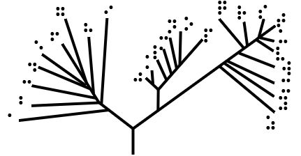
A phylogenetic tree (also phylogeny or evolutionary tree Felsenstein J. (2004). ''Inferring Phylogenies'' Sinauer Associates: Sunderland, MA.) is a branching diagram or a tree showing the evolutionary relationships among various biological species or other entities based upon similarities and differences in their physical or genetic characteristics. All life on Earth is part of a single phylogenetic tree, indicating common ancestry. In a ''rooted'' phylogenetic tree, each node with descendants represents the inferred most recent common ancestor of those descendants, and the edge lengths in some trees may be interpreted as time estimates. Each node is called a taxonomic unit. Internal nodes are generally called hypothetical taxonomic units, as they cannot be directly observed. Trees are useful in fields of biology such as bioinformatics, systematics, and phylogenetics. ''Unrooted'' trees illustrate only the relatedness of the leaf nodes and do not require the ancestral root to b ... [...More Info...] [...Related Items...] OR: [Wikipedia] [Google] [Baidu] |
C-terminus
The C-terminus (also known as the carboxyl-terminus, carboxy-terminus, C-terminal tail, C-terminal end, or COOH-terminus) is the end of an amino acid chain (protein or polypeptide), terminated by a free carboxyl group (-COOH). When the protein is translated from messenger RNA, it is created from N-terminus to C-terminus. The convention for writing peptide sequences is to put the C-terminal end on the right and write the sequence from N- to C-terminus. Chemistry Each amino acid has a carboxyl group and an amine group. Amino acids link to one another to form a chain by a dehydration reaction which joins the amine group of one amino acid to the carboxyl group of the next. Thus polypeptide chains have an end with an unbound carboxyl group, the C-terminus, and an end with an unbound amine group, the N-terminus. Proteins are naturally synthesized starting from the N-terminus and ending at the C-terminus. Function C-terminal retention signals While the N-terminus of a protein often c ... [...More Info...] [...Related Items...] OR: [Wikipedia] [Google] [Baidu] |
Coiled Coil
A coiled coil is a structural motif in proteins in which 2–7 alpha-helices are coiled together like the strands of a rope. (Dimers and trimers are the most common types.) Many coiled coil-type proteins are involved in important biological functions, such as the regulation of gene expression — e.g., transcription factors. Notable examples are the oncoproteins c-Fos and c-Jun, as well as the muscle protein tropomyosin. Discovery The possibility of coiled coils for α-keratin was initially somewhat controversial. Linus Pauling and Francis Crick independently came to the conclusion that this was possible at about the same time. In the summer of 1952, Pauling visited the laboratory in England where Crick worked. Pauling and Crick met and spoke about various topics; at one point, Crick asked whether Pauling had considered "coiled coils" (Crick came up with the term), to which Pauling said he had. Upon returning to the United States, Pauling resumed research on the topic. He conc ... [...More Info...] [...Related Items...] OR: [Wikipedia] [Google] [Baidu] |
EF Hand
The EF hand is a helix–loop–helix structural domain or ''motif'' found in a large family of calcium-binding proteins. The EF-hand motif contains a helix–loop–helix topology, much like the spread thumb and forefinger of the human hand, in which the Ca2+ ions are coordinated by ligands within the loop. The motif takes its name from traditional nomenclature used in describing the protein parvalbumin, which contains three such motifs and is probably involved in muscle relaxation via its calcium-binding activity. The EF-hand consists of two alpha helices linked by a short loop region (usually about 12 amino acids) that usually binds calcium ions. EF-hands also appear in each structural domain of the signaling protein calmodulin and in the muscle protein troponin-C. Calcium ion binding site The calcium ion is coordinated in a pentagonal bipyramidal configuration. The six residues involved in the binding are in positions 1, 3, 5, 7, 9 and 12; these residues are denoted by X ... [...More Info...] [...Related Items...] OR: [Wikipedia] [Google] [Baidu] |
Zinc Finger
A zinc finger is a small protein structural motif that is characterized by the coordination of one or more zinc ions (Zn2+) in order to stabilize the fold. It was originally coined to describe the finger-like appearance of a hypothesized structure from the African clawed frog (''Xenopus laevis'') transcription factor IIIA. However, it has been found to encompass a wide variety of differing protein structures in eukaryotic cells. ''Xenopus laevis'' TFIIIA was originally demonstrated to contain zinc and require the metal for function in 1983, the first such reported zinc requirement for a gene regulatory protein followed soon thereafter by the Krüppel factor in ''Drosophila''. It often appears as a metal-binding domain in multi-domain proteins. Proteins that contain zinc fingers (zinc finger proteins) are classified into several different structural families. Unlike many other clearly defined supersecondary structures such as Greek keys or β hairpins, there are a number of t ... [...More Info...] [...Related Items...] OR: [Wikipedia] [Google] [Baidu] |
Alternative Splicing
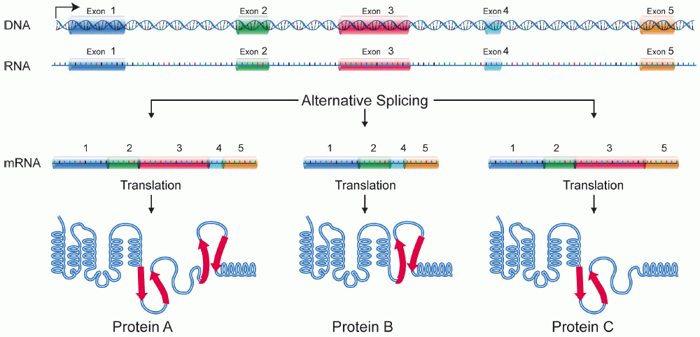
Alternative splicing, or alternative RNA splicing, or differential splicing, is an alternative splicing process during gene expression that allows a single gene to code for multiple proteins. In this process, particular exons of a gene may be included within or excluded from the final, processed messenger RNA (mRNA) produced from that gene. This means the exons are joined in different combinations, leading to different (alternative) mRNA strands. Consequently, the proteins translated from alternatively spliced mRNAs will contain differences in their amino acid sequence and, often, in their biological functions (see Figure). Biologically relevant alternative splicing occurs as a normal phenomenon in eukaryotes, where it increases the number of proteins that can be encoded by the genome. In humans, it is widely believed that ~95% of multi-exonic genes are alternatively spliced to produce functional alternative products from the same gene but many scientists believe that most o ... [...More Info...] [...Related Items...] OR: [Wikipedia] [Google] [Baidu] |
Protein Dimer
In biochemistry, a protein dimer is a macromolecular complex formed by two protein monomers, or single proteins, which are usually non-covalently bound. Many macromolecules, such as proteins or nucleic acids, form dimers. The word ''dimer'' has roots meaning "two parts", '' di-'' + '' -mer''. A protein dimer is a type of protein quaternary structure. A protein homodimer is formed by two identical proteins. A protein heterodimer is formed by two different proteins. Most protein dimers in biochemistry are not connected by covalent bonds. An example of a non-covalent heterodimer is the enzyme reverse transcriptase, which is composed of two different amino acid chains. An exception is dimers that are linked by disulfide bridges such as the homodimeric protein NEMO. Some proteins contain specialized domains to ensure dimerization (dimerization domains) and specificity. The G protein-coupled cannabinoid receptors have the ability to form both homo- and heterodimers with several ... [...More Info...] [...Related Items...] OR: [Wikipedia] [Google] [Baidu] |
Utrophin
Utrophin is a protein that in humans is encoded by the ''UTRN'' gene. The protein encoded by this gene is a component of the cytoskeleton. Utrophin was found during research into Duchenne's muscular dystrophy. The name is a contraction for ''ubiquitous dystrophin''. The 900 kb gene for utrophin is found on the long arm of human chromosome 6. Utrophin was discovered due to its homology with dystrophin. It was found by screening a peptide containing the C-terminal domain of dystrophin against cDNA libraries. The homology varies over its full length from less than 30% in regions of the central rod structural domain to 85% (identity 73%) for the actin binding domain. The tertiary structure of utrophin contains a C-terminus that consists of protein–protein interaction motifs that interact with dystroglycan, a central rod region consisting of a triple coiled-coil repeat, and an actin-binding N-terminus. In normal muscle cells, utrophin is located at the neuromuscular synapse and my ... [...More Info...] [...Related Items...] OR: [Wikipedia] [Google] [Baidu] |


