|
Ductus Venosus
In the fetus, the ''ductus venosus'' (Arantius' duct after Julius Caesar Aranzi) shunts a portion of umbilical vein blood flow directly to the inferior vena cava. Thus, it allows oxygenated blood from the placenta to bypass the liver. Compared to the 50% shunting of umbilical blood through the ''ductus venosus'' found in animal experiments, the degree of shunting in the human fetus under physiological conditions is considerably less, 30% at 20 weeks, which decreases to 18% at 32 weeks, suggesting a higher priority of the fetal liver than previously realized. In conjunction with the other fetal shunts, the ''foramen ovale'' and ''ductus arteriosus'', it plays a critical role in preferentially shunting oxygenated blood to the fetal brain. It is a part of fetal circulation. Anatomic course The pathway of fetal umbilical venous flow is umbilical vein to left portal vein to ''ductus venosus'' to ''inferior vena cava'' and eventually the right atrium. This anatomic course is important ... [...More Info...] [...Related Items...] OR: [Wikipedia] [Google] [Baidu] |
Fetal Circulation
In humans, the circulatory system is different before and after birth. The fetal circulation is composed of the placenta, umbilical blood vessels encapsulated by the umbilical cord, heart and systemic blood vessels. A major difference between the fetal circulation and postnatal circulation is that the lungs are not used during the fetal stage resulting in the presence of shunts to move oxygenated blood and nutrients from the placenta to the fetal tissue. At birth, the start of breathing and the severance of the umbilical cord prompt various changes that quickly transforms fetal circulation into postnatal circulation. Oxygenation, nutrient, and waste exchange Placenta The placenta functions as the exchange site of nutrients and wastes between the maternal and fetal circulation. Water, glucose, amino acids, vitamins, and inorganic salts freely diffuse across the placenta along with oxygen. Two umbilical arteries carry deoxygenated blood and waste from the fetus to the placenta ... [...More Info...] [...Related Items...] OR: [Wikipedia] [Google] [Baidu] |
Foramen Ovale (heart)
In the fetal heart, the foramen ovale (), also foramen Botalli, or the ostium secundum of Born, allows blood to enter the left atrium from the right atrium. It is one of two fetal cardiac shunts, the other being the ductus arteriosus (which allows blood that still escapes to the right ventricle to bypass the pulmonary circulation). Another similar adaptation in the fetus is the ductus venosus. In most individuals, the foramen ovale closes at birth. It later forms the fossa ovalis. Development The foramen ovale () forms in the late fourth week of gestation, as a small passageway between the septum secundum and the ostium secundum. Initially the atria are separated from one another by the septum primum except for a small opening below the septum, the ostium primum. As the septum primum grows, the ostium primum narrows and eventually closes. Before it does so, bloodflow from the inferior vena cava wears down a portion of the septum primum, forming the ostium secundum. So ... [...More Info...] [...Related Items...] OR: [Wikipedia] [Google] [Baidu] |
Preterm Infant
Preterm birth, also known as premature birth, is the birth of a baby at fewer than 37 weeks gestational age, as opposed to full-term delivery at approximately 40 weeks. Extreme preterm is less than 28 weeks, very early preterm birth is between 28 and 32 weeks, early preterm birth occurs between 32 and 36 weeks, late preterm birth is between 34 and 36 weeks' gestation. These babies are also known as premature babies or colloquially preemies (American English) or premmies (Australian English). Symptoms of preterm labor include uterine contractions which occur more often than every ten minutes and/or the leaking of fluid from the vagina before 37 weeks. Premature infants are at greater risk for cerebral palsy, delays in development, hearing problems and problems with their vision. The earlier a baby is born, the greater these risks will be. The cause of spontaneous preterm birth is often not known. Risk factors include diabetes, high blood pressure, multiple gestation (being preg ... [...More Info...] [...Related Items...] OR: [Wikipedia] [Google] [Baidu] |
Portosystemic Shunt
A portosystemic shunt or portasystemic shunt (medical subject heading term; PSS), also known as a liver shunt, is a bypass of the liver by the body's circulatory system. It can be either a congenital (present at birth) or acquired condition and occurs in humans as well as in other species of animals. Congenital PSS are extremely rare in humans but are relatively common in dogs. Thus a large part of medical and scientific literature on the subject is grounded in veterinary medicine. Background Blood leaving the digestive tract is rich in nutrients, as well as in toxins, which under normal conditions undergo processing and detoxification in the liver. The liver's position downstream to the intestines in the body's circulatory system - the hepatic portal vein conveys blood from the intestines to the liver - allows it to filter this nutrient rich blood before it passes to the rest of the body. The presence of a shunt, a bypass of the liver, causes blood to flow directly to the hear ... [...More Info...] [...Related Items...] OR: [Wikipedia] [Google] [Baidu] |
Ligamentum Venosum
The ligamentum venosum, also known as Arantius' ligament, is the fibrous remnant of the ductus venosus of the fetal circulation. Usually, it is attached to the left branch of the portal vein within the porta hepatis. It may be continuous with the round ligament of liver. It is invested by the peritoneal folds of the lesser omentum within a fissure on the visceral/posterior surface of the liver between the caudate and main parts of the left lobe. It is grouped with the liver in ''Terminologia Anatomica''. See also * Ligamentum teres * Ligamentum arteriosum The ligamentum arteriosum (arterial ligament), also known as the Ligament of Botallo or Harvey's ligament, is a small ligament attaching the aorta to the pulmonary artery. It serves no function in adults but is the remnant of the ductus arteriosus ... References External links * () {{Authority control Abdomen Ligaments ... [...More Info...] [...Related Items...] OR: [Wikipedia] [Google] [Baidu] |
Catheterization
In medicine, a catheter (/ˈkæθətər/) is a thin tube made from medical grade materials serving a broad range of functions. Catheters are medical devices that can be inserted in the body to treat diseases or perform a surgical procedure. Catheters are manufactured for specific applications, such as cardiovascular, urological, gastrointestinal, neurovascular and ophthalmic procedures. The process of inserting a catheter is ''catheterization''. In most uses, a catheter is a thin, flexible tube (''soft'' catheter) though catheters are available in varying levels of stiffness depending on the application. A catheter left inside the body, either temporarily or permanently, may be referred to as an "indwelling catheter" (for example, a peripherally inserted central catheter). A permanently inserted catheter may be referred to as a "permcath" (originally a trademark). Catheters can be inserted into a body cavity, duct, or vessel, brain, skin or adipose tissue. Functionally, they a ... [...More Info...] [...Related Items...] OR: [Wikipedia] [Google] [Baidu] |
Fetal Circulation
In humans, the circulatory system is different before and after birth. The fetal circulation is composed of the placenta, umbilical blood vessels encapsulated by the umbilical cord, heart and systemic blood vessels. A major difference between the fetal circulation and postnatal circulation is that the lungs are not used during the fetal stage resulting in the presence of shunts to move oxygenated blood and nutrients from the placenta to the fetal tissue. At birth, the start of breathing and the severance of the umbilical cord prompt various changes that quickly transforms fetal circulation into postnatal circulation. Oxygenation, nutrient, and waste exchange Placenta The placenta functions as the exchange site of nutrients and wastes between the maternal and fetal circulation. Water, glucose, amino acids, vitamins, and inorganic salts freely diffuse across the placenta along with oxygen. Two umbilical arteries carry deoxygenated blood and waste from the fetus to the placenta ... [...More Info...] [...Related Items...] OR: [Wikipedia] [Google] [Baidu] |
Ductus Arteriosus
The ''ductus arteriosus'', also called the ''ductus Botalli'', named after the Italian physiologist Leonardo Botallo, is a blood vessel in the developing fetus connecting the trunk of the pulmonary artery to the proximal descending aorta. It allows most of the blood from the right ventricle to bypass the fetus's fluid-filled non-functioning lungs. Upon closure at birth, it becomes the ''ligamentum arteriosum''. Development and structure The ''ductus arteriosus'' is formed from the left 6th aortic arch during embryonic development and attaches to the final part of the aortic arch (the isthmus of aorta) and the first part of the pulmonary artery. Disorder: Patent ductus arteriosus Consequences Failure of the ''ductus arteriosus'' to close after birth results in a condition called ''patent ductus arteriosus'', which results in the abnormal flow of blood from the aorta to the pulmonary artery: a left-to-right shunt. If left uncorrected, this usually leads to pulmonary hyperten ... [...More Info...] [...Related Items...] OR: [Wikipedia] [Google] [Baidu] |
Placenta
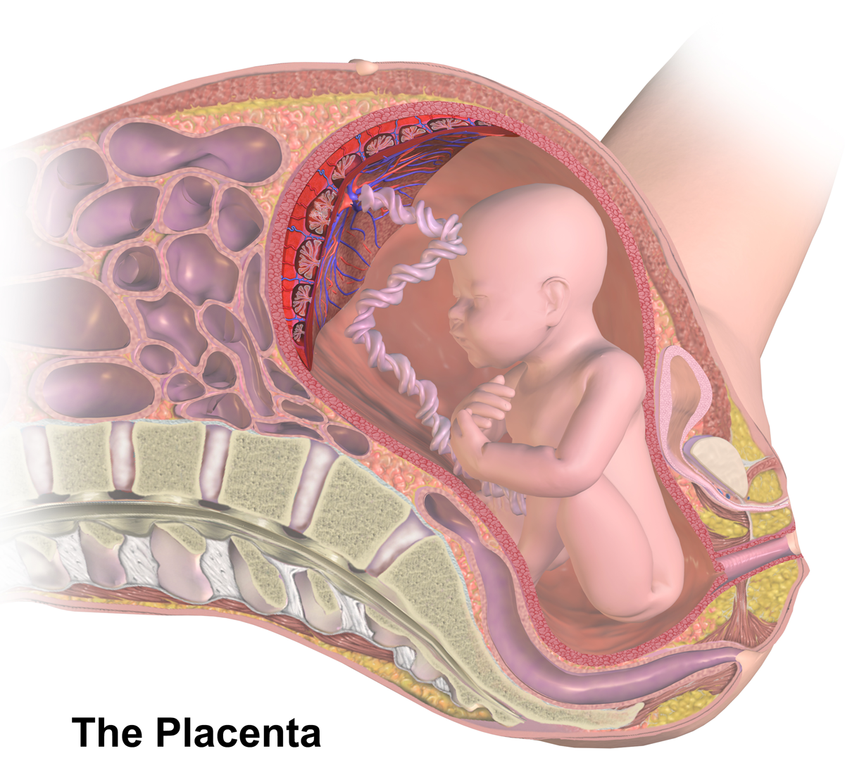
The placenta is a temporary embryonic and later fetal organ (anatomy), organ that begins embryonic development, developing from the blastocyst shortly after implantation (embryology), implantation. It plays critical roles in facilitating nutrient, gas and waste exchange between the physically separate maternal and fetal circulations, and is an important Endocrine system, endocrine organ, producing hormones that regulate both Maternal physiological changes in pregnancy, maternal and fetal physiology during pregnancy. The placenta connects to the fetus via the umbilical cord, and on the opposite aspect to the maternal uterus in a species-dependent manner. In humans, a thin layer of maternal decidual (Endometrium, endometrial) tissue comes away with the placenta when it is expelled from the uterus following birth (sometimes incorrectly referred to as the 'maternal part' of the placenta). Placentas are a defining characteristic of placental mammals, but are also found in marsupials an ... [...More Info...] [...Related Items...] OR: [Wikipedia] [Google] [Baidu] |
Umbilical Vein
The umbilical vein is a vein present during fetal development that carries oxygenated blood from the placenta into the growing fetus. The umbilical vein provides convenient access to the central circulation of a neonate for restoration of blood volume and for administration of glucose and drugs. The blood pressure inside the umbilical vein is approximately 20 mmHg.Wang, Y. Vascular biology of the placenta. in Colloquium Series on Integrated Systems Physiology: from Molecule to Function. 2010. Morgan & Claypool Life Sciences. Fetal circulation The unpaired umbilical vein carries oxygen and nutrient rich blood derived from fetal-maternal blood exchange at the chorionic villi. More than two-thirds of fetal hepatic circulation is via the main portal vein, while the remainder is shunted from the left portal vein via the ductus venosus to the inferior vena cava, eventually being delivered to the fetal right atrium. Closure Closure of the umbilical vein usually occurs after the umbi ... [...More Info...] [...Related Items...] OR: [Wikipedia] [Google] [Baidu] |
Julius Caesar Aranzi
Julius Caesar Aranzi (Giulio Cesare Aranzio, Arantius) (1529/1530 – April 7, 1589) was a leading figure in the history of the science of human anatomy. He was born in Bologna, the son of Ottaviano di Jacopo and Maria Maggi. Owing to the poverty of the family, he studied with his uncle Bartolomeo Maggi (1477–1552), a famous surgeon who was a lecturer at the University of Bologna as well as court physician to Julius III. He held this uncle in such high esteem that he assumed his surname, calling himself Giulio Cesare Aranzio Maggio. He was admitted to the University of Padua, where he made his first discovery in 1548, at the age of nineteen, when he described the elevator muscle of the upper eyelid. Later, at the University of Bologna, he received a doctorate in medicine in 1556 and was appointed a lecturer in medicine and surgery shortly thereafter at the age of twenty-seven. In 1570, surgery and anatomy were separated into separate professorships at his instigation and he hel ... [...More Info...] [...Related Items...] OR: [Wikipedia] [Google] [Baidu] |


