|
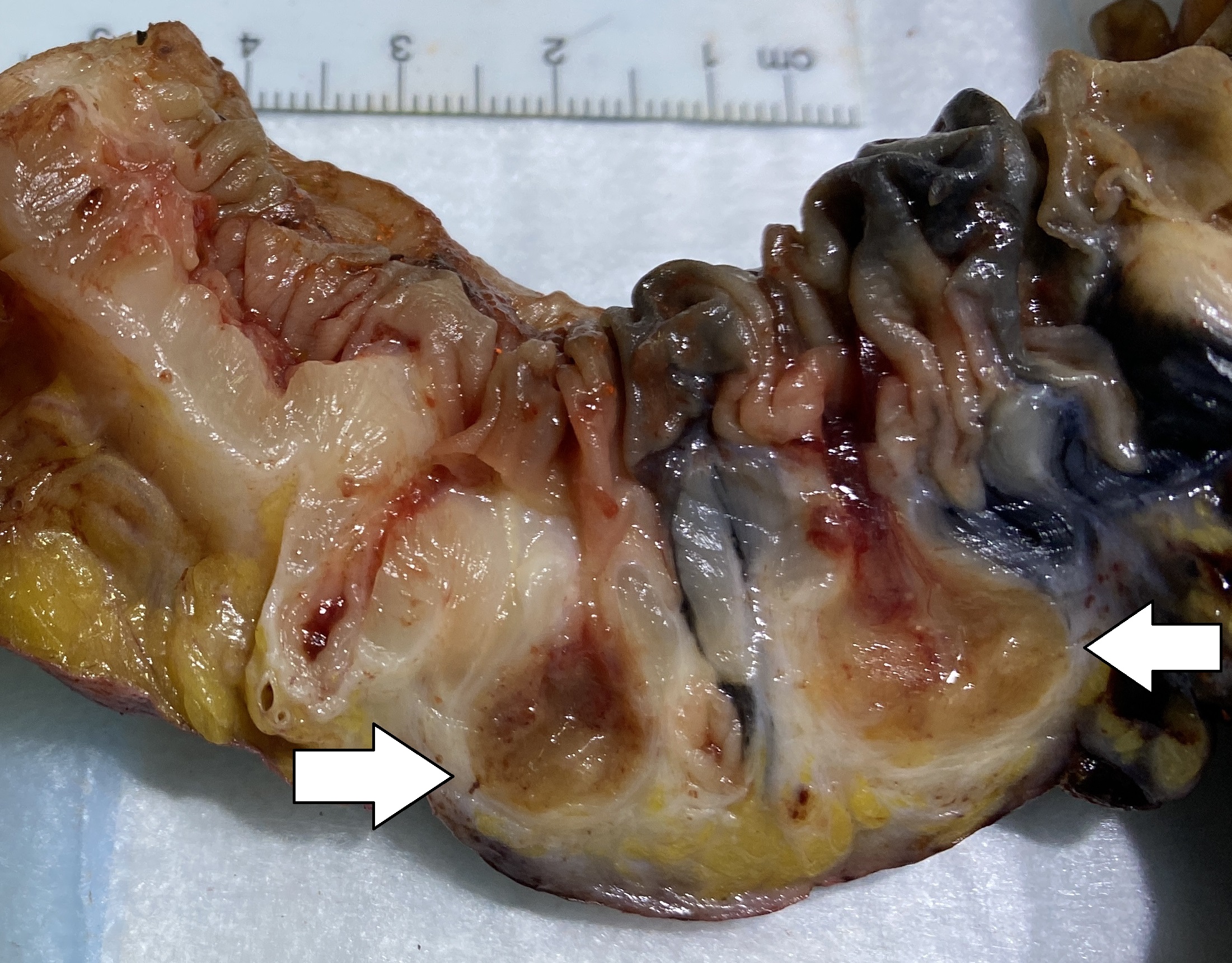
Diverticula
In medicine or biology, a diverticulum is an outpouching of a hollow (or a fluid-filled) structure in the body. Depending upon which layers of the structure are involved, diverticula are described as being either true or false. In medicine, the term usually implies the structure is not normally present, but in embryology, the term is used for some normal structures arising from others, as for instance the thyroid diverticulum, which arises from the tongue. The word comes from Latin ''dīverticulum'', "bypath" or "byway". Classification Diverticula are described as being true or false depending upon the layers involved: *False diverticula (also known as "pseudodiverticula") do not involve muscular layers or adventitia. False diverticula, in the gastrointestinal tract for instance, involve only the submucosa and mucosa. *True diverticula involve all layers of the structure, including muscularis propria and adventitia, such as Meckel's diverticulum. Embryology *The kidney ... [...More Info...] [...Related Items...] OR: [Wikipedia] [Google] [Baidu] |
Diverticulosis
Diverticulosis is the condition of having multiple pouches ( diverticula) in the colon that are not inflamed. These are outpockets of the colonic mucosa and submucosa through weaknesses of muscle layers in the colon wall. Diverticula do not cause symptoms in most people. Diverticular disease occurs when diverticula become clinically inflamed, a condition known as diverticulitis. Diverticula typically occur in the sigmoid colon, which is commonplace for increased pressure. The left side of the colon is more commonly affected in the United States while the right side is more commonly affected in Asia. Diagnosis is often during routine colonoscopy or as an incidental finding during CT scan. It is common in Western countries with about half of those over the age of 60 affected in Canada and the United States. Diverticula are uncommon before the age of 40, and increase in incidence beyond that age. Rates are lower in Africa; the reasons for this remain unclear but may involve th ... [...More Info...] [...Related Items...] OR: [Wikipedia] [Google] [Baidu] |
Diverticulitis
Diverticulitis, specifically colonic diverticulitis, is a gastrointestinal disease characterized by inflammation of abnormal pouches— diverticula—which can develop in the wall of the large intestine. Symptoms typically include lower abdominal pain of sudden onset, but the onset may also occur over a few days. There may also be nausea; and diarrhea or constipation. Fever or blood in the stool suggests a complication. Repeated attacks may occur. The causes of diverticulitis are unclear. Risk factors may include obesity, lack of exercise, smoking, a family history of the disease, and use of nonsteroidal anti-inflammatory drugs (NSAIDs). The role of a low fiber diet as a risk factor is unclear. Having pouches in the large intestine that are not inflamed is known as diverticulosis. Inflammation occurs in between 10% and 25% at some point in time, and is due to a bacterial infection. Diagnosis is typically by CT scan, though blood tests, colonoscopy, or a lower gastrointesti ... [...More Info...] [...Related Items...] OR: [Wikipedia] [Google] [Baidu] |
Meckel's Diverticulum
A Meckel's diverticulum, a true congenital diverticulum, is a slight bulge in the small intestine present at birth and a vestigial remnant of the omphalomesenteric duct (also called the vitelline duct or yolk stalk). It is the most common malformation of the Human gastrointestinal tract, gastrointestinal tract and is present in approximately 2% of the population, with males more frequently experiencing symptoms. Meckel's diverticulum was first explained by Fabricius Hildanus in the sixteenth century and later named after Johann Friedrich Meckel, who described the embryological origin of this type of diverticulum in 1809. Signs and symptoms The majority of people with a Meckel's diverticulum are asymptomatic. An asymptomatic Meckel's diverticulum is called a ''silent'' Meckel's diverticulum. If symptoms do occur, they typically appear before the age of two years. The most common presenting symptom is painless rectal bleeding such as melaena-like black offensive stools, followed by i ... [...More Info...] [...Related Items...] OR: [Wikipedia] [Google] [Baidu] |
Rokitansky-Aschoff Sinuses
Adenomyomatosis is a benign condition characterized by hyperplastic changes of unknown cause involving the wall of the gallbladder. Adenomyomatosis is caused by an overgrowth of the mucosa, thickening of the muscular wall, and formation of intramural diverticula or sinus tracts termed Rokitansky–Aschoff sinuses, also called entrapped epithelial crypts. Signs and symptoms Pathophysiology Rokitansky–Aschoff sinuses Rokitansky–Aschoff sinuses are pseudodiverticula or pockets in the wall of the gallbladder. They may be microscopic or macroscopic. Histologically, they are outpouchings of gallbladder mucosa into the gallbladder muscle layer and subserosal tissue as a result of hyperplasia and herniation of epithelial cells through the fibromuscular layer of the gallbladder wall. Rokitansky–Aschoff sinuses are not of themselves considered abnormal but they can be associated with cholecystitis. They form as a result of increased pressure in the gallbladder and recurrent dama ... [...More Info...] [...Related Items...] OR: [Wikipedia] [Google] [Baidu] |
Zenker's Diverticulum
A Zenker's diverticulum, also pharyngeal pouch, is a diverticulum of the mucosa of the human pharynx, just above the cricopharyngeal muscle (i.e. above the upper sphincter of the esophagus). It is a pseudo diverticulum (not involving all layers of the esophageal wall). It was named in 1877 after German pathologist Friedrich Albert von Zenker. Signs and symptoms In simple words, when there is excessive pressure within the lower pharynx, the weakest portion of the pharyngeal wall balloons out, forming a diverticulum which may reach several centimetres in diameter. More precisely, while traction and pulsion mechanisms have long been deemed the main factors promoting development of a Zenker's diverticulum, current consensus considers occlusive mechanisms to be most important: uncoordinated swallowing, impaired relaxation and spasm of the cricopharyngeus muscle lead to an increase in pressure within the distal pharynx, so that its wall herniates through the point of least resist ... [...More Info...] [...Related Items...] OR: [Wikipedia] [Google] [Baidu] |
Vitelline Duct
In the human embryo, the vitelline duct, also known as the vitellointestinal duct, the yolk stalk, the omphaloenteric duct, or the omphalomesenteric duct, is a long narrow tube that joins the yolk sac to the midgut lumen of the developing fetus. It appears at the end of the fourth week, when the yolk sac presents the appearance of a small pear-shaped vesicle (the umbilical vesicle). Function Obliteration Generally, the duct fully obliterates (narrows and disappears) during the 5–6th week of fertilization age (9th week of gestational age), but a failure of the duct to close is termed a vitelline fistula. This results in discharge of meconium from the umbilicus. About two percent of fetuses exhibit a type of vitelline fistula characterized by persistence of the proximal part of the vitelline duct as a diverticulum protruding from the small intestine, Meckel's diverticulum, which is typically situated within two feet of the ileocecal junction and may be attached by a fibrous co ... [...More Info...] [...Related Items...] OR: [Wikipedia] [Google] [Baidu] |
Development Of The Urinary And Reproductive Organs
The development of the urinary system begins during prenatal development, and relates to the development of the urogenital system – both the organs of the urinary system and the sex organs of the reproductive system. The development continues as a part of sexual differentiation. The urinary and reproductive organs are developed from the intermediate mesoderm. The permanent organs of the adult are preceded by a set of structures which are purely embryonic, and which with the exception of the ducts disappear almost entirely before birth. These embryonic structures are on either side; the pronephros, the mesonephros and the metanephros of the kidney, and the Wolffian and Müllerian ducts of the sex organ. The pronephros disappears very early; the structural elements of the mesonephros mostly degenerate, but the gonad is developed in their place, with which the Wolffian duct remains as the duct in males, and the Müllerian as that of the female. Some of the tubules of the mesone ... [...More Info...] [...Related Items...] OR: [Wikipedia] [Google] [Baidu] |
Oesophageal Diverticula
The esophagus (American English) or oesophagus (British English; both ), non-technically known also as the food pipe or gullet, is an organ in vertebrates through which food passes, aided by peristaltic contractions, from the pharynx to the stomach. The esophagus is a fibromuscular tube, about long in adults, that travels behind the trachea and heart, passes through the diaphragm, and empties into the uppermost region of the stomach. During swallowing, the epiglottis tilts backwards to prevent food from going down the larynx and lungs. The word ''oesophagus'' is from Ancient Greek οἰσοφάγος (oisophágos), from οἴσω (oísō), future form of φέρω (phérō, “I carry”) + ἔφαγον (éphagon, “I ate”). The wall of the esophagus from the lumen outwards consists of mucosa, submucosa (connective tissue), layers of muscle fibers between layers of fibrous tissue, and an outer layer of connective tissue. The mucosa is a stratified squamous epithelium ... [...More Info...] [...Related Items...] OR: [Wikipedia] [Google] [Baidu] |
Histopathology Of A False Diverticulum Of The Gallbladder
Histopathology (compound of three Greek words: ''histos'' "tissue", πάθος ''pathos'' "suffering", and -λογία ''-logia'' "study of") refers to the microscopic examination of tissue in order to study the manifestations of disease. Specifically, in clinical medicine, histopathology refers to the examination of a biopsy or surgical specimen by a pathologist, after the specimen has been processed and histological sections have been placed onto glass slides. In contrast, cytopathology examines free cells or tissue micro-fragments (as "cell blocks"). Collection of tissues Histopathological examination of tissues starts with surgery, biopsy, or autopsy. The tissue is removed from the body or plant, and then, often following expert dissection in the fresh state, placed in a fixative which stabilizes the tissues to prevent decay. The most common fixative is 10% neutral buffered formalin (corresponding to 3.7% w/v formaldehyde in neutral buffered water, such as phosphate buf ... [...More Info...] [...Related Items...] OR: [Wikipedia] [Google] [Baidu] |
Windsock
A windsock (also called a wind cone) is a conical textile tube that resembles a giant sock. It can be used as a basic indicator of wind speed and direction, or as decoration. They are typically used at airports to show the direction and strength of the wind to pilots, and at chemical plants where there is risk of gaseous leakage. They are also sometimes located alongside highways at windy locations. At many airports, windsocks are externally or internally lit at night. Function Wind direction is the direction in which the windsock is pointing. (Wind directions are conventionally specified as the compass point from which the wind originates; so a windsock pointing due north indicates a southerly wind). Wind speed is indicated by the windsock's angle relative to the mounting pole; in low winds it droops; in high winds it flies horizontally. Alternating stripes of high visibility orange and white were initially used to help to estimate wind speed, with each stripe adding 3 kn ... [...More Info...] [...Related Items...] OR: [Wikipedia] [Google] [Baidu] |
Ampulla Of Vater
The ampulla of Vater, also known as the or the hepatopancreatic duct, is formed by the union of the pancreatic duct and the common bile duct. The ampulla is specifically located at the major duodenal papilla. The ampulla of Vater is an important landmark halfway along the second part of the duodenum that marks the anatomical transition from foregut to midgut, and hence the point where the celiac trunk stops supplying the gut and the superior mesenteric artery takes over. Structure The cystic duct leaves the gallbladder and joins with the common hepatic duct to form the common bile duct. This duct subsequently joins with the pancreatic duct; this junction is known as the ampulla of Vater. The pancreatic duct delivers substances such as bicarbonate and digestive enzymes to the duodenum. The bile from the gallbladder contains salts which emulsify large fat droplets into much smaller units. This provides a large surface area for the lipase enzymes to act on. The bicarbonate n ... [...More Info...] [...Related Items...] OR: [Wikipedia] [Google] [Baidu] |


