|
Rokitansky-Aschoff Sinuses
Adenomyomatosis is a benign condition characterized by hyperplastic changes of unknown cause involving the wall of the gallbladder. Adenomyomatosis is caused by an overgrowth of the mucosa, thickening of the muscular wall, and formation of intramural diverticula or sinus tracts termed Rokitansky–Aschoff sinuses, also called entrapped epithelial crypts. Signs and symptoms Pathophysiology Rokitansky–Aschoff sinuses Rokitansky–Aschoff sinuses are pseudodiverticula or pockets in the wall of the gallbladder. They may be microscopic or macroscopic. Histologically, they are outpouchings of gallbladder mucosa into the gallbladder muscle layer and subserosal tissue as a result of hyperplasia and herniation of epithelial cells through the fibromuscular layer of the gallbladder wall. Rokitansky–Aschoff sinuses are not of themselves considered abnormal but they can be associated with cholecystitis. They form as a result of increased pressure in the gallbladder and recurrent dam ... [...More Info...] [...Related Items...] OR: [Wikipedia] [Google] [Baidu] |
Uterus
The uterus (from Latin ''uterus'', plural ''uteri'') or womb () is the organ in the reproductive system of most female mammals, including humans that accommodates the embryonic and fetal development of one or more embryos until birth. The uterus is a hormone-responsive sex organ that contains glands in its lining that secrete uterine milk for embryonic nourishment. In the human, the lower end of the uterus, is a narrow part known as the isthmus that connects to the cervix, leading to the vagina. The upper end, the body of the uterus, is connected to the fallopian tubes, at the uterine horns, and the rounded part above the openings to the fallopian tubes is the fundus. The connection of the uterine cavity with a fallopian tube is called the uterotubal junction. The fertilized egg is carried to the uterus along the fallopian tube. It will have divided on its journey to form a blastocyst that will implant itself into the lining of the uterus – the endometrium, where it will ... [...More Info...] [...Related Items...] OR: [Wikipedia] [Google] [Baidu] |
Abdominal Ultrasound
Abdominal ultrasonography (also called abdominal ultrasound imaging or abdominal sonography) is a form of medical ultrasonography (medical application of ultrasound technology) to visualise abdominal anatomical structures. It uses transmission and reflection of ultrasound waves to visualise internal organs through the abdominal wall (with the help of gel, which helps transmission of the sound waves). For this reason, the procedure is also called a transabdominal ultrasound, in contrast to endoscopic ultrasound, the latter combining ultrasound with endoscopy through visualize internal structures from within hollow organs. Abdominal ultrasound examinations are performed by gastroenterologists or other specialists in internal medicine, radiologists, or sonographers trained for this procedure. Medical uses Abdominal ultrasound can be used to diagnose abnormalities in various internal organs, such as the kidneys, liver, gallbladder, pancreas, spleen and abdominal aorta. If Doppler ... [...More Info...] [...Related Items...] OR: [Wikipedia] [Google] [Baidu] |
Gallbladder
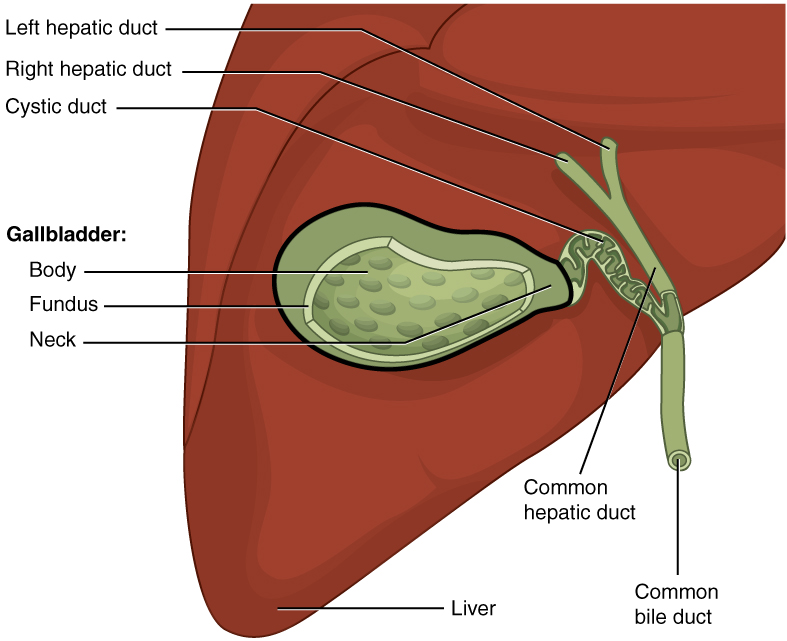
In vertebrates, the gallbladder, also known as the cholecyst, is a small hollow organ where bile is stored and concentrated before it is released into the small intestine. In humans, the pear-shaped gallbladder lies beneath the liver, although the structure and position of the gallbladder can vary significantly among animal species. It receives and stores bile, produced by the liver, via the common hepatic duct, and releases it via the common bile duct into the duodenum, where the bile helps in the digestion of fats. The gallbladder can be affected by gallstones, formed by material that cannot be dissolved – usually cholesterol or bilirubin, a product of haemoglobin breakdown. These may cause significant pain, particularly in the upper-right corner of the abdomen, and are often treated with removal of the gallbladder (called a cholecystectomy). Cholecystitis, inflammation of the gallbladder, has a wide range of causes, including result from the impaction of gallstones, inf ... [...More Info...] [...Related Items...] OR: [Wikipedia] [Google] [Baidu] |
Diverticulum
In medicine or biology, a diverticulum is an outpouching of a hollow (or a fluid-filled) structure in the body. Depending upon which layers of the structure are involved, diverticula are described as being either true or false. In medicine, the term usually implies the structure is not normally present, but in embryology, the term is used for some normal structures arising from others, as for instance the thyroid diverticulum, which arises from the tongue. The word comes from Latin ''dīverticulum'', "bypath" or "byway". Classification Diverticula are described as being true or false depending upon the layers involved: *False diverticula (also known as "pseudodiverticula") do not involve muscular layers or adventitia. False diverticula, in the gastrointestinal tract for instance, involve only the submucosa and mucosa. *True diverticula involve all layers of the structure, including muscularis propria and adventitia, such as Meckel's diverticulum. Embryology *The kidneys are ... [...More Info...] [...Related Items...] OR: [Wikipedia] [Google] [Baidu] |
Strawberry Gallbladder
In surgical pathology, strawberry gallbladder, more formally cholesterolosis of the gallbladder and gallbladder cholesterolosis, is a change in the gallbladder wall due to excess cholesterol. - cancerweb.ncl.ac.uk. The name ''strawberry gallbladder'' comes from the typically stippled appearance of the l surface on , which resembles a . Cholesterolosis results from abnormal deposits of cholesterol esters in macrophages with ... [...More Info...] [...Related Items...] OR: [Wikipedia] [Google] [Baidu] |
Cholecystectomy
Cholecystectomy is the surgical removal of the gallbladder. Cholecystectomy is a common treatment of symptomatic gallstones and other gallbladder conditions. In 2011, cholecystectomy was the eighth most common operating room procedure performed in hospitals in the United States. Cholecystectomy can be performed either laparoscopically, or via an open surgical technique. The surgery is usually successful in relieving symptoms, but up to 10 percent of people may continue to experience similar symptoms after cholecystectomy, a condition called postcholecystectomy syndrome. Complications of cholecystectomy include bile duct injury, wound infection, bleeding, retained gallstones, abscess formation and stenosis (narrowing) of the bile duct. Medical use Pain and complications caused by gallstones are the most common reasons for removal of the gallbladder. The gallbladder can also be removed in order to treat biliary dyskinesia or gallbladder cancer. Gallstones are very common but ... [...More Info...] [...Related Items...] OR: [Wikipedia] [Google] [Baidu] |
Ludwig Aschoff
Karl Albert Ludwig Aschoff (10 January 1866 – 24 June 1942) was a German physician and pathologist. He is considered to be one of the most influential pathologists of the early 20th century and is regarded as the most important German pathologist after Rudolf Virchow. Early life and education Aschoff was born in Berlin, Prussia on 10 January 1866. He studied medicine at the University of Bonn, University of Strasbourg, and the University of Würzburg. Career After his habilitation in 1894, Ludwig Aschoff was appointed professor for pathology at the University of Göttingen in 1901. Aschoff transferred to the University of Marburg in 1903 to head the department for pathological anatomy. In 1906, he accepted a position as ordinarius at the University of Freiburg, where he remained until his death. Aschoff was interested in the pathology and pathophysiology of the heart. He discovered nodules in the myocardium present during rheumatic fever, the so-called Aschoff bodies. Aschoff ... [...More Info...] [...Related Items...] OR: [Wikipedia] [Google] [Baidu] |
Carl Freiherr Von Rokitansky
Baron Carl von Rokitansky (german: Carl Freiherr von Rokitansky, cs, Karel Rokytanský; 19 February 1804 – 23 July 1878) was a Bohemian physician, pathologist, humanist philosopher and liberal politician, founder of the Viennese School of Medicine of the 19th century. Founder of science-based diagnostics. Early life Carl Joseph Wenzel Prokop Rokitansky, usually known as Carl Rokitansky, was born in Hradec Králové (german: Königgrätz), Bohemia. His father Prokop Rokitansky (1871–1813) was a civil servant in Leitmeritz. His mother Theresia (1772–1827) was the daughter of Václav Lodgman Ritter von Auen, the first regional commissioner of Hradec Králové. Carl was the eldest of four children (Prokop, Marie, Theresie). Due to the early death of his father († 1812), Carl and his siblings grew up in straitened circumstances. Despite this, his mother made sure that he attended grammar school in Hradec Králové and made it possible for him to study at the Charles Univer ... [...More Info...] [...Related Items...] OR: [Wikipedia] [Google] [Baidu] |
MRI Sequence
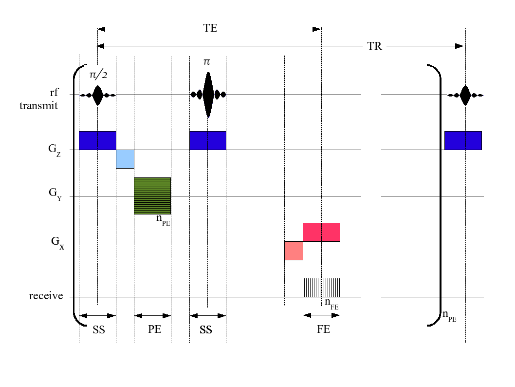
An MRI sequence in magnetic resonance imaging (MRI) is a particular setting of pulse sequences and pulsed field gradients, resulting in a particular image appearance. A multiparametric MRI is a combination of two or more sequences, and/or including other specialized MRI configurations such as spectroscopy. Spin echo T1 and T2 Each tissue returns to its equilibrium state after excitation by the independent relaxation processes of T1 ( spin-lattice; that is, magnetization in the same direction as the static magnetic field) and T2 ( spin-spin; transverse to the static magnetic field). To create a T1-weighted image, magnetization is allowed to recover before measuring the MR signal by changing the repetition time (TR). This image weighting is useful for assessing the cerebral cortex, identifying fatty tissue, characterizing focal liver lesions, and in general, obtaining morphological information, as well as for post-contrast imaging. To create a T2-weighted image, magnetizati ... [...More Info...] [...Related Items...] OR: [Wikipedia] [Google] [Baidu] |
Magnetic Resonance Imaging
Magnetic resonance imaging (MRI) is a medical imaging technique used in radiology to form pictures of the anatomy and the physiological processes of the body. MRI scanners use strong magnetic fields, magnetic field gradients, and radio waves to generate images of the organs in the body. MRI does not involve X-rays or the use of ionizing radiation, which distinguishes it from CT and PET scans. MRI is a medical application of nuclear magnetic resonance (NMR) which can also be used for imaging in other NMR applications, such as NMR spectroscopy. MRI is widely used in hospitals and clinics for medical diagnosis, staging and follow-up of disease. Compared to CT, MRI provides better contrast in images of soft-tissues, e.g. in the brain or abdomen. However, it may be perceived as less comfortable by patients, due to the usually longer and louder measurements with the subject in a long, confining tube, though "Open" MRI designs mostly relieve this. Additionally, implants and oth ... [...More Info...] [...Related Items...] OR: [Wikipedia] [Google] [Baidu] |
Cholelithiasis
A gallstone is a stone formed within the gallbladder from precipitated bile components. The term cholelithiasis may refer to the presence of gallstones or to any disease caused by gallstones, and choledocholithiasis refers to the presence of migrated gallstones within bile ducts. Most people with gallstones (about 80%) are asymptomatic. However, when a gallstone obstructs the bile duct and causes acute cholestasis, a reflexive smooth muscle spasm often occurs, resulting in an intense cramp-like visceral pain in the right upper part of the abdomen known as a biliary colic (or "gallbladder attack"). This happens in 1–4% of those with gallstones each year. Complications from gallstones may include inflammation of the gallbladder (cholecystitis), inflammation of the pancreas (pancreatitis), obstructive jaundice, and infection in bile ducts ( cholangitis). Symptoms of these complications may include pain that lasts longer than five hours, fever, yellowish skin, vomiting, dark urin ... [...More Info...] [...Related Items...] OR: [Wikipedia] [Google] [Baidu] |
Micrograph
A micrograph or photomicrograph is a photograph or digital image taken through a microscope or similar device to show a magnified image of an object. This is opposed to a macrograph or photomacrograph, an image which is also taken on a microscope but is only slightly magnified, usually less than 10 times. Micrography is the practice or art of using microscopes to make photographs. A micrograph contains extensive details of microstructure. A wealth of information can be obtained from a simple micrograph like behavior of the material under different conditions, the phases found in the system, failure analysis, grain size estimation, elemental analysis and so on. Micrographs are widely used in all fields of microscopy. Types Photomicrograph A light micrograph or photomicrograph is a micrograph prepared using an optical microscope, a process referred to as ''photomicroscopy''. At a basic level, photomicroscopy may be performed simply by connecting a camera to a microscope, th ... [...More Info...] [...Related Items...] OR: [Wikipedia] [Google] [Baidu] |


_–_Gerd_Hruška.png)


