|
Digastric
The digastric muscle (also digastricus) (named ''digastric'' as it has two 'bellies') is a small muscle located under the jaw. The term "digastric muscle" refers to this specific muscle. However, other muscles that have two separate muscle bellies include the suspensory muscle of duodenum, omohyoid, occipitofrontalis. It lies below the body of the mandible, and extends, in a curved form, from the mastoid notch to the mandibular symphysis. It belongs to the suprahyoid muscles group. A broad aponeurotic layer is given off from the tendon of the digastric muscle on either side, to be attached to the body and greater cornu of the hyoid bone; this is termed the suprahyoid aponeurosis. Structure The digastricus (digastric muscle) consists of two muscular bellies united by an intermediate rounded tendon. The two bellies of the digastric muscle have different embryological origins, and are supplied by different cranial nerves. Each person has a right and left digastric muscle. I ... [...More Info...] [...Related Items...] OR: [Wikipedia] [Google] [Baidu] |
Suprahyoid Muscles
The suprahyoid muscles are four muscles located above the hyoid bone in the neck. They are the digastric, stylohyoid, geniohyoid, and mylohyoid muscle, mylohyoid muscles. They are all pharyngeal muscles, with the exception of the geniohyoid muscle. The digastric is uniquely named for its two bellies. Its posterior belly rises from the mastoid process of the human skull, cranium and slopes downward and forward. The anterior belly arises from the digastric fossa on the inner surface of the mandibular body, which slopes downward and backward. The two bellies connect at the digastric muscle#intermediate tendon, intermediate tendon. The intermediate tendon passes through a connective tissue loop attached to the hyoid bone. The mylohyoid muscles are thin, flat muscles that form a sling inferior to the tongue supporting the floor of the mouth. The geniohyoids are short, narrow muscles that contact each other in the midline. The stylohyoids are long, thin muscles that are nearly parallel wit ... [...More Info...] [...Related Items...] OR: [Wikipedia] [Google] [Baidu] |
Suprahyoid Aponeurosis
The suprahyoid muscles are four muscles located above the hyoid bone in the neck. They are the digastric, stylohyoid, geniohyoid, and mylohyoid muscles. They are all pharyngeal muscles, with the exception of the geniohyoid muscle. The digastric is uniquely named for its two bellies. Its posterior belly rises from the mastoid process of the cranium and slopes downward and forward. The anterior belly arises from the digastric fossa on the inner surface of the mandibular body, which slopes downward and backward. The two bellies connect at the intermediate tendon. The intermediate tendon passes through a connective tissue loop attached to the hyoid bone. The mylohyoid muscles are thin, flat muscles that form a sling inferior to the tongue supporting the floor of the mouth. The geniohyoids are short, narrow muscles that contact each other in the midline. The stylohyoids are long, thin muscles that are nearly parallel with the posterior belly of the digastric muscle. Function These fo ... [...More Info...] [...Related Items...] OR: [Wikipedia] [Google] [Baidu] |
Digastric Fossa Of Mandible
The digastric muscle (also digastricus) (named ''digastric'' as it has two 'bellies') is a small muscle located under the jaw. The term "digastric muscle" refers to this specific muscle. However, other muscles that have two separate muscle bellies include the suspensory muscle of duodenum, omohyoid, occipitofrontalis. It lies below the body of the mandible, and extends, in a curved form, from the mastoid notch to the mandibular symphysis. It belongs to the suprahyoid muscles group. A broad aponeurotic layer is given off from the tendon of the digastric muscle on either side, to be attached to the body and greater cornu of the hyoid bone; this is termed the suprahyoid aponeurosis. Structure The digastricus (digastric muscle) consists of two muscular bellies united by an intermediate rounded tendon. The two bellies of the digastric muscle have different embryological origins, and are supplied by different cranial nerves. Each person has a right and left digastric muscle. In mo ... [...More Info...] [...Related Items...] OR: [Wikipedia] [Google] [Baidu] |
Trigeminal Nerve
In neuroanatomy, the trigeminal nerve ( lit. ''triplet'' nerve), also known as the fifth cranial nerve, cranial nerve V, or simply CN V, is a cranial nerve responsible for sensation in the face and motor functions such as biting and chewing; it is the most complex of the cranial nerves. Its name ("trigeminal", ) derives from each of the two nerves (one on each side of the pons) having three major branches: the ophthalmic nerve (V), the maxillary nerve (V), and the mandibular nerve (V). The ophthalmic and maxillary nerves are purely sensory, whereas the mandibular nerve supplies motor as well as sensory (or "cutaneous") functions. Adding to the complexity of this nerve is that autonomic nerve fibers as well as special sensory fibers (taste) are contained within it. The motor division of the trigeminal nerve derives from the basal plate of the embryonic pons, and the sensory division originates in the cranial neural crest. Sensory information from the face and body is proc ... [...More Info...] [...Related Items...] OR: [Wikipedia] [Google] [Baidu] |
Trigeminal
In neuroanatomy, the trigeminal nerve ( lit. ''triplet'' nerve), also known as the fifth cranial nerve, cranial nerve V, or simply CN V, is a cranial nerve responsible for sensation in the face and motor functions such as biting and chewing; it is the most complex of the cranial nerves. Its name ("trigeminal", ) derives from each of the two nerves (one on each side of the pons) having three major branches: the ophthalmic nerve (V), the maxillary nerve (V), and the mandibular nerve (V). The ophthalmic and maxillary nerves are purely sensory, whereas the mandibular nerve supplies motor as well as sensory (or "cutaneous") functions. Adding to the complexity of this nerve is that autonomic nerve fibers as well as special sensory fibers (taste) are contained within it. The motor division of the trigeminal nerve derives from the basal plate of the embryonic pons, and the sensory division originates in the cranial neural crest. Sensory information from the face and body is proces ... [...More Info...] [...Related Items...] OR: [Wikipedia] [Google] [Baidu] |
Digastric Branch Of Facial Nerve
The digastric branch of facial nerve arises close to the stylomastoid foramen, and divides into several filaments, which supply the posterior belly of the Digastricus; one of these filaments joins the glossopharyngeal nerve The glossopharyngeal nerve (), also known as the ninth cranial nerve, cranial nerve IX, or simply CN IX, is a cranial nerve that exits the brainstem from the sides of the upper Medulla oblongata, medulla, just anterior (closer to the nose) to t .... References External links * () * () * http://www.dartmouth.edu/~humananatomy/figures/chapter_47/47-5.HTM Facial nerve {{neuroanatomy-stub ... [...More Info...] [...Related Items...] OR: [Wikipedia] [Google] [Baidu] |
Greater Cornu
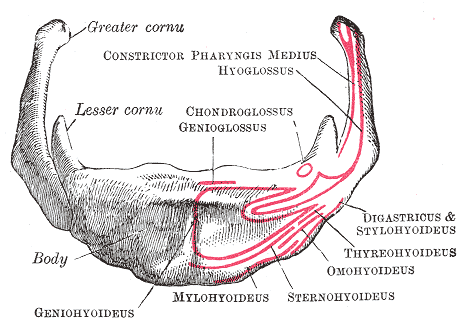
The hyoid bone (lingual bone or tongue-bone) () is a horseshoe-shaped bone situated in the anterior midline of the neck between the chin and the thyroid cartilage. At rest, it lies between the base of the mandible and the third cervical vertebra. Unlike other bones, the hyoid is only distantly articulated to other bones by muscles or ligaments. It is the only bone in the human body that is not connected to any other bones nearby. The hyoid is anchored by muscles from the anterior, posterior and inferior directions, and aids in tongue movement and swallowing. The hyoid bone provides attachment to the muscles of the floor of the mouth and the tongue above, the larynx below, and the epiglottis and pharynx behind. Its name is derived . Structure The hyoid bone is classed as an irregular bone and consists of a central part called the body, and two pairs of horns, the greater and lesser horns. Body The body of the hyoid bone is the central part of the hyoid bone. *At the front, ... [...More Info...] [...Related Items...] OR: [Wikipedia] [Google] [Baidu] |
Mandibular Nerve
In neuroanatomy, the mandibular nerve (V) is the largest of the three divisions of the trigeminal nerve, the fifth cranial nerve (CN V). Unlike the other divisions of the trigeminal nerve (ophthalmic nerve, maxillary nerve) which contain only afferent fibers, the mandibular nerve contains both afferent and efferent fibers. These nerve fibers innervate structures of the lower jaw and face, such as the tongue, lower lip, and chin. The mandibular nerve also innervates the muscles of mastication. Structure The large sensory root emerges from the lateral part of the trigeminal ganglion and exits the cranial cavity through the foramen ovale. Portio minor, the small motor root of the trigeminal nerve, passes under the trigeminal ganglion and through the foramen ovale to unite with the sensory root just outside the skull. The mandibular nerve immediately passes between tensor veli palatini, which is medial, and lateral pterygoid, which is lateral, and gives off a meningeal branch (n ... [...More Info...] [...Related Items...] OR: [Wikipedia] [Google] [Baidu] |
Hyoid Bone
The hyoid bone (lingual bone or tongue-bone) () is a horseshoe-shaped bone situated in the anterior midline of the neck between the chin and the thyroid cartilage. At rest, it lies between the base of the mandible and the third cervical vertebra. Unlike other bones, the hyoid is only distantly articulated to other bones by muscles or ligaments. It is the only bone in the human body that is not connected to any other bones nearby. The hyoid is anchored by muscles from the anterior, posterior and inferior directions, and aids in tongue movement and swallowing. The hyoid bone provides attachment to the muscles of the floor of the mouth and the tongue above, the larynx below, and the epiglottis and pharynx behind. Its name is derived . Structure The hyoid bone is classed as an irregular bone and consists of a central part called the body, and two pairs of horns, the greater and lesser horns. Body The body of the hyoid bone is the central part of the hyoid bone. *At the fro ... [...More Info...] [...Related Items...] OR: [Wikipedia] [Google] [Baidu] |
Mandibular Division
In neuroanatomy, the mandibular nerve (V) is the largest of the three divisions of the trigeminal nerve, the fifth Cranial nerves, cranial nerve (CN V). Unlike the other divisions of the trigeminal nerve (Ophthalmic nerve, ophthalmic nerve, maxillary nerve) which contain only Afferent nerve fiber, afferent fibers, the mandibular nerve contains both afferent and Efferent nerve fiber, efferent fibers. These nerve fibers innervate structures of the lower jaw and face, such as the tongue, lower lip, and chin. The mandibular nerve also innervates the muscles of mastication. Structure The large sensory root emerges from the lateral part of the trigeminal ganglion and exits the cranial cavity through the Foramen ovale (skull), foramen ovale. Portio minor, the small motor root of the trigeminal nerve, passes under the trigeminal ganglion and through the Foramen ovale (skull), foramen ovale to unite with the sensory root just outside the skull. The mandibular nerve immediately passes betw ... [...More Info...] [...Related Items...] OR: [Wikipedia] [Google] [Baidu] |
Cranial Nerve
Cranial nerves are the nerves that emerge directly from the brain (including the brainstem), of which there are conventionally considered twelve pairs. Cranial nerves relay information between the brain and parts of the body, primarily to and from regions of the head and neck, including the special senses of vision, taste, smell, and hearing. The cranial nerves emerge from the central nervous system above the level of the first vertebra of the vertebral column. Each cranial nerve is paired and is present on both sides. There are conventionally twelve pairs of cranial nerves, which are described with Roman numerals I–XII. Some considered there to be thirteen pairs of cranial nerves, including cranial nerve zero. The numbering of the cranial nerves is based on the order in which they emerge from the brain and brainstem, from front to back. The terminal nerves (0), olfactory nerves (I) and optic nerves (II) emerge from the cerebrum, and the remaining ten pairs arise from t ... [...More Info...] [...Related Items...] OR: [Wikipedia] [Google] [Baidu] |
Embryology
Embryology (from Greek ἔμβρυον, ''embryon'', "the unborn, embryo"; and -λογία, '' -logia'') is the branch of animal biology that studies the prenatal development of gametes (sex cells), fertilization, and development of embryos and fetuses. Additionally, embryology encompasses the study of congenital disorders that occur before birth, known as teratology. Early embryology was proposed by Marcello Malpighi, and known as preformationism, the theory that organisms develop from pre-existing miniature versions of themselves. Aristotle proposed the theory that is now accepted, epigenesis. Epigenesis is the idea that organisms develop from seed or egg in a sequence of steps. Modern embryology, developed from the work of Karl Ernst von Baer, though accurate observations had been made in Italy by anatomists such as Aldrovandi and Leonardo da Vinci in the Renaissance. Comparative embryology Preformationism and epigenesis As recently as the 18th century, the prevailin ... [...More Info...] [...Related Items...] OR: [Wikipedia] [Google] [Baidu] |


