|
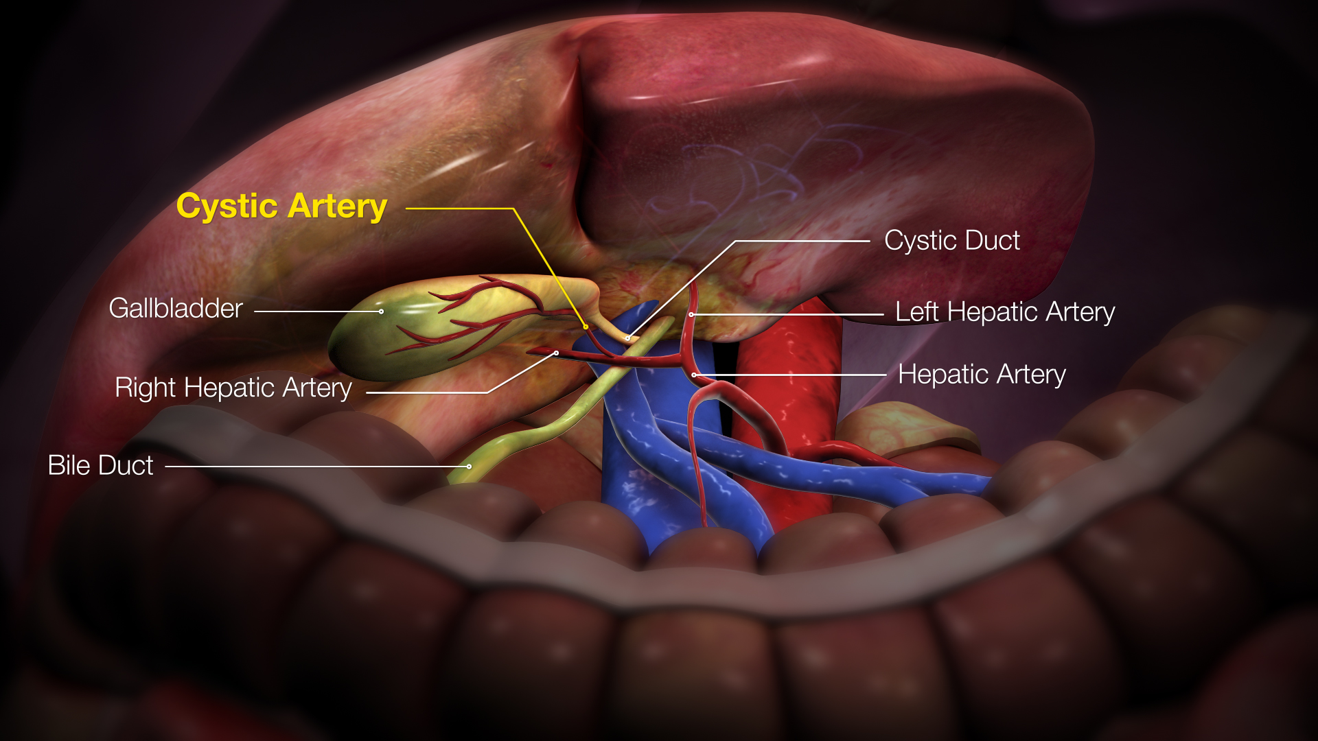
Cystic Artery
The cystic artery (also known as bachelor artery) supplies oxygenated blood to the gallbladder and cystic duct. Most common arrangement In the classic arrangement, occurring with a frequency of approximately 70%, a singular cystic artery originates from the geniculate flexure of the right hepatic artery in the upper portion of the hepatobiliary triangle. A site of origin from a more proximal or distal portion of the right hepatic artery is also considered relatively normal. After separating from the right hepatic artery, the cystic artery travels superiorly to the cystic duct and produces 2 to 4 minor branches, known as ''Calot’s arteries'', that supply part of the cystic duct and cervix of the gallbladder before dividing into the major superficial and deep branches at the superior aspect of the gallbladder neck: * The ''superficial branch'' (or ''anterior branch'') passes subserously over the left aspect of the gallbladder. * The ''deep branch'' (or ''posterior branch'') runs b ... [...More Info...] [...Related Items...] OR: [Wikipedia] [Google] [Baidu] |
Right Hepatic Artery
The hepatic artery proper (also proper hepatic artery) is the artery that supplies the liver and gallbladder. It raises from the common hepatic artery, a branch of the celiac artery. Structure The hepatic artery proper arises from the common hepatic artery and runs alongside the portal vein and the common bile duct to form the portal triad. A branch of the common hepatic artery –the gastroduodenal artery gives off the small supraduodenal artery to the duodenal bulb. Then the right gastric artery comes off and runs to the left along the lesser curvature of the stomach to meet the left gastric artery, which is a branch of the celiac trunk. It subsequently bifurcates into the right and left hepatic arteries. Variant anatomy Of note, the right and left hepatic arteries may demonstrate variant anatomy. A misplaced right hepatic artery may arise from the superior mesenteric artery (SMA) and a misplaced left hepatic artery may arise from the left gastric artery. The cystic ar ... [...More Info...] [...Related Items...] OR: [Wikipedia] [Google] [Baidu] |
Hepatic Artery
The common hepatic artery is a short blood vessel that supplies oxygenated blood to the liver, pylorus of the stomach, duodenum, pancreas, and gallbladder. It arises from the celiac artery and has the following branches: Additional images File:Common hepatic artery.jpg, Common hepatic artery and its branches including hepatic artery proper and right gastric artery (pyloric artery) References External links * - "Stomach, Spleen and Liver: Contents of the Hepatoduodenal ligament The hepatoduodenal ligament is the portion of the lesser omentum extending between the porta hepatis of the liver and the superior part of the duodenum. Running inside it are the following structures collectively known as the portal triad: * hep ..." * {{Authority control Arteries of the abdomen ... [...More Info...] [...Related Items...] OR: [Wikipedia] [Google] [Baidu] |
Cystic Vein
When present the cystic vein drains the blood from the gall-bladder, and, accompanying the cystic duct, usually ends in the right branch of the portal vein The portal vein or hepatic portal vein (HPV) is a blood vessel that carries blood from the gastrointestinal tract, gallbladder, pancreas and spleen to the liver. This blood contains nutrients and toxins extracted from digested contents. Approxima .... It is usually not present, and the blood drains via small veins in the gall-bladder bed directly to the parenchyma of the liver. References External links * Veins of the torso {{circulatory-stub ... [...More Info...] [...Related Items...] OR: [Wikipedia] [Google] [Baidu] |
Gall Bladder
In vertebrates, the gallbladder, also known as the cholecyst, is a small hollow organ where bile is stored and concentrated before it is released into the small intestine. In humans, the pear-shaped gallbladder lies beneath the liver, although the structure and position of the gallbladder can vary significantly among animal species. It receives and stores bile, produced by the liver, via the common hepatic duct, and releases it via the common bile duct into the duodenum, where the bile helps in the digestion of fats. The gallbladder can be affected by gallstones, formed by material that cannot be dissolved – usually cholesterol or bilirubin, a product of haemoglobin breakdown. These may cause significant pain, particularly in the upper-right corner of the abdomen, and are often treated with removal of the gallbladder (called a cholecystectomy). Cholecystitis, inflammation of the gallbladder, has a wide range of causes, including result from the impaction of gallstones, infec ... [...More Info...] [...Related Items...] OR: [Wikipedia] [Google] [Baidu] |
Cystic Duct
The cystic duct is the short duct that joins the gallbladder to the common hepatic duct. It usually lies next to the cystic artery. It is of variable length. It contains 'spiral valves of Heister', which do not provide much resistance to the flow of bile. Function Bile can flow in both directions between the gallbladder and the common bile duct and the hepatic duct. In this way, bile is stored in the gallbladder in between meal times. The hormone cholecystokinin, when stimulated by a fatty meal, promotes bile secretion by increased production of hepatic bile, contraction of the gall bladder, and relaxation of the Sphincter of Oddi. Clinical significance Gallstones can enter and obstruct the cystic duct, preventing the flow of bile. The increased pressure in the gallbladder leads to swelling and pain. This pain, known as biliary colic, is sometimes referred to as a gallbladder "attack" because of its sudden onset. During a cholecystectomy, the cystic duct is clipped two or ... [...More Info...] [...Related Items...] OR: [Wikipedia] [Google] [Baidu] |
3D Medical Animation Still Shot Cystic Artery
3-D, 3D, or 3d may refer to: Science, technology, and mathematics Relating to three-dimensionality * Three-dimensional space ** 3D computer graphics, computer graphics that use a three-dimensional representation of geometric data ** 3D film, a motion picture that gives the illusion of three-dimensional perception ** 3D modeling, developing a representation of any three-dimensional surface or object ** 3D printing, making a three-dimensional solid object of a shape from a digital model ** 3D display, a type of information display that conveys depth to the viewer ** 3D television, television that conveys depth perception to the viewer ** Stereoscopy, any technique capable of recording three-dimensional visual information or creating the illusion of depth in an image Other uses in science and technology or commercial products * 3D projection * 3D rendering * 3D scanning, making a digital representation of three-dimensional objects * 3D video game (other) * 3-D Secure, a ... [...More Info...] [...Related Items...] OR: [Wikipedia] [Google] [Baidu] |
Gallbladder
In vertebrates, the gallbladder, also known as the cholecyst, is a small hollow organ where bile is stored and concentrated before it is released into the small intestine. In humans, the pear-shaped gallbladder lies beneath the liver, although the structure and position of the gallbladder can vary significantly among animal species. It receives and stores bile, produced by the liver, via the common hepatic duct, and releases it via the common bile duct into the duodenum, where the bile helps in the digestion of fats. The gallbladder can be affected by gallstones, formed by material that cannot be dissolved – usually cholesterol or bilirubin, a product of haemoglobin breakdown. These may cause significant pain, particularly in the upper-right corner of the abdomen, and are often treated with removal of the gallbladder (called a cholecystectomy). Cholecystitis, inflammation of the gallbladder, has a wide range of causes, including result from the impaction of gallstones, inf ... [...More Info...] [...Related Items...] OR: [Wikipedia] [Google] [Baidu] |
Hepatobiliary Triangle
The cystohepatic triangle (or hepatobiliary triangle) is an anatomic space bordered by the cystic duct inferiorly, the common hepatic duct medially, and the inferior surface of the liver superiorly. The cystic artery lies within the hepatobiliary triangle, which is used to locate it during a laparoscopic cholecystectomy. Structure The hepatobiliary triangle is the area bound by the: * cystic duct inferiorly. * common hepatic duct medially. * inferior margin of the liver superiorly.Schwartz's Manual of Surgery BRUNICARDI C.F 10th edition It is covered in peritoneum both anteriorly and posteriorly. It contains the cystic artery and cystic lymph nodes. The right hepatic artery may also pass through the hepatobiliary triangle. Clinical significance General surgeons frequently quiz medical students on this term and the name for the lymph node located within the triangle, Mascagni's lymph node or Lund's node, however many often erroneously refer to it as "Calot's node". The latter ... [...More Info...] [...Related Items...] OR: [Wikipedia] [Google] [Baidu] |
Superior Mesenteric Artery
In human anatomy, the superior mesenteric artery (SMA) is an artery which arises from the anterior surface of the abdominal aorta, just inferior to the origin of the celiac trunk, and supplies blood to the intestine from the lower part of the duodenum through two-thirds of the transverse colon, as well as the pancreas. Structure It arises anterior to lower border of vertebra L1 in an adult. It is usually 1 cm lower than the celiac trunk. It initially travels in an anterior/inferior direction, passing behind/under the neck of the pancreas and the splenic vein. Located under this portion of the superior mesenteric artery, between it and the aorta, are the following: * left renal vein - travels between the left kidney and the inferior vena cava (can be compressed between the SMA and the abdominal aorta at this location, leading to nutcracker syndrome). * the third part of the duodenum, a segment of the small intestines (can be compressed by the SMA at this location, lea ... [...More Info...] [...Related Items...] OR: [Wikipedia] [Google] [Baidu] |
Gastroduodenal Artery
In anatomy, the gastroduodenal artery is a small blood vessel in the abdomen. It supplies blood directly to the pylorus (distal part of the stomach) and proximal part of the duodenum. It also indirectly supplies the pancreatic head (via the anterior and posterior superior pancreaticoduodenal arteries). Structure The gastroduodenal artery most commonly arises from either the left hepatic artery or the right hepatic artery instead. It may also arise from the common hepatic artery of the coeliac trunk in a trifork arrangement with the two other arteries, but there are numerous variations of the origin.Bergman RA, Afifi AK, Miyauchi R. Variations in Origin of Gastroduodenal Artery. from Anatomy Atlases. (http://www.anatomyatlases.org/AnatomicVariants/Cardiovascular/Images0001/0017.shtml) It first gives rise to the supraduodenal artery, followed by the posterior superior pancreaticoduodenal artery. It terminates in a bifurcation when it splits into the right gastroepiploic artery a ... [...More Info...] [...Related Items...] OR: [Wikipedia] [Google] [Baidu] |
Parenchyma
Parenchyma () is the bulk of functional substance in an animal organ or structure such as a tumour. In zoology it is the name for the tissue that fills the interior of flatworms. Etymology The term ''parenchyma'' is New Latin from the word παρέγχυμα ''parenchyma'' meaning 'visceral flesh', and from παρεγχεῖν ''parenchyma'' meaning 'to pour in' from παρα- ''para-'' 'beside' + ἐν ''en-'' 'in' + χεῖν ''chyma'' 'to pour'. Originally, Erasistratus and other anatomists used it to refer to certain human tissues. Later, it was also applied to plant tissues by Nehemiah Grew. Structure The parenchyma is the ''functional'' parts of an organ (anatomy), organ, or of a structure such as a tumour in the body. This is in contrast to the Stroma (animal tissue), stroma, which refers to the ''structural'' tissue of organs or of structures, namely, the connective tissues. Brain The brain parenchyma refers to the functional tissue in the brain that is made up of t ... [...More Info...] [...Related Items...] OR: [Wikipedia] [Google] [Baidu] |


