|
Coronary Sulcus
The coronary sulcus (also called coronary groove, auriculoventricular groove, atrioventricular groove, AV groove) is a groove on the surface of the heart at the base of right auricle that separates the atria from the ventricles. The structure contains the trunks of the nutrient vessels of the heart, and is deficient in front, where it is crossed by the root of the pulmonary trunk. On the posterior surface of the heart, the coronary sulcus contains the coronary sinus. Structure In relation to the rib cage, the coronary sulcus spans from the medial side of the 3rd left costal cartilage, to the middle of the right 6th costal cartilage. Epicardial fat tends to be concentrated along the coronary sulcus. There are two coronary sulci in the heart, including left and right coronary sulci. Left coronary sulcus The left coronary sulcus originates posterior to the pulmonary trunk, and travels inferiorly separating the left atrium and left ventricle. The location of the left coronary ... [...More Info...] [...Related Items...] OR: [Wikipedia] [Google] [Baidu] |
Sulcus (morphology)
In biological morphology and anatomy, a sulcus (pl. ''sulci'') is a furrow or fissure (Latin ''fissura'', plural ''fissurae''). It may be a groove, natural division, deep furrow, elongated cleft, or tear in the surface of a limb or an organ, most notably on the surface of the brain, but also in the lungs, certain muscles (including the heart), as well as in bones, and elsewhere. Many sulci are the product of a surface fold or junction, such as in the gums, where they fold around the neck of the tooth. In invertebrate zoology, a sulcus is a fold, groove, or boundary, especially at the edges of sclerites or between segments. In pollen a grain that is grooved by a sulcus is termed sulcate. Examples in anatomy Liver *Ligamentum teres hepatis fissure *Ligamentum venosum fissure *Portal fissure, found in the under-surface of the liver *Transverse fissure of liver, found in the lower surface of the liver *Umbilical fissure, found in front of the liver Lung *Azygos fissure, of ri ... [...More Info...] [...Related Items...] OR: [Wikipedia] [Google] [Baidu] |
Adipose Tissue
Adipose tissue, body fat, or simply fat is a loose connective tissue composed mostly of adipocytes. In addition to adipocytes, adipose tissue contains the stromal vascular fraction (SVF) of cells including preadipocytes, fibroblasts, vascular endothelial cells and a variety of immune cells such as adipose tissue macrophages. Adipose tissue is derived from preadipocytes. Its main role is to store energy in the form of lipids, although it also cushions and insulates the body. Far from being hormonally inert, adipose tissue has, in recent years, been recognized as a major endocrine organ, as it produces hormones such as leptin, estrogen, resistin, and cytokines (especially TNFα). In obesity, adipose tissue is also implicated in the chronic release of pro-inflammatory markers known as adipokines, which are responsible for the development of metabolic syndrome, a constellation of diseases including, but not limited to, type 2 diabetes, cardiovascular disease and atherosclerosis. T ... [...More Info...] [...Related Items...] OR: [Wikipedia] [Google] [Baidu] |
Posterior Interventricular Sulcus
The posterior interventricular sulcus or posterior longitudinal sulcus is one of the two grooves that separates the ventricles of the heart and is on the diaphragmatic surface of the heart near the right margin. The other groove is the anterior interventricular sulcus, situated on the sternocostal surface of the heart, close to its left margin. In it runs the posterior interventricular artery and middle cardiac vein The middle cardiac vein commences at the apex of the heart; ascends in the posterior longitudinal sulcus, and ends in the coronary sinus In anatomy, the coronary sinus () is a collection of veins joined together to form a large vessel that c .... References External links * Cardiac anatomy {{circulatory-stub ... [...More Info...] [...Related Items...] OR: [Wikipedia] [Google] [Baidu] |
Right Atrial Appendage
The atrium ( la, ātrium, , entry hall) is one of two upper chambers in the heart that receives blood from the circulatory system. The blood in the atria is pumped into the heart ventricles through the atrioventricular valves. There are two atria in the human heart – the left atrium receives blood from the pulmonary circulation, and the right atrium receives blood from the venae cavae of the systemic circulation. During the cardiac cycle the atria receive blood while relaxed in diastole, then contract in systole to move blood to the ventricles. Each atrium is roughly cube-shaped except for an ear-shaped projection called an atrial appendage, sometimes known as an auricle. All animals with a closed circulatory system have at least one atrium. The atrium was formerly called the 'auricle'. That term is still used to describe this chamber in some other animals, such as the ''Mollusca''. They have thicker muscular walls than the atria do. Structure Humans have a four-chambered h ... [...More Info...] [...Related Items...] OR: [Wikipedia] [Google] [Baidu] |
Small Cardiac Vein
The small cardiac vein, also known as the right coronary vein, is a coronary vein that drains the right atrium and right ventricle of the heart. Despite its size, it is one of the major drainage vessels for the heart. Location The small cardiac vein runs in the coronary sulcus between the right atrium and right ventricle, and opens into the right extremity of the coronary sinus. Function The small cardiac vein receives blood from the posterior portion of the right atrium and ventricle. Variations The small cardiac vein may drain to the coronary sinus, right atrium, middle cardiac vein The middle cardiac vein commences at the apex of the heart; ascends in the posterior longitudinal sulcus, and ends in the coronary sinus In anatomy, the coronary sinus () is a collection of veins joined together to form a large vessel that c ..., or be absent. References External links * - "Anterior view of the heart." {{Authority control Veins of the torso ... [...More Info...] [...Related Items...] OR: [Wikipedia] [Google] [Baidu] |
Right Coronary Artery
In the blood supply of the heart, the right coronary artery (RCA) is an artery originating above the right cusp of the aortic valve, at the right aortic sinus in the heart. It travels down the right coronary sulcus, towards the crux of the heart. It supplies the right side of the heart, and the interventricular septum. Structure The right coronary artery originates above the right aortic sinus above the aortic valve. It passes through the right coronary sulcus (right atrioventricular groove), towards the crux of the heart. It gives off many branches, including the posterior interventricular artery, the right marginal artery, the conus artery, and the sinoatrial nodal artery. Segments * Proximal: starting at RCA origin, spanning half the distance to the acute margin * Middle: from proximal segment to the acute margin * Distal: from middle segment to origination point of the posterior interventricular artery, where the posterior interventricular sulcus meets the atrioven ... [...More Info...] [...Related Items...] OR: [Wikipedia] [Google] [Baidu] |
Circumflex Branch Of Left Coronary Artery
The circumflex branch of left coronary artery, or left circumflex artery or circumflex artery, is a branch of the left coronary artery. Description The left circumflex artery follows the left part of the coronary sulcus, running first to the left and then to the right, reaching nearly as far as the posterior longitudinal sulcus. There have been multiple anomalies described, for example the left circumflex having an aberrant course from the right coronary artery. Branches The circumflex artery curves to the left around the heart within the coronary sulcus, giving rise to one or more left marginal arteries (also called obtuse marginal branches) as it curves toward the posterior surface of the heart. It helps form the posterior left ''ventricular branch'' or posterolateral artery. The circumflex artery ends at the point where it joins to form to the posterior interventricular artery in 15% of all cases, which lies in the posterior interventricular sulcus. In the other 85% of all ... [...More Info...] [...Related Items...] OR: [Wikipedia] [Google] [Baidu] |
Pulmonary Artery
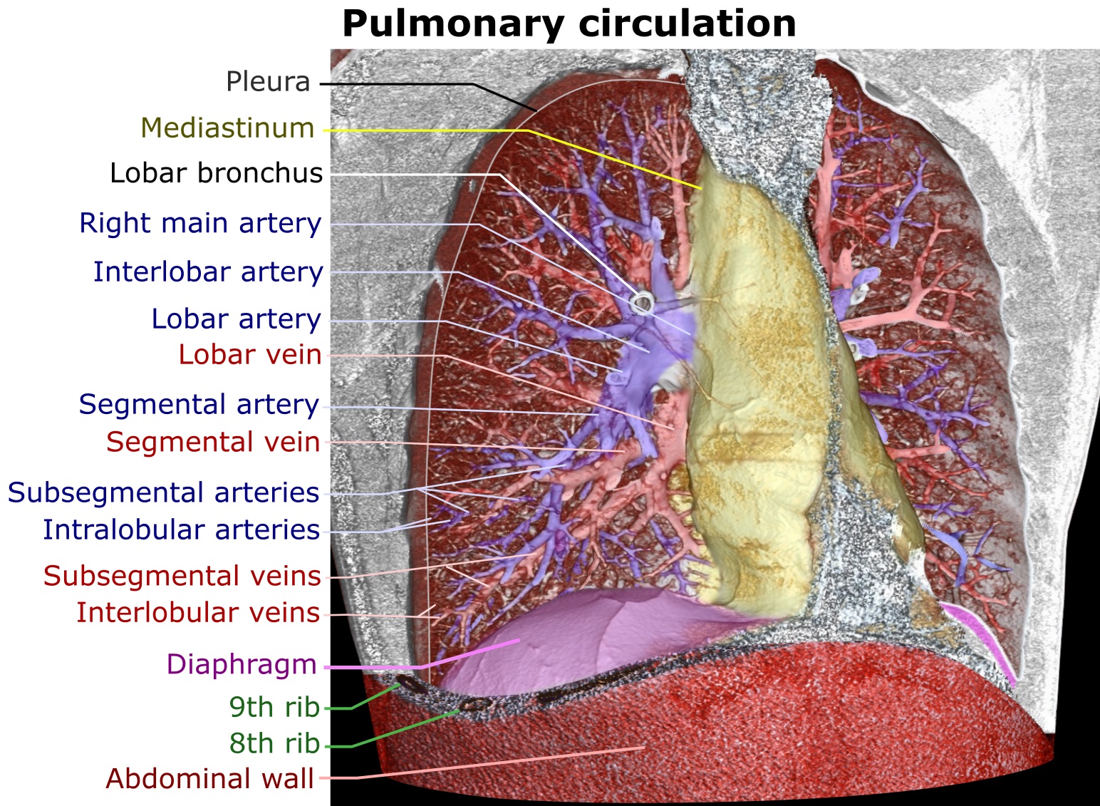
A pulmonary artery is an artery in the pulmonary circulation that carries deoxygenated blood from the right side of the heart to the lungs. The largest pulmonary artery is the ''main pulmonary artery'' or ''pulmonary trunk'' from the heart, and the smallest ones are the arterioles, which lead to the capillaries that surround the pulmonary alveoli. Structure The pulmonary arteries are blood vessels that carry systemic venous blood from the right ventricle of the heart to the microcirculation of the lungs. Unlike in other organs where arteries supply oxygenated blood, the blood carried by the pulmonary arteries is deoxygenated, as it is venous blood returning to the heart. The main pulmonary arteries emerge from the right side of the heart, and then split into smaller arteries that progressively divide and become arterioles, eventually narrowing into the capillary microcirculation of the lungs where gas exchange occurs. Pulmonary trunk In order of blood flow, the pulmonary art ... [...More Info...] [...Related Items...] OR: [Wikipedia] [Google] [Baidu] |
Pericardium
The pericardium, also called pericardial sac, is a double-walled sac containing the heart and the roots of the great vessels. It has two layers, an outer layer made of strong connective tissue (fibrous pericardium), and an inner layer made of serous membrane (serous pericardium). It encloses the pericardial cavity, which contains pericardial fluid, and defines the middle mediastinum. It separates the heart from interference of other structures, protects it against infection and blunt trauma, and lubricates the heart's movements. The English name originates from the Ancient Greek prefix "''peri-''" (περί; "around") and the suffix "''-cardion''" (κάρδιον; "heart"). Anatomy The pericardium is a tough fibroelastic sac which covers the heart from all sides except at the cardiac root (where the great vessels join the heart) and the bottom (where only the serous pericardium exists to cover the upper surface of the central tendon of diaphragm). The fibrous pericardiu ... [...More Info...] [...Related Items...] OR: [Wikipedia] [Google] [Baidu] |
Heart
The heart is a muscular organ in most animals. This organ pumps blood through the blood vessels of the circulatory system. The pumped blood carries oxygen and nutrients to the body, while carrying metabolic waste such as carbon dioxide to the lungs. In humans, the heart is approximately the size of a closed fist and is located between the lungs, in the middle compartment of the chest. In humans, other mammals, and birds, the heart is divided into four chambers: upper left and right atria and lower left and right ventricles. Commonly the right atrium and ventricle are referred together as the right heart and their left counterparts as the left heart. Fish, in contrast, have two chambers, an atrium and a ventricle, while most reptiles have three chambers. In a healthy heart blood flows one way through the heart due to heart valves, which prevent backflow. The heart is enclosed in a protective sac, the pericardium, which also contains a small amount of fluid. The wall ... [...More Info...] [...Related Items...] OR: [Wikipedia] [Google] [Baidu] |
Costal Cartilage
The costal cartilages are bars of hyaline cartilage that serve to prolong the ribs forward and contribute to the elasticity of the walls of the thorax. Costal cartilage is only found at the anterior ends of the ribs, providing medial extension. Differences from Ribs 1-12 The first seven pairs are connected with the sternum; the next three are each articulated with the lower border of the cartilage of the preceding rib; the last two have pointed extremities, which end in the wall of the abdomen. Like the ribs, the costal cartilages vary in their length, breadth, and direction. They increase in length from the first to the seventh, then gradually decrease to the twelfth. Their breadth, as well as that of the intervals between them, diminishes from the first to the last. They are broad at their attachments to the ribs, and taper toward their sternal extremities, excepting the first two, which are of the same breadth throughout, and the sixth, seventh, and eighth, which are enlarge ... [...More Info...] [...Related Items...] OR: [Wikipedia] [Google] [Baidu] |
Rib Cage
The rib cage, as an enclosure that comprises the ribs, vertebral column and sternum in the thorax of most vertebrates, protects vital organs such as the heart, lungs and great vessels. The sternum, together known as the thoracic cage, is a semi-rigid bony and cartilaginous structure which surrounds the thoracic cavity and supports the shoulder girdle to form the core part of the human skeleton. A typical human thoracic cage consists of 12 pairs of ribs and the adjoining costal cartilages, the sternum (along with the manubrium and xiphoid process), and the 12 thoracic vertebrae articulating with the ribs. Together with the skin and associated fascia and muscles, the thoracic cage makes up the thoracic wall and provides attachments for extrinsic skeletal muscles of the neck, upper limbs, upper abdomen and back. The rib cage intrinsically holds the muscles of respiration ( diaphragm, intercostal muscles, etc.) that are crucial for active inhalation and forced exhalation, and ... [...More Info...] [...Related Items...] OR: [Wikipedia] [Google] [Baidu] |


