|
Condyloid Fossa
Behind either condyle of the lateral parts of occipital bone is a depression, the condyloid fossa (or condylar fossa), which receives the posterior margin of the superior facet of the atlas when the head is bent backward; the floor of this fossa is sometimes perforated by the condyloid canal, through which an emissary vein passes from the transverse sinus. Additional images File:Condyloid fossa - animation02.gif, Human skull seen from below. Position of condyloid fossa shown in red. File:Condyloid fossa - animation04.gif, Skull and cervical vertebra. Position of condyloid fossa shown in red. File:Cervical XRayFlexionExtension.jpg, X-ray of cervical spine (neck) in flexion and extension (bending backwards) See also * Occipital condyle * Atlas An atlas is a collection of maps; it is typically a bundle of maps of Earth or of a region of Earth. Atlases have traditionally been bound into book form, but today many atlases are in multimedia formats. In addition to presenting ... [...More Info...] [...Related Items...] OR: [Wikipedia] [Google] [Baidu] |
Occipital Bone
The occipital bone () is a neurocranium, cranial dermal bone and the main bone of the occiput (back and lower part of the skull). It is trapezoidal in shape and curved on itself like a shallow dish. The occipital bone overlies the occipital lobes of the cerebrum. At the base of skull in the occipital bone, there is a large oval opening called the foramen magnum, which allows the passage of the spinal cord. Like the other cranial bones, it is classed as a flat bone. Due to its many attachments and features, the occipital bone is described in terms of separate parts. From its front to the back is the basilar part of occipital bone, basilar part, also called the basioccipital, at the sides of the foramen magnum are the lateral parts of occipital bone, lateral parts, also called the exoccipitals, and the back is named as the squamous part of occipital bone, squamous part. The basilar part is a thick, somewhat quadrilateral piece in front of the foramen magnum and directed towards the ... [...More Info...] [...Related Items...] OR: [Wikipedia] [Google] [Baidu] |
Cervical Vertebra
In tetrapods, cervical vertebrae (singular: vertebra) are the vertebrae of the neck, immediately below the skull. Truncal vertebrae (divided into thoracic and lumbar vertebrae in mammals) lie caudal (toward the tail) of cervical vertebrae. In sauropsid species, the cervical vertebrae bear cervical ribs. In lizards and saurischian dinosaurs, the cervical ribs are large; in birds, they are small and completely fused to the vertebrae. The vertebral transverse processes of mammals are homologous to the cervical ribs of other amniotes. Most mammals have seven cervical vertebrae, with the only three known exceptions being the manatee with six, the two-toed sloth with five or six, and the three-toed sloth with nine. In humans, cervical vertebrae are the smallest of the true vertebrae and can be readily distinguished from those of the thoracic or lumbar regions by the presence of a foramen (hole) in each transverse process, through which the vertebral artery, vertebral veins, and inferio ... [...More Info...] [...Related Items...] OR: [Wikipedia] [Google] [Baidu] |
Occipital Condyle
The occipital condyles are undersurface protuberances of the occipital bone in vertebrates, which function in articulation with the superior facets of the atlas vertebra. The condyles are oval or reniform (kidney-shaped) in shape, and their anterior extremities, directed forward and medialward, are closer together than their posterior, and encroach on the basilar portion of the bone; the posterior extremities extend back to the level of the middle of the foramen magnum. The articular surfaces of the condyles are convex from before backward and from side to side, and look downward and lateralward. To their margins are attached the capsules of the atlanto-occipital joints, and on the medial side of each is a rough impression or tubercle for the alar ligament. At the base of either condyle the bone is tunnelled by a short canal, the hypoglossal canal. Clinical significance Fracture of an occipital condyle may occur in isolation, or as part of a more extended basilar skull fracture ... [...More Info...] [...Related Items...] OR: [Wikipedia] [Google] [Baidu] |
Lateral Parts Of Occipital Bone
The lateral parts of the occipital bone (also called the exoccipitals) are situated at the sides of the foramen magnum; on their under surfaces are the condyles for articulation with the superior facets of the atlas. Description The condyles are oval or reniform (kidney-shaped) in shape, and their anterior extremities, directed forward and medialward, are closer together than their posterior, and encroach on the basilar portion of the bone; the posterior extremities extend back to the level of the middle of the foramen magnum. The articular surfaces of the condyles are convex from before backward and from side to side, and look downward and lateralward. To their margins are attached the capsules of the atlantoöccipital articulations, and on the medial side of each is a rough impression or tubercle for the alar ligament. At the base of either condyle the bone is tunnelled by a short canal, the hypoglossal canal (anterior condyloid foramen). This begins on the cranial surface o ... [...More Info...] [...Related Items...] OR: [Wikipedia] [Google] [Baidu] |
Atlas (anatomy)
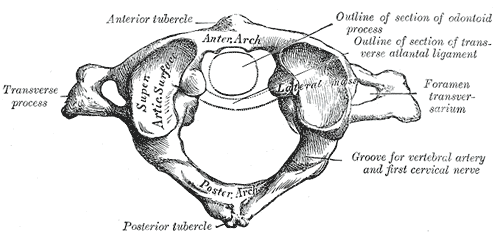
In anatomy, the atlas (C1) is the most superior (first) cervical vertebra of the spine and is located in the neck. It is named for Atlas of Greek mythology because, just as Atlas supported the globe, it supports the entire head. The atlas is the topmost vertebra and, with the axis (the vertebra below it), forms the joint connecting the skull and spine. The atlas and axis are specialized to allow a greater range of motion than normal vertebrae. They are responsible for the nodding and rotation movements of the head. The atlanto-occipital joint allows the head to nod up and down on the vertebral column. The dens acts as a pivot that allows the atlas and attached head to rotate on the axis, side to side. The atlas's chief peculiarity is that it has no body. It is ring-like and consists of an anterior and a posterior arch and two lateral masses. The atlas and axis are important neurologically because the brainstem extends down to the axis. Structure Anterior arch The anterio ... [...More Info...] [...Related Items...] OR: [Wikipedia] [Google] [Baidu] |
Condyloid Canal
The condylar canal (or condyloid canal) is a canal in the condyloid fossa of the lateral parts of occipital bone behind the occipital condyle. Resection of the rectus capitis posterior major and minor muscles reveals the bony recess leading to the condylar canal, which is situated posterior and lateral to the occipital condyle. It is immediately superior to the extradural vertebral artery, which makes a loop above the posterior C1 ring to enter the foramen magnum. The anteriomedial wall of the condylar canal thickens to join the foramen magnum rim and connect to the occipital condyle. Through the condylar canal, the occipital emissary vein connects to the venous system including the suboccipital venous plexus, occipital sinus and sigmoid sinus The sigmoid sinuses (sigma- or s-shaped hollow curve), also known as the , are venous sinuses within the skull that receive blood from posterior dural venous sinus veins. Structure The sigmoid sinus is a dural venous sinus situated wit ... [...More Info...] [...Related Items...] OR: [Wikipedia] [Google] [Baidu] |
Emissary Vein
The emissary veins connect the extracranial venous system with the intracranial venous sinuses. They connect the veins outside the cranium to the venous sinuses inside the cranium. They drain from the scalp, through the human skull, skull, into the larger meningeal veins and dural venous sinuses. Emissary veins have an important role in selective cooling of the head. They also serve as routes where infections are carried into the cranial cavity from the extracranial veins to the intracranial veins. There are several types of emissary veins including posterior condyloid, mastoid, occipital and parietal emissary vein. Structure There are also emissary veins passing through the Foramen ovale (skull), foramen ovale, jugular foramen, foramen lacerum, and hypoglossal canal. Function Because the emissary veins are valveless, they are an important part in selective brain cooling through bidirectional flow of cooler blood from the evaporating surface of the head. In general, blood flow ... [...More Info...] [...Related Items...] OR: [Wikipedia] [Google] [Baidu] |
Transverse Sinus
The transverse sinuses (left and right lateral sinuses), within the human head, are two areas beneath the brain which allow blood to drain from the back of the head. They run laterally in a groove along the interior surface of the occipital bone. They drain from the confluence of sinuses (by the internal occipital protuberance) to the sigmoid sinuses, which ultimately connect to the internal jugular vein. ''See diagram (at right)'': labeled under the brain as "" (for Latin: ''sinus transversus''). Structure The transverse sinuses are of large size and begin at the internal occipital protuberance; one, generally the right, being the direct continuation of the superior sagittal sinus, the other of the straight sinus. Each transverse sinus passes lateral and forward, describing a slight curve with its convexity upward, to the base of the petrous portion of the temporal bone, and lies, in this part of its course, in the attached margin of the tentorium cerebelli; it then leaves th ... [...More Info...] [...Related Items...] OR: [Wikipedia] [Google] [Baidu] |


