|
Common Palmar Digital Branch
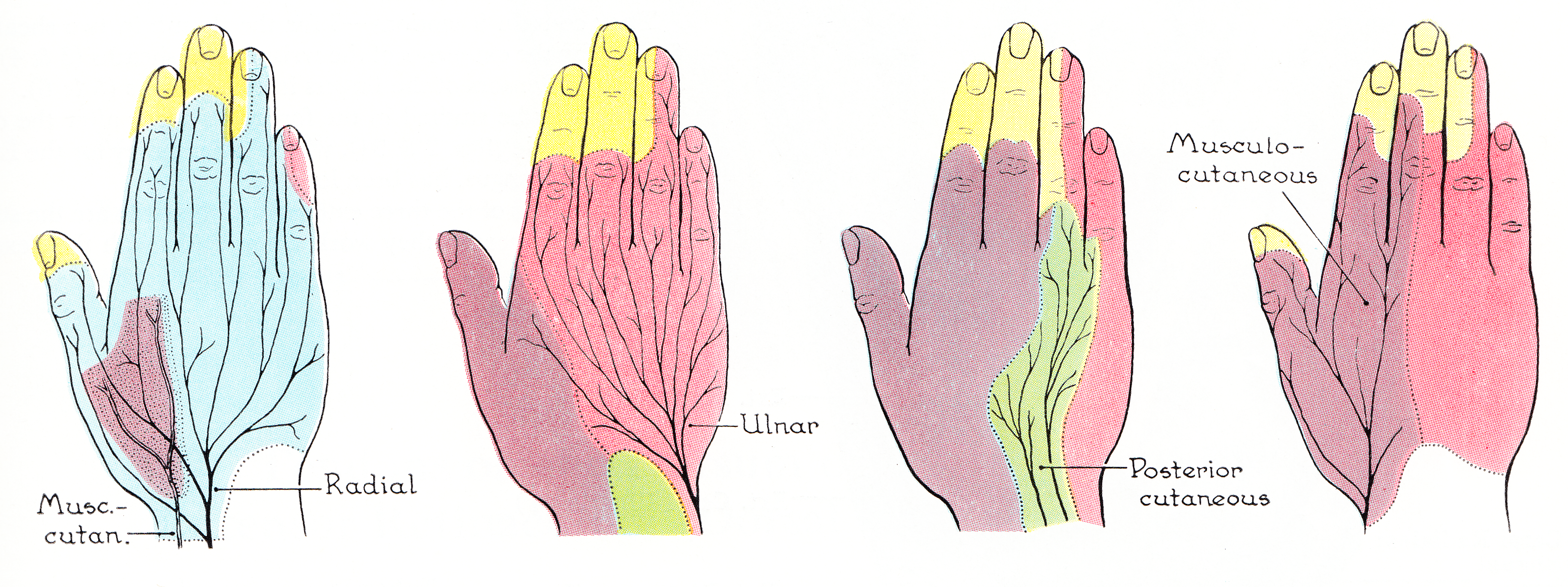
In the palm of the hand the median nerve is covered by the skin and the palmar aponeurosis, and rests on the tendons of the Flexor muscles. Immediately after emerging from under the transverse carpal ligament the median nerve becomes enlarged and flattened and splits into a smaller, lateral, and a larger, medial portion. The ''medial portion'' of the nerve divides into two Common palmar digital nerves (common volar digital nerves). * The first of these gives a twig to the second Lumbricalis and runs toward the cleft between the index finger, index and middle fingers, where it divides into two proper digital nerves for the adjoining sides of these digits; * the second runs toward the cleft between the middle finger, middle and ring fingers, and splits into two proper digital nerves for the adjoining sides of these digits; it communicates with a branch from the ulnar nerve and sometimes sends a twig to the third Lumbricalis. Additional images File:Gray811and813.PNG, Cutaneous nerve ... [...More Info...] [...Related Items...] OR: [Wikipedia] [Google] [Baidu] |
Median Nerve
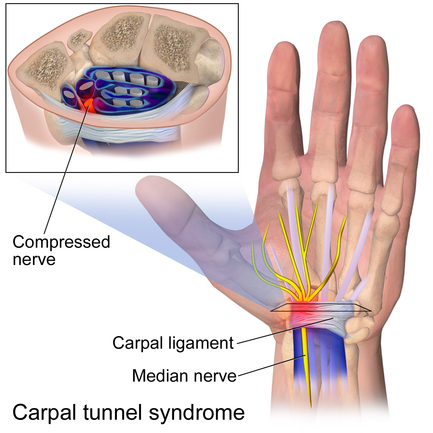
The median nerve is a nerve in humans and other animals in the upper limb. It is one of the five main nerves originating from the brachial plexus. The median nerve originates from the lateral and medial cords of the brachial plexus, and has contributions from ventral roots of C5-C7 (lateral cord) and C8 and T1 (medial cord). The median nerve is the only nerve that passes through the carpal tunnel. Carpal tunnel syndrome is the disability that results from the median nerve being pressed in the carpal tunnel. Structure The median nerve arises from the branches from lateral and medial cords of the brachial plexus, courses through the anterior part of arm, forearm, and hand, and terminates by supplying the muscles of the hand. Arm After receiving inputs from both the lateral and medial cords of the brachial plexus, the median nerve enters the arm from the axilla at the inferior margin of the teres major muscle. It then passes vertically down and courses lateral to the brachial ar ... [...More Info...] [...Related Items...] OR: [Wikipedia] [Google] [Baidu] |
Hand
A hand is a prehensile, multi-fingered appendage located at the end of the forearm or forelimb of primates such as humans, chimpanzees, monkeys, and lemurs. A few other vertebrates such as the koala (which has two opposable thumbs on each "hand" and fingerprints extremely similar to human fingerprints) are often described as having "hands" instead of paws on their front limbs. The raccoon is usually described as having "hands" though opposable thumbs are lacking. Some evolutionary anatomists use the term ''hand'' to refer to the appendage of digits on the forelimb more generally—for example, in the context of whether the three digits of the bird hand involved the same homologous loss of two digits as in the dinosaur hand. The human hand usually has five digits: four fingers plus one thumb; these are often referred to collectively as five fingers, however, whereby the thumb is included as one of the fingers. It has 27 bones, not including the sesamoid bone, the number o ... [...More Info...] [...Related Items...] OR: [Wikipedia] [Google] [Baidu] |
Palmar Aponeurosis
The palmar aponeurosis (palmar fascia) invests the muscles of the palm, and consists of central, lateral, and medial portions. Structure The central portion occupies the middle of the palm, is triangular in shape, and of great strength Its apex is continuous with the lower margin of the transverse carpal ligament, and receives the expanded tendon of the palmaris longus. Its base divides below into four slips, one for each finger. Each slip gives off superficial fibers to the skin of the palm and finger, those to the palm joining the skin at the furrow corresponding to the metacarpophalangeal articulations, and those to the fingers passing into the skin at the transverse fold at the bases of the fingers. The deeper part of each slip subdivides into two processes, which are inserted into the fibrous sheaths of the flexor tendons. From the sides of these processes offsets are attached to the transverse metacarpal ligament. By this arrangement short channels are formed on the front ... [...More Info...] [...Related Items...] OR: [Wikipedia] [Google] [Baidu] |
Transverse Carpal Ligament
The flexor retinaculum (transverse carpal ligament, or anterior annular ligament) is a fibrous band on the palmar side of the hand near the wrist. It arches over the carpal bones of the hands, covering them and forming the carpal tunnel. Structure The flexor retinaculum is a strong, fibrous band that covers the carpal bones on the palmar side of the hand near the wrist. It attaches to the bones near the radius (bone), radius and ulna. On the ulnar side, the flexor retinaculum attaches to the pisiform bone and the hamate, hook of the hamate bone. On the radial side, it attaches to the tubercle of the scaphoid bone, and to the medial part of the palmar surface and the ridge of the trapezium (bone), trapezium bone. The flexor retinaculum is continuous with the palmar carpal ligament, and deeper with the palmar aponeurosis. The ulnar artery and ulnar nerve, and the cutaneous branches of the median and ulnar nerves, pass on top of the flexor retinaculum. On the radial side of the retin ... [...More Info...] [...Related Items...] OR: [Wikipedia] [Google] [Baidu] |
Index Finger
The index finger (also referred to as forefinger, first finger, second finger, pointer finger, trigger finger, digitus secundus, digitus II, and many other terms) is the second digit of a human hand. It is located between the thumb and the middle finger. It is usually the most dextrous and sensitive digit of the hand, though not the longest. It is shorter than the middle finger, and may be shorter or longer than the ring finger (see digit ratio). Anatomy "Index finger" literally means "pointing finger", from the same Latin source as '' indicate;'' its anatomical names are "index finger" and "second digit". The index finger has three phalanges. It does not contain any muscles, but is controlled by muscles in the hand by attachments of tendons to the bones. Uses A lone index finger held vertically is often used to represent the number 1 (but finger counting differs across cultures), or when held up or moved side to side (finger-wagging), it can be an admonitory gesture. Wit ... [...More Info...] [...Related Items...] OR: [Wikipedia] [Google] [Baidu] |
Middle Finger
The middle finger, long finger, second finger, third finger, toll finger or tall man is the third digit of the human hand, located between the index finger and the ring finger. It is typically the longest digit. In anatomy, it is also called ''the third finger'', ''digitus medius'', ''digitus tertius'' or ''digitus III''. In Western countries, The finger, extending the middle finger (either by itself, or along with the index finger The index finger (also referred to as forefinger, first finger, second finger, pointer finger, trigger finger, digitus secundus, digitus II, and many other terms) is the second digit of a human hand. It is located between the thumb and the mid ... in the United Kingdom: see V sign) is an offensive and obscene gesture, widely recognized as a form of insult, due to its resemblance of an Erection, erect penis, It is known, colloquially, as "flipping the bird", "flipping (someone) off", or "giving (someone) the finger". The middle finger is ofte ... [...More Info...] [...Related Items...] OR: [Wikipedia] [Google] [Baidu] |
Ring Finger
The ring finger, third finger, fourth finger, leech finger, or annulary is the fourth digit of the human hand, located between the middle finger and the little finger. Sometimes the term ring finger only refers to the fourth digit of a left-hand, so named for its traditional association with wedding rings in many societies, although not all use this digit as the ring finger. Traditionally, a wedding ring was worn only by the bride or wife, but in recent times more men also wear a wedding ring. It is also the custom in some societies to wear an engagement ring on the ring finger. In anatomy, the ring finger is called ''digitus medicinalis'', ''the fourth digit'', ''digitus annularis'', ''digitus quartus'', or ''digitus IV''. In Latin, the word ''anulus'' means "ring", ''digitus'' means "digit", and ''quartus'' means "fourth". Etymology The origin of the selection of the fourth digit as the ring finger is not definitively known. According to László A. Magyar, the names of the ... [...More Info...] [...Related Items...] OR: [Wikipedia] [Google] [Baidu] |
Ulnar Nerve
In human anatomy, the ulnar nerve is a nerve that runs near the ulna bone. The ulnar collateral ligament of elbow joint is in relation with the ulnar nerve. The nerve is the largest in the human body unprotected by muscle or bone, so injury is common. This nerve is directly connected to the little finger, and the adjacent half of the ring finger, innervating the palmar aspect of these fingers, including both front and back of the tips, perhaps as far back as the fingernail beds. This nerve can cause an electric shock-like sensation by striking the medial epicondyle of the humerus posteriorly, or inferiorly with the elbow flexed. The ulnar nerve is trapped between the bone and the overlying skin at this point. This is commonly referred to as bumping one's "funny bone". This name is thought to be a pun, based on the sound resemblance between the name of the bone of the upper arm, the humerus, and the word "humorous". Alternatively, according to the Oxford English Dictionary, i ... [...More Info...] [...Related Items...] OR: [Wikipedia] [Google] [Baidu] |
Proper Palmar Digital Nerves Of Median Nerve
In the palm of the hand the median nerve is covered by the skin and the palmar aponeurosis, and rests on the tendons of the flexor muscles. Immediately after emerging from under the transverse carpal ligament the median nerve becomes enlarged and flattened and splits into a smaller, lateral, and a larger, medial portion. The ''lateral portion'' supplies a short, stout branch to certain of the muscles of the ball of the thumb, viz., the abductor pollicis brevis, the opponens pollicis, and the superficial head of the flexor brevis, and then divides into three proper palmar digital nerves of median nerve (proper volar digital nerves): * two of these supply the sides of the thumb, * while the third gives a twig to the first lumbricalis and is distributed to the radial side of the index finger. It also divides into two common palmar digital nerves: * The first of these divides into two proper digital nerves for the adjoining sides of the index and middle fingers; * the second common ... [...More Info...] [...Related Items...] OR: [Wikipedia] [Google] [Baidu] |


