|
Common Extensor Tendon
The common extensor tendon is a tendon that attaches to the lateral epicondyle of the humerus. Structure The common extensor tendon serves as the upper attachment (in part) for the superficial muscles that are located on the posterior aspect of the forearm: * Extensor carpi radialis brevis * Extensor digitorum * Extensor digiti minimi * Extensor carpi ulnaris The tendon of extensor carpi radialis brevis is usually the most major tendon to which the other tendons merge. Function The common extensor tendon is the major attachment point for extensor muscles of the forearm. This enables finger extension and aids in forearm supination. Clinical significance Lateral elbow pain can be caused by various pathologies of the common extensor tendon. Overuse injuries can lead to inflammation. Tennis elbow is a common issue with the common extensor tendon. See also * Common flexor tendon The common flexor tendon is a tendon that attaches to the medial epicondyle of the humerus (lowe ... [...More Info...] [...Related Items...] OR: [Wikipedia] [Google] [Baidu] |
Tendon
A tendon or sinew is a tough, high-tensile-strength band of dense fibrous connective tissue that connects muscle to bone. It is able to transmit the mechanical forces of muscle contraction to the skeletal system without sacrificing its ability to withstand significant amounts of tension. Tendons are similar to ligaments; both are made of collagen. Ligaments connect one bone to another, while tendons connect muscle to bone. Structure Histologically, tendons consist of dense regular connective tissue. The main cellular component of tendons are specialized fibroblasts called tendon cells (tenocytes). Tenocytes synthesize the extracellular matrix of tendons, abundant in densely packed collagen fibers. The collagen fibers are parallel to each other and organized into tendon fascicles. Individual fascicles are bound by the endotendineum, which is a delicate loose connective tissue containing thin collagen fibrils and elastic fibres. Groups of fascicles are bounded by the epitenon, ... [...More Info...] [...Related Items...] OR: [Wikipedia] [Google] [Baidu] |
Lateral Epicondyle Of The Humerus
The lateral epicondyle of the humerus is a large, tuberculated eminence, curved a little forward, and giving attachment to the radial collateral ligament of the elbow joint, and to a tendon common to the origin of the supinator and some of the extensor muscles. Specifically, these extensor muscles include the anconeus muscle, the supinator, extensor carpi radialis brevis, extensor digitorum, extensor digiti minimi, and extensor carpi ulnaris. In birds, where the arm is somewhat rotated compared to other tetrapods, it is termed dorsal epicondyle of the humerus. In comparative anatomy, the term ''ectepicondyle'' is sometimes used. A common injury associated with the lateral epicondyle of the humerus is lateral epicondylitis also known as tennis elbow. Repetitive overuse of the forearm, as seen in tennis or other sports, can result in inflammation of "the tendons that join the forearm muscles on the outside of the elbow. The forearm muscles and tendons become damaged from overus ... [...More Info...] [...Related Items...] OR: [Wikipedia] [Google] [Baidu] |
Humerus
The humerus (; ) is a long bone in the arm that runs from the shoulder to the elbow. It connects the scapula and the two bones of the lower arm, the radius and ulna, and consists of three sections. The humeral upper extremity consists of a rounded head, a narrow neck, and two short processes (tubercles, sometimes called tuberosities). The body is cylindrical in its upper portion, and more prismatic below. The lower extremity consists of 2 epicondyles, 2 processes (trochlea & capitulum), and 3 fossae (radial fossa, coronoid fossa, and olecranon fossa). As well as its true anatomical neck, the constriction below the greater and lesser tubercles of the humerus is referred to as its surgical neck due to its tendency to fracture, thus often becoming the focus of surgeons. Etymology The word "humerus" is derived from la, humerus, umerus meaning upper arm, shoulder, and is linguistically related to Gothic ''ams'' shoulder and Greek ''ōmos''. Structure Upper extremity The upper or pr ... [...More Info...] [...Related Items...] OR: [Wikipedia] [Google] [Baidu] |
Forearm
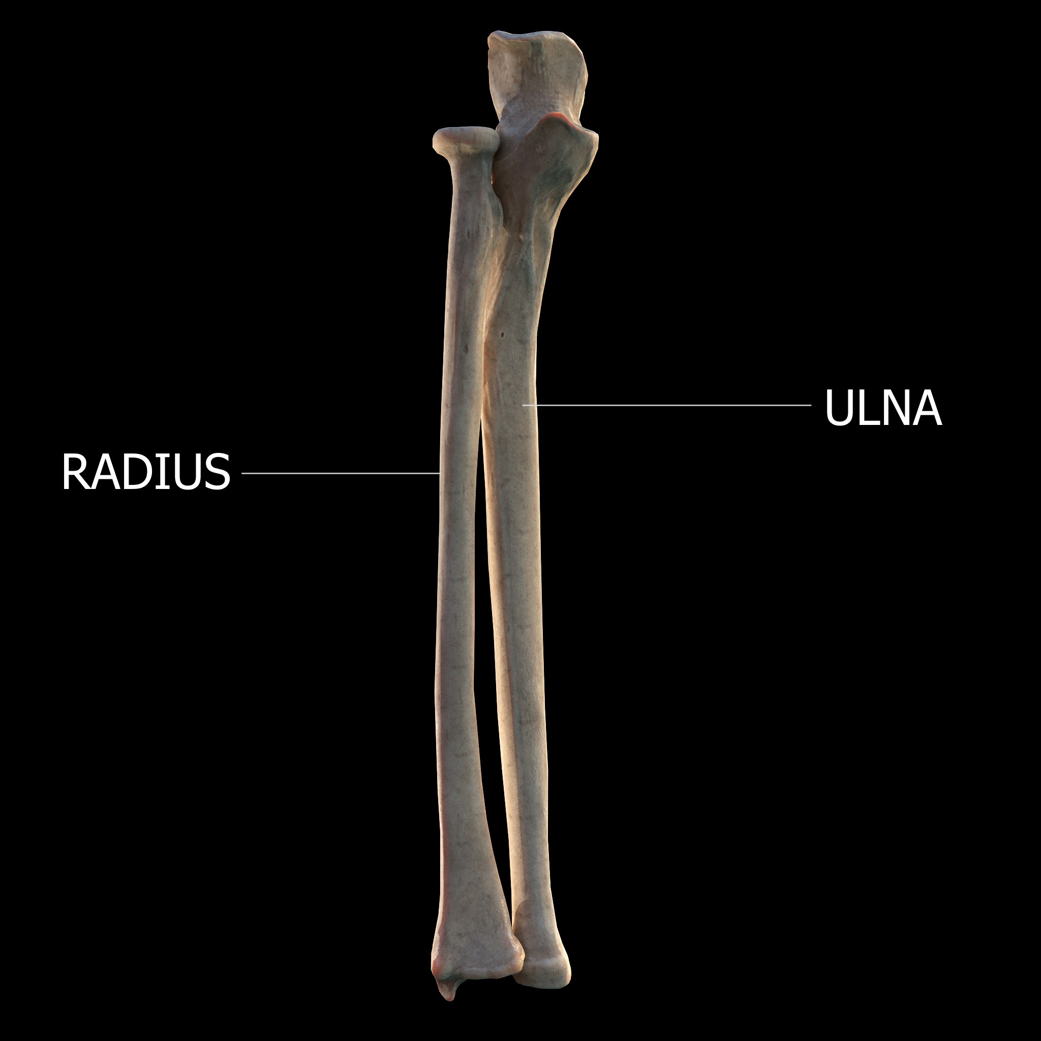
The forearm is the region of the upper limb between the elbow and the wrist. The term forearm is used in anatomy to distinguish it from the arm, a word which is most often used to describe the entire appendage of the upper limb, but which in anatomy, technically, means only the region of the upper arm, whereas the lower "arm" is called the forearm. It is homologous to the region of the leg that lies between the knee and the ankle joints, the crus. The forearm contains two long bones, the radius and the ulna, forming the two radioulnar joints. The interosseous membrane connects these bones. Ultimately, the forearm is covered by skin, the anterior surface usually being less hairy than the posterior surface. The forearm contains many muscles, including the flexors and extensors of the wrist, flexors and extensors of the digits, a flexor of the elbow (brachioradialis), and pronators and supinators that turn the hand to face down or upwards, respectively. In cross-section, the for ... [...More Info...] [...Related Items...] OR: [Wikipedia] [Google] [Baidu] |
Extensor Carpi Radialis Brevis
In human anatomy, extensor carpi radialis brevis is a muscle in the forearm that acts to extend and abduct the wrist. It is shorter and thicker than its namesake extensor carpi radialis longus which can be found above the proximal end of the extensor carpi radialis brevis. Origin and insertion It arises from the lateral epicondyle of the humerus, by the common extensor tendon; from the radial collateral ligament of the elbow-joint; from a strong aponeurosis which covers its surface; and from the intermuscular septa between it and the adjacent muscles.''Gray's Anatomy'' 1918, see infobox The fibres end approximately at the middle of the forearm in the form of a flat tendon, which is closely connected with that of the extensor carpi radialis longus, and accompanies it to the wrist; it passes beneath the abductor pollicis longus and extensor pollicis brevis, beneath the extensor retinaculum, and inserts into the lateral dorsal surface of the base of the third metacarpal bone, with ... [...More Info...] [...Related Items...] OR: [Wikipedia] [Google] [Baidu] |
Extensor Digitorum
The extensor digitorum muscle (also known as extensor digitorum communis) is a muscle of the posterior forearm present in humans and other animals. It extends the medial four digits of the hand. Extensor digitorum is innervated by the posterior interosseous nerve, which is a branch of the radial nerve. Structure The extensor digitorum muscle arises from the lateral epicondyle of the humerus, by the common tendon; from the intermuscular septa between it and the adjacent muscles, and from the antebrachial fascia. It divides below into four tendons, which pass, together with that of the extensor indicis proprius, through a separate compartment of the dorsal carpal ligament, within a mucous sheath. The tendons then diverge on the back of the hand, and are inserted into the middle and distal phalanges of the fingers in the following manner.''Gray's anatomy'' (1918), see infobox Opposite the metacarpophalangeal articulation each tendon is bound by fasciculi to the collateral ligaments an ... [...More Info...] [...Related Items...] OR: [Wikipedia] [Google] [Baidu] |
Extensor Digiti Minimi
The extensor digiti minimi (extensor digiti quinti proprius) is a slender muscle of the forearm, placed on the ulnar side of the extensor digitorum communis, with which it is generally connected. It arises from the common extensor tendon by a thin tendinous slip and frequently from the intermuscular septa between it and the adjacent muscles. Its tendon passes through a compartment of the extensor retinaculum, posterior to distal radio-ulnar joint, then divides into two as it crosses the dorsum of the hand, and finally joins the extensor digitorum tendon. All three tendons attach to the dorsal digital expansion of the fifth digit (little finger). There may be a slip of tendon to the fourth digit. Variations An additional fibrous slip from the lateral epicondyle; the tendon of insertion may not divide or may send a slip to the ring finger. Absence of muscle rare; fusion of the belly with the extensor digitorum communis The extensor digitorum muscle (also known as extensor dig ... [...More Info...] [...Related Items...] OR: [Wikipedia] [Google] [Baidu] |
Extensor Carpi Ulnaris
In human anatomy, the extensor carpi ulnaris is a skeletal muscle located on the ulnar side of the forearm. The extensor carpi ulnaris acts to extend and adduct at the carpus/wrist from anatomical position. Being an extensor muscle, extensor carpi ulnaris is located on the posterior side of the forearm. Origin and insertion It originates from the lateral epicondyle of the humerus and the posterior border of the ulna, and crosses the forearm to the ulnar (medial) side to insert at the base of the 5th metacarpal. Action The extensor carpi ulnaris extends the wrist, but when acting alone inclines the hand toward the ulnar side; by its continued action it extends the elbow-joint. The muscle is a minor extensor of the carpus in carnivores, but has become a flexor in ungulates. In this case it would be described as ''ulnaris lateralis''. Innervation Despite its name, the extensor carpi ulnaris is innervated by the posterior interosseous nerve (C7 and C8), the continuation of the dee ... [...More Info...] [...Related Items...] OR: [Wikipedia] [Google] [Baidu] |
Disease
A disease is a particular abnormal condition that negatively affects the structure or function of all or part of an organism, and that is not immediately due to any external injury. Diseases are often known to be medical conditions that are associated with specific signs and symptoms. A disease may be caused by external factors such as pathogens or by internal dysfunctions. For example, internal dysfunctions of the immune system can produce a variety of different diseases, including various forms of immunodeficiency, hypersensitivity, allergies and autoimmune disorders. In humans, ''disease'' is often used more broadly to refer to any condition that causes pain, dysfunction, distress, social problems, or death to the person affected, or similar problems for those in contact with the person. In this broader sense, it sometimes includes injuries, disabilities, disorders, syndromes, infections, isolated symptoms, deviant behaviors, and atypical variations of structur ... [...More Info...] [...Related Items...] OR: [Wikipedia] [Google] [Baidu] |
Repetitive Strain Injury
A repetitive strain injury (RSI) is an injury to part of the musculoskeletal or nervous system caused by repetitive use, vibrations, compression or long periods in a fixed position. Other common names include repetitive stress disorders, cumulative trauma disorders (CTDs), and overuse syndrome. Signs and symptoms Some examples of symptoms experienced by patients with RSI are aching, pulsing pain, tingling and extremity weakness, initially presenting with intermittent discomfort and then with a higher degree of frequency. Definition Repetitive strain injury (RSI) and associative trauma orders are umbrella terms used to refer to several discrete conditions that can be associated with repetitive tasks, forceful exertions, vibrations, mechanical compression, sustained or awkward positions, or repetitive eccentric contractions. The exact terminology is controversial, but the terms now used by the United States Department of Labor and the National Institute of Occupational Safe ... [...More Info...] [...Related Items...] OR: [Wikipedia] [Google] [Baidu] |
Inflammation
Inflammation (from la, wikt:en:inflammatio#Latin, inflammatio) is part of the complex biological response of body tissues to harmful stimuli, such as pathogens, damaged cells, or Irritation, irritants, and is a protective response involving immune cells, blood vessels, and molecular mediators. The function of inflammation is to eliminate the initial cause of cell injury, clear out necrotic cells and tissues damaged from the original insult and the inflammatory process, and initiate tissue repair. The five cardinal signs are heat, pain, redness, swelling, and Functio laesa, loss of function (Latin ''calor'', ''dolor'', ''rubor'', ''tumor'', and ''functio laesa''). Inflammation is a generic response, and therefore it is considered as a mechanism of innate immune system, innate immunity, as compared to adaptive immune system, adaptive immunity, which is specific for each pathogen. Too little inflammation could lead to progressive tissue destruction by the harmful stimulus (e.g. b ... [...More Info...] [...Related Items...] OR: [Wikipedia] [Google] [Baidu] |
Tennis Elbow
Tennis elbow, also known as lateral epicondylitis or enthesopathy of the extensor carpi radialis origin, is a condition in which the outer part of the elbow becomes painful and tender. The pain may also extend into the back of the forearm. Onset of symptoms is generally gradual although they can seem sudden and be misinterpreted as an injury. Golfer's elbow is a similar condition that affects the inside of the elbow. Enthesopathies are idiopathic, meaning science has not yet determined the cause. Enthesopathies are most common in middle age (ages 35 to 60). It is often stated that the condition is caused by excessive use of the muscles of the back of the forearm, but this is not supported by experimental evidence and is a common misinterpretation or unhelpful thought about symptoms. It may be associated with work or sports, classically racquet sports, but most people with the condition are not exposed to these activities. The diagnosis is based on the symptoms and examination ... [...More Info...] [...Related Items...] OR: [Wikipedia] [Google] [Baidu] |


